AP Images for Neck/Face
1/30
Earn XP
Description and Tags
do flashcards
Name | Mastery | Learn | Test | Matching | Spaced | Call with Kai |
|---|
No analytics yet
Send a link to your students to track their progress
31 Terms
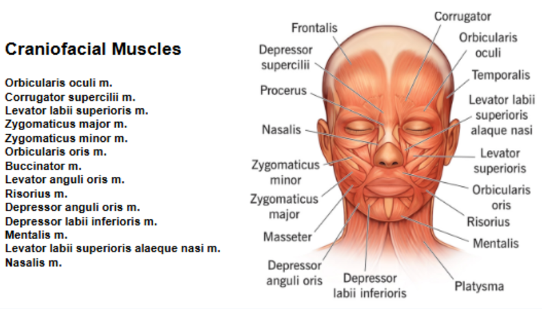
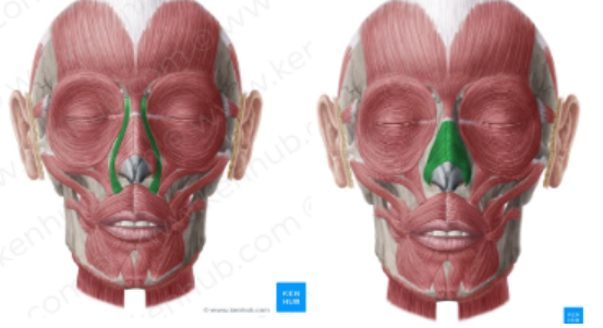
green =
red =
Orbicularis oris = green
Buccinator = red
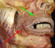
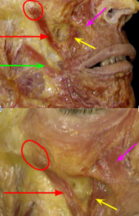
Red circle =
Red arrow:
What is the point:
Green arrow:
Pink:
yellow:
Red circle = zygomatic arch
Red arrow: zygomaticus major
What is the point: moteolus (central spot where a bunch of muscles for facial expression/elevators and depressors meet)
Green arrow: risorius
Pink: levator labii superioris
yellow: levator anguli oris
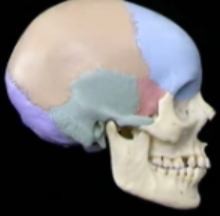
green, red, purple, orange, blue, and zygomatic is where
Temporal (green),
sphenoid (red),
occipital (purple),
parietal (orange),
frontal (blue),
zygomatic (in front of red)
what is this
foramen magnum

what is the “crest” and surrounding
crest: crysta galli
around: cribiform plate
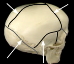
what are these suture lines
Sagittal (left side)
Coronal (Front)
Squamous (right side on image)
Lamboid (back)
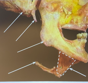
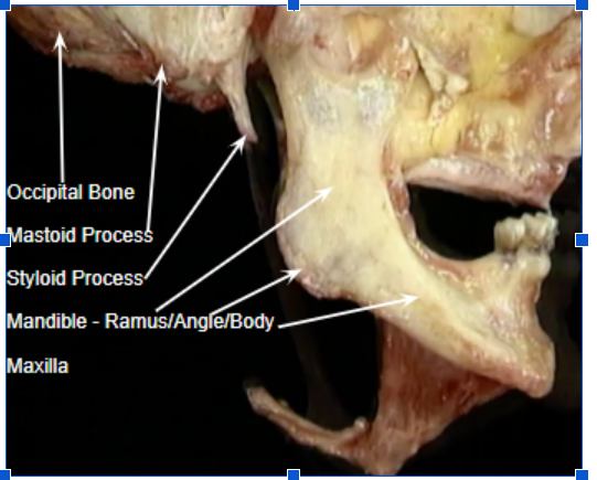
Thing sticking out: hyoid bone
Jaw bone: mandible
A little in front of the mandible (no arrow): body
Back of mandible: angle
“Top stem” of mandible: ramus
Pointy thing that is high: styloid process
Behind the styloid process: mastoid process
Muscle: mylohyoid muscle
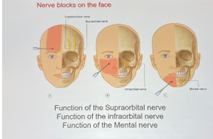
what are these holes
L → R
supraorbital foramen
infraorbital foramen
mental foramen
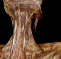
what muscle
semispinalis muscle
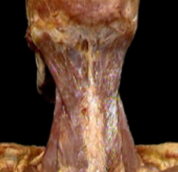
what muscle
splenius muscle
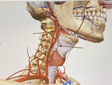
left arrow
right arrow
What is the tiny branch artery
left arrow: internal carotid
right arrow: superior thyroid branch (external carotid)
tiny branch a: thyroid a → thyrocervical trunk
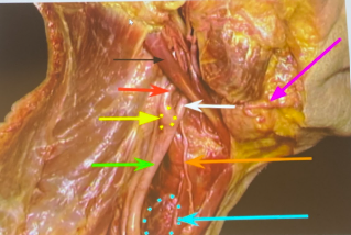
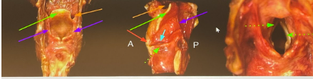
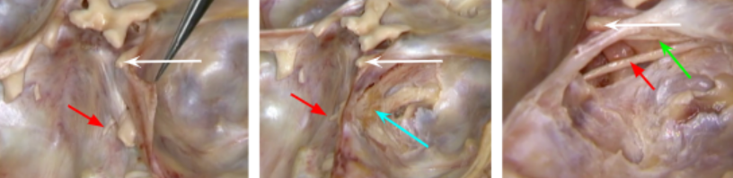
Muscle far behind the black, red, yellow, green arrows:
Muscle right behind black/red/yellow/green arrows: s
Green arrow:
Yellow circle:
Red arrow:
White arrow:
Orange arrow:
Blue circle:
Muscle next to pink arrow:
Black arrow:
Muscle to the right of the digastric muscle that joins:
Together if you keep going to the right =
Pink arrow:
Bulb under the facial artery:
Muscle far behind the black, red, yellow, green arrows: splenius (wide and wraps around)
Muscle right behind black/red/yellow/green arrows: scalene muscles
Green arrow: common carotid arter
Yellow circle: carotid sinus
Red arrow: internal carotid artery
White arrow: external carotid
Orange arrow: superior thyroid a
Blue circle: thyroid gland
Muscle next to pink arrow: masseter muscle
Black arrow: posterior belly of digastric muscle
Muscle to the right of the digastric muscle that joins: stylohyoid muscle
Together if you keep going to the right = Anterior belly of digastric muscles
Pink arrow: facial artery
Bulb under the facial artery: submandibular gland
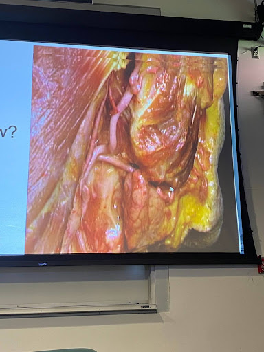
Middle upper muscle:
Bulbous looking thing:
Artery between them:
Middle upper muscle: masster muscle (if you contract masster muscle, you feel facial artery)
Bulbous looking thing: submandibular gland
Artery between them: facial artery
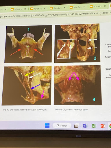
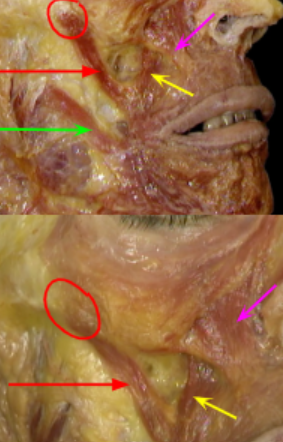
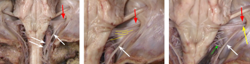
Red arrow:
Yellow:
Pink:
Bottom bone:
White arrow:
Red arrow: myohyoid (deeper) [image is posterior]
Yellow: posterior belly digastric
Pink: anterior belly
Bottom bone: hyoid
White arrow: styloid process
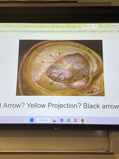
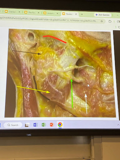
Black: t
The hole near the black arrow will eventually lead to: foramen magnum, but before that, under that layer =
Yellow:
Red:
Black: tentorium cerebelli
The hole near the black arrow will eventually lead to: foramen magnum, but before that, under that layer = cerebellum
Yellow: crysta galli
Red: falx cerebri
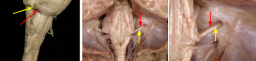
Red =
Yellow =
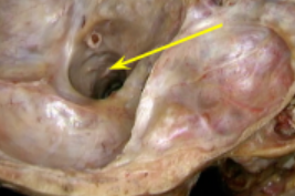
Exit point:
Red = facial nerve
Yellow = vestibulocochlear nerve
Exit point: internal acoustic meatus
exit point around the tip of the 2 white arrows (the sapce):
Below the hole:
Other white arrow:
Exit point/hole:
yellow arrow:
the nerves through the hole:
red:
exit point around the tip of the 2 white arrows (the space): foramen magnum
Remember: above is brainstem (you can see the medulla “triangle” and the pons is above that)
Below the hole = cervical spinal cord
Other white arrow: accessory nerve (coming off near spinal cord from cervical region)
Exit point: jugular foramen
Which means the 3 nerves (yellow) near the hole: accessory, glossopharyngeal, vagus nerve
Yellow arrow: vagus nerve
red? = glossopharyngeal?
White circle:
Red arrow:
Orange:
Black arrow:
pink:
White circle: where parotid gland is (superficial), if tucked in would be deep
Red arrow: sternocleidomastoid
Orange: anterior digastric muscle
Black arrow: buccinator muscle
pink: masseter
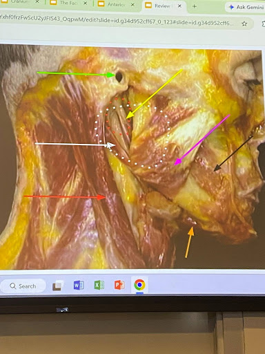
black:
Yellow:
Nerves comes through:
Green arrow:
White arrow:
Yellow:
Artery on top of masseter:
Red:
A little in front of that muscle =
black: parotid gland
Nerves comes through: facial nerve
Green arrow: parotid duct (piercing the buccinator muscle)
White arrow: buccinator muscle
Yellow: masseter muscle
Artery on top of masseter: facial artery
Red: zygomatic arch
A little in front of that muscle = zygomaticus major (has to have involvement of zygomatic arch)
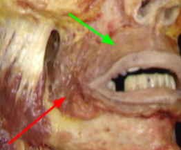
Red circle =
Red arrow:
What is the central point of muscles:
Green arrow:
Pink:
yellow:
Red circle = zygomatic arch
Red arrow: zygomaticus major
What is the point: modiolus (central spot where a bunch of muscles for facial expression/elevators and depressors meet)
Green arrow: risorius
Pink: levator labii superioris
yellow: (prolly) levator anguli oris
green =
red =
Orbicularis oris = green
Buccinator = red
left
right
2 parts
left: Levator labii superioris alaeque nasi m
right: nasalis muscle (transverse and alar)
Green dotted:
Green =
Orange:
Green dotted: true vocal cords
False is the membrane before
Green = epiglottis
Orange: vallecula
White:
Green:
Red:
Blue:
White: Oculomotor
Green: trochlear
Red: abducens
Blue: trigeminal ganglion
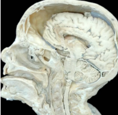
Space =
Line down =
Right above =
Space = foramen magnum
Line down = spinal cord
Right above = medulla
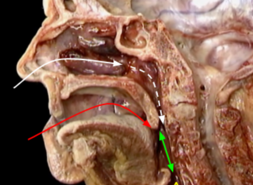
white dotted:
White:
Red:
Green:
Yellow:
white dotted: nasopharynx
White: nasal cavity
Red: oral cavity
Green: oropharynx
Yellow: hypopharynx
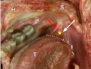
Red:
White:
Yellow star:
Red: palatoglossal arch
White: palatopharyngeal arch
Yellow star: palatine tonsils
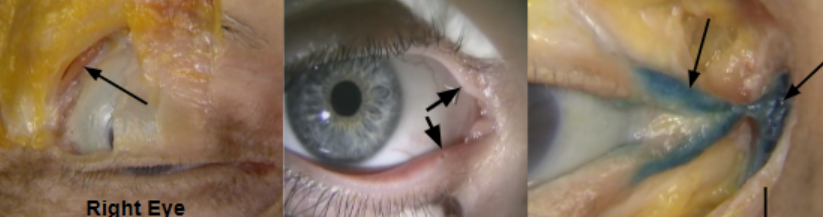
Left:
middle arrows
Last image arrows (left and right)
Left: lacrimal gland
Punctum (middle images)
Last image: canaliculus (left) and orbicularis oculi muscle (right)
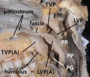
tensor palati
levator palati
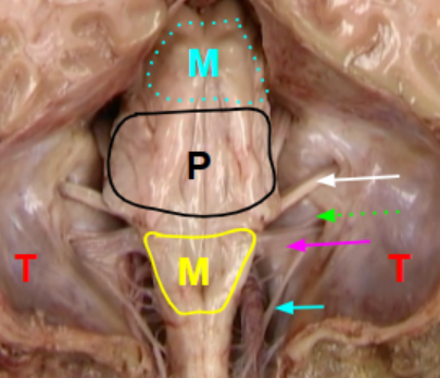
Blue:
Pink:
Blue dotted:
White:
green:
Blue: accessory nerve
Pink: vagus nerve
White: vestibulocochlear nerve
green: glossopharyngeal