BSCI201 Lab practical exam 3 study guide
1/218
There's no tags or description
Looks like no tags are added yet.
Name | Mastery | Learn | Test | Matching | Spaced | Call with Kai |
|---|
No analytics yet
Send a link to your students to track their progress
219 Terms
What is the difference between a nerve and a tract?
tracts are inside the Central Nervous System (CNS - brain and spinal cord), while nerves are in the Peripheral Nervous System (PNS)
What are the three structural classifications of neurons?
Unipolar / pseudounipolar, bipolar, and multipolar
Where would you find unipolar neurons?
Unipolar: In the CNS
Where would you find bipolar neurons?
Part of the receptor apparatus of the eye, ear, and olfactory mucosa
Where would you find multipolar neurons?
In the brain and spinal cord
Label the structures on the presynaptic cell
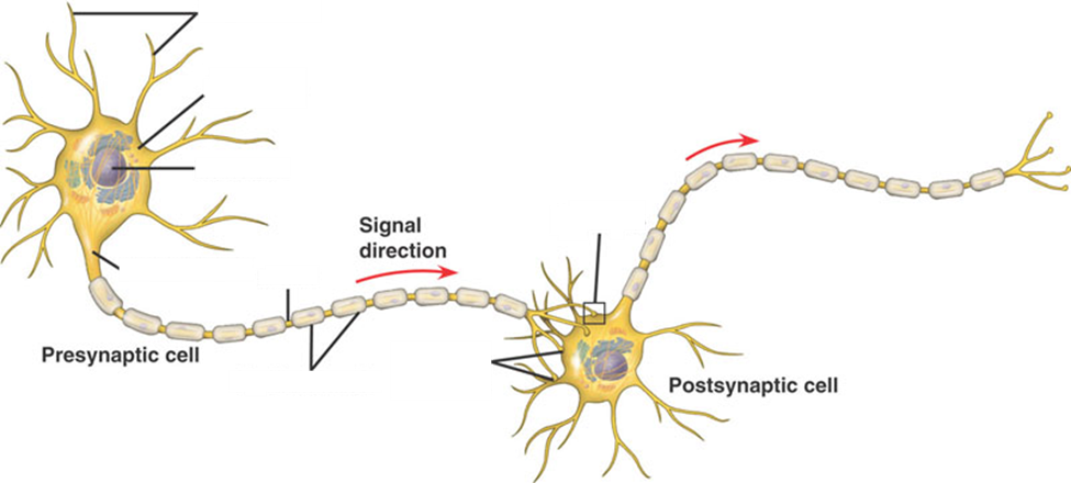
Dendrites
Cell body
Nucleus
Axon hillock
Axon
Schwann cell
Terminal branches
Axon terminals
What is the difference between afferent and efferent neurons?
Afferent= Sensory. Unipolar. Carry impulses FROM internal organs (viscera), the skin, skeletal muscles, joints, or special sensory organs.
Efferent= Motor. Multipolar. Carry impulses TO viscera and/or body muscles and glands from CNS.
Label the parts of the nerve in the figure below.
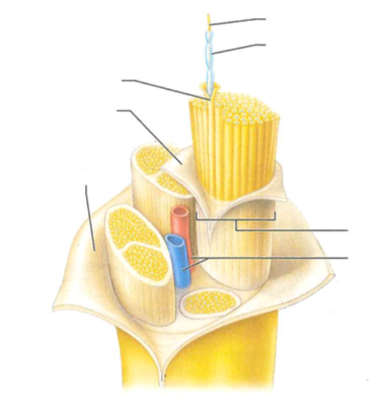
Axon
Myelin sheath
Endoneurium
Perineurium
Epineurium
Fascicle
Blood vessels
What are the four major regions of the brain?
Cerebrum, diencephalon, brain stem, and cerebellum
Label the lobes, structures, sulci, and gyri visible on the brain in the figure below.
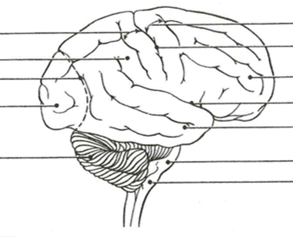
Postcentral gyrus
Parietal lobe
Parieto-occipital sulcus
Occipital lobe
Cerebellum
Precentral gyrus
Central sulcus
Frontal lobe
Lateral sulcus
Temporal lobe
Pons
Medulla
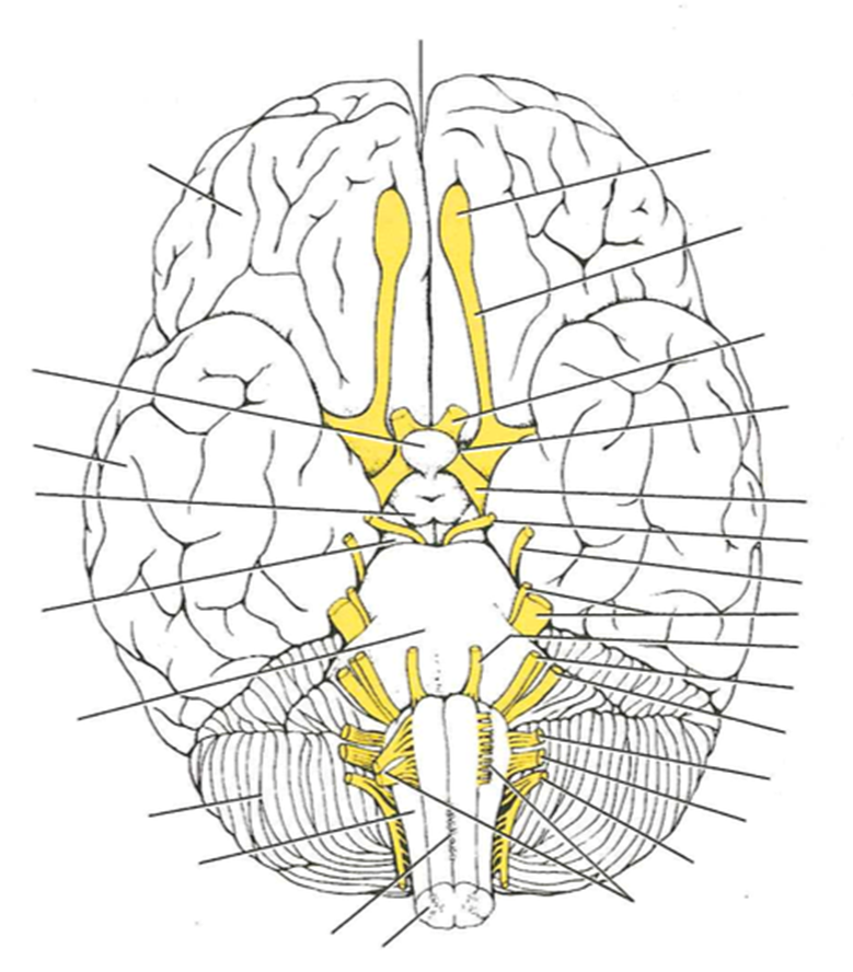
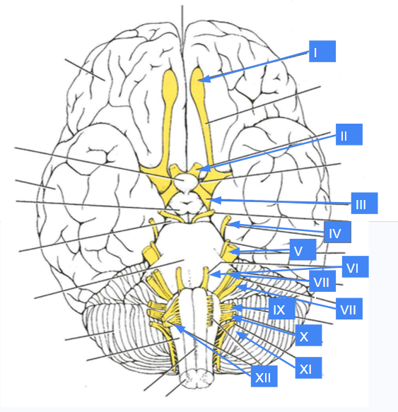
Label the cranial nerves and brain structures on the figure below.
What are the four structures that provide protection to the brain?
Cranium: Bony helmet composed of 8 cranial bones.
Meninges: Three connective tissue membranes surrounding the brain
Cerebrospinal fluid (CSF): Fluid cushion in the subarachnoid space and ventricles.
Blood-brain barrier: Selective barrier that prevents harmful substances harmful substances in blood from cross into the brain.
Name the three meninges and write them in order from outermost to innermost.
(outermost): Dura mater
Arachnoid mater
(innermost) Pia mater
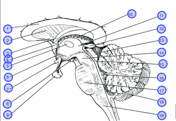
Label the parts of the brain stem and diencephalon on the figure below.
Corpus callosum
Septum pellucidum
Fornix
Anterior commissure
Interthalamic adhesion
Hypothalamus
Optic chiasma
Mammillary bodies
Pituitary gland
Choroid plexus
Thalamus
Pineal gland
Corpora quadrigemina
cerebral peduncle
Cerebral aqueduct
fourth ventricle
Pons
Cerebellum
Medulla oblongata
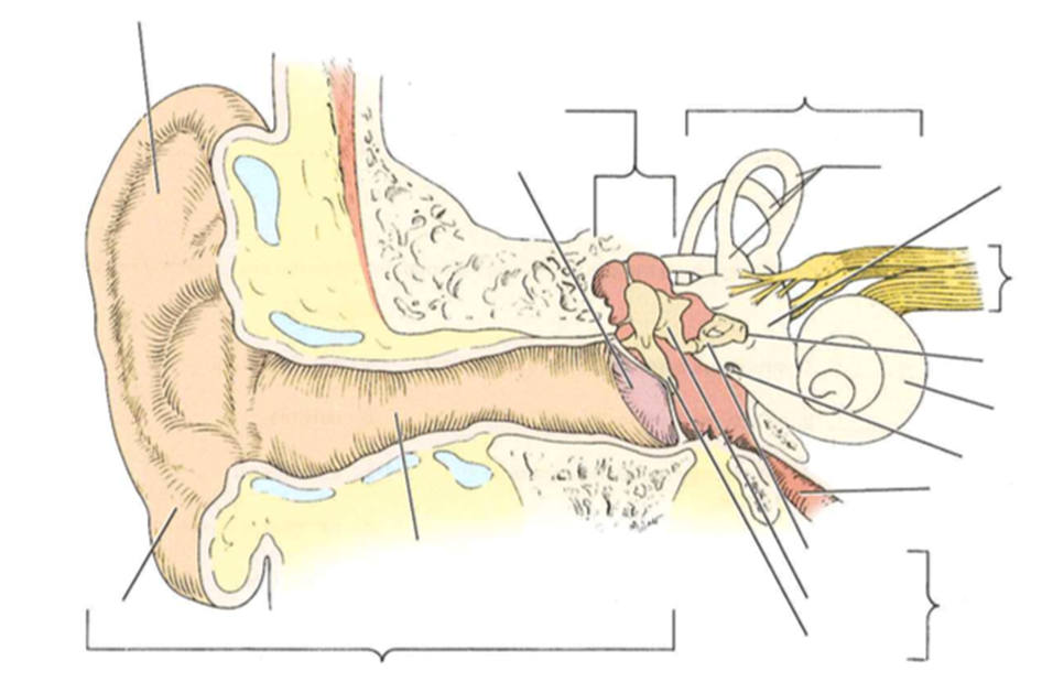
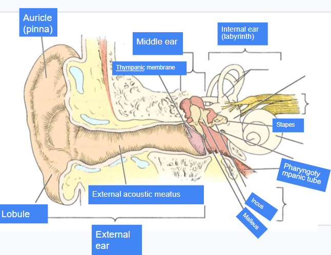
Label the structures of the ear in the figure below.
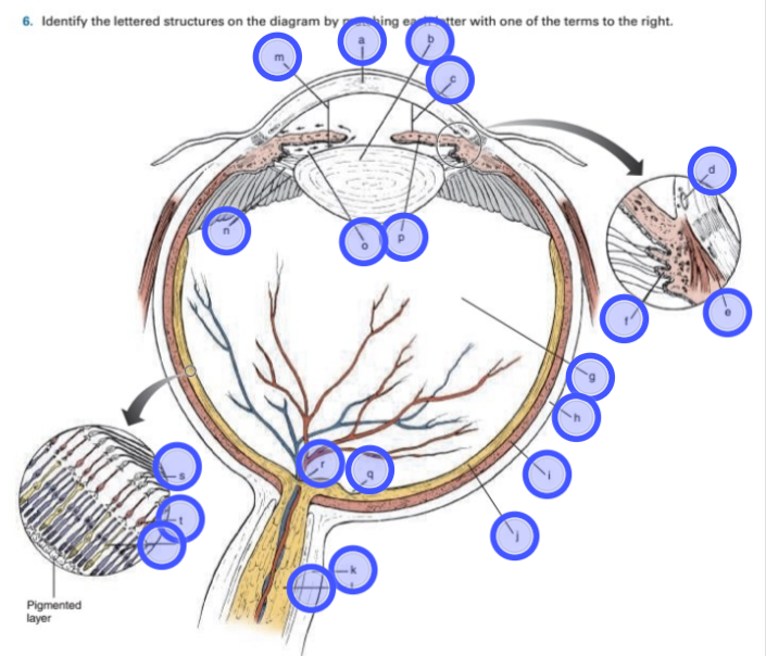
Label the structures of the eye in the figure below.
a. Cornea: Anterior transparent part of the fibrous tunic.
b. Lens: Important light bending structure of the eye; shape can be modified.
c. Anterior chamber: between cornea and iris
d. Scleral venous sinus: Drains the aqueous humor from the eye.
e. Ciliary muscle: Smooth muscle portion of the ciliary body. Contracts to assist in near vision.
f. Ciliary process: Produces aqueous humor.
g. Vitreous body (humor): Substance occupying the posterior segment of the eyeball.
h. Sclera: Composed of tough, white, opaque, fibrous connective tissue.
i. Choroid: Forms most of the pigmented vascular tunic.
j. Retina: Layer containing the rods and cones.
k. Dura mater: Outermost layer of the meninges.
l. Optic nerve: Carries neural impulses from the eye to the brain.
m. Anterior segment: Makes up the front one-third of the eyeball.
o. Posterior chamber: Space between the back of the front of the iris and the front of the vitreous chamber; filled with aqueous humor.
p. Iris: Contains muscle that controls size of pupil.
q. Fovea centralis: Tiny pit in the macula lutea; contains only cones.
r. Optic disc: Blind spot.
s. Ganglion cells: In the retina, the specialized neurons that connect to the bipolar cells; the bundled axons of the ganglion cells form the optic nerve.
t. Bipolar cells: In the retina, the specialized neurons that connect the rods and cones with the ganglion cells.
u. Photoreceptors: Respond to light rods and cones.
What is the function of the tapetum lucidum in the cow eye?
Specialized surface that reflects the light within the eye to enhance low-light vision (seeing in the dark)
Match the eye muscle to the appropriate action:
Inferior oblique: Elevates eye and turns it laterally.
Lateral rectus= Moves eye laterally.
Inferior rectus= Depresses eye and turn it medially.
Superior rectus= Elevates eye and turns it medially.
Superior oblique= Depresses eye and turns it laterally.
Medial rectus= Moves eye medially.
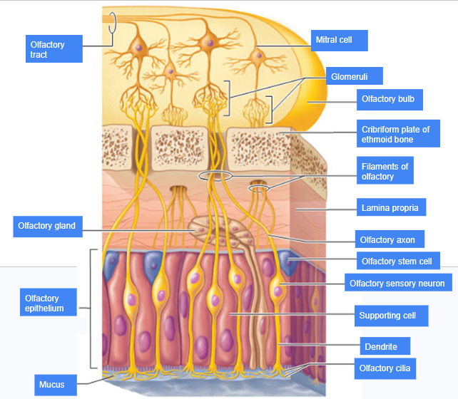
What is pictured in the figure in the figure below?
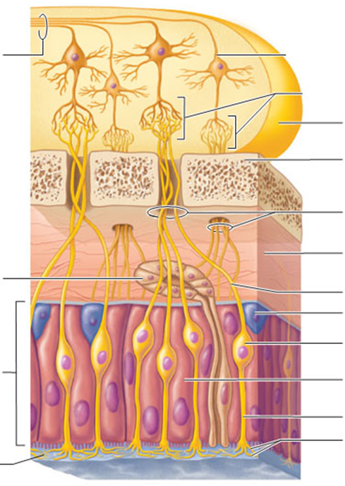
Olfactory epithelium
Label the cell types and structures in the figure below

Name all eight glands of the endocrine glands; where they are located, and one hormone that is released by each
Pituitary gland, pineal gland, thyroid gland, parathyroid gland, thymus, adrenal gland
What diagnosis was given to Ellie, the patient in the case study used in class?
What endocrine gland was affected by this Ellie’s condition?
What symptoms did Ellie exhibit?
What hormones are produced by the thyroid?
What interaction do these hormones have with TSH?
What is the difference between a hyperactive and a hypoactive thyroid?
What condition would result if the thyroid was destroyed by radiation or removed by surgery?
PNS
CNS
Motor neuron
Association/interneuron
Unipolar neuron
Bipolar neuron
Multiplar neuron
Cell body of neuron
Dendrite of neuron
Axon of neuron
Axon hillock of neuron
Axon terminal of neuron
Myelin sheath of neuron
Neurofibrils of neuron
Chromatophilic substance of neuron
Myeline sheath gap of neuron
Astrocytes (Neuroglia and their functions)
Microglia (Neuroglia and their functions)
Ependymal cell (Neuroglia and their functions)
Oligodendrocyte (Neuroglia and their functions)
Schwann cell (Neuroglia and their functions)
Satellite cell (Neuroglia and their functions)
Cerebrum
Longitudinal fissure
Corpus collosum
Septum pellucidum
Central sulcus
Frontal lobe
Temporal lobe
Insula lobe
Occipital lobe
Parietal lobe
Central sulcus
Parieto-occipital sulcus
Precentral gyrus
Postcentral gyrus
Lateral sulcus
Cerebral cortex
White matter
Basal nuclei
Diencephalon
Thalamus
Hypothalamus
Epithalamus
Pineal gland
Brain stem
Pons
Midbrain
Corpora quadrigemina
distinguish superior and inferior
Cerebral aqueduct
C
Cerebral peduncles
Medulla oblgonta
Cerebellum
Superior colliculi of corpora quadrigemina
Inferior colliculi of corpora quadrigemina
Cerebral hemisphere (Sagittal view)
Corpus collosum (Sagittal view)
Fornix (Sagittal view)
Intermediate mass of thalamus / thalamus (Sagittal view)
Optic chiasma (Sagittal view)
Cerebellum (Sagittal view)
Pineal gland (Sagittal view)
Arbor vitae (Sagittal view)
Corpora quadrigemina / midbrain (Sagittal view)
Pons (Sagittal view)
Medulla oblongata (Sagittal view)
Nuclei
Cell body clusters and location
Ganglia
Cell body clusters and location
Nerve
Fascicle
E
Epineurium
Perineurium