Lab 11: EENT (models)
1/86
There's no tags or description
Looks like no tags are added yet.
Name | Mastery | Learn | Test | Matching | Spaced | Call with Kai |
|---|
No analytics yet
Send a link to your students to track their progress
87 Terms
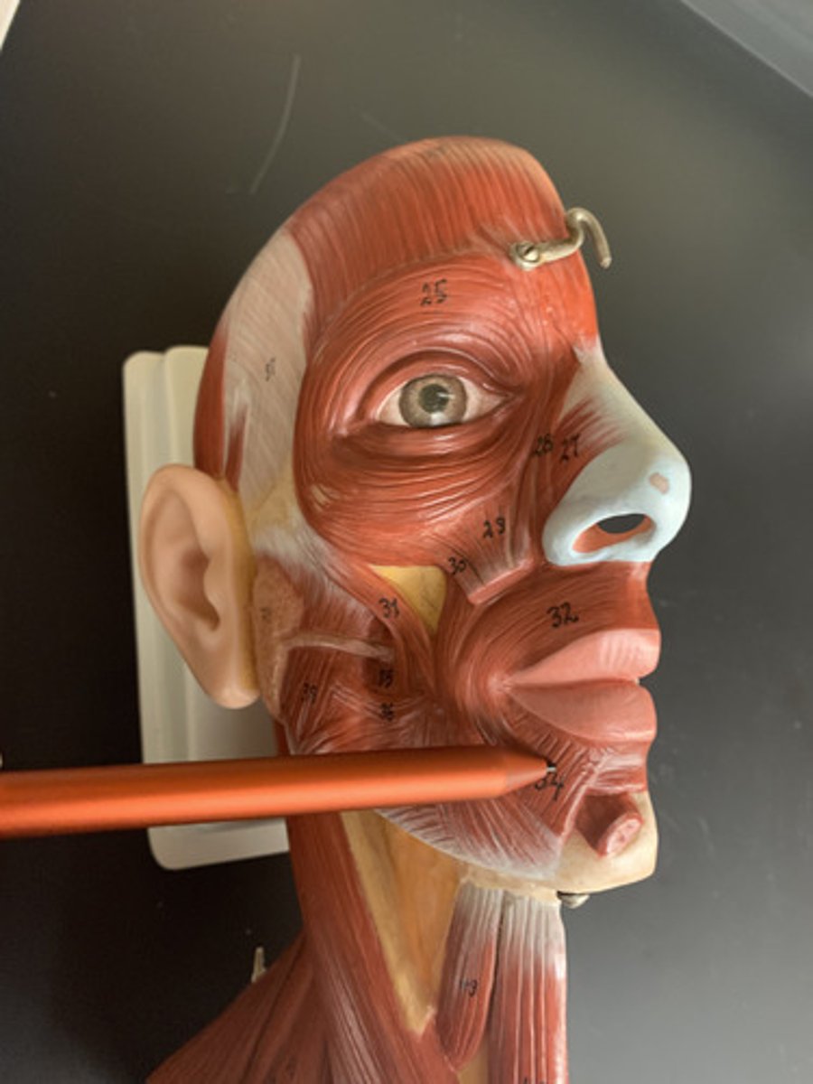
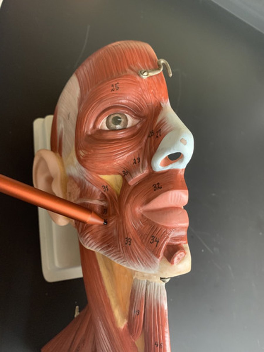
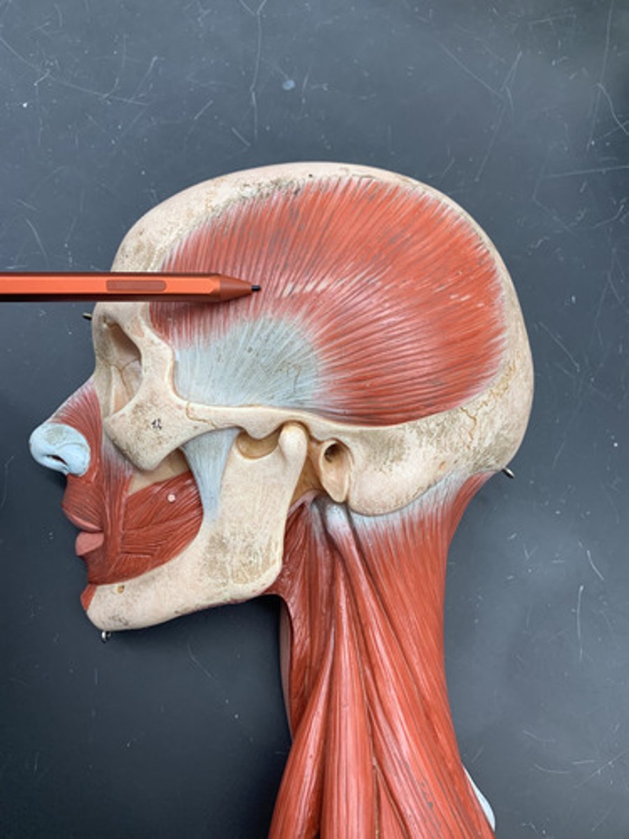
Frontalis
O: Cranial aponeurosis
I: Skin of eyebrows
A: Raises eyebrows
Inn: Facial n. (CN VII)
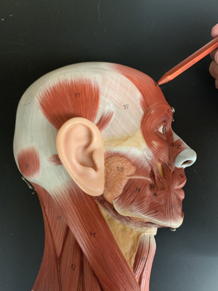
Orbicularis oculi
Origin: Medial margin of orbit
Insertion: Skin around eyelids
Action: Closes eye
Inn: Facial nerve (CN VII)
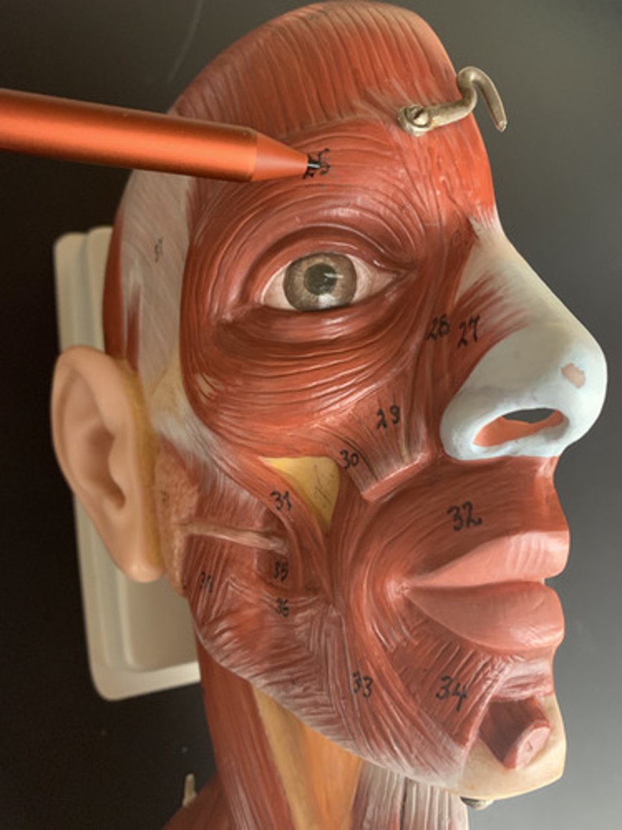
Nasalis
Origin: superior maxilla
Insertion: nasal cartilage
Action: dilates/contracts nostrils
Inn: Facial nerve (CN VII)
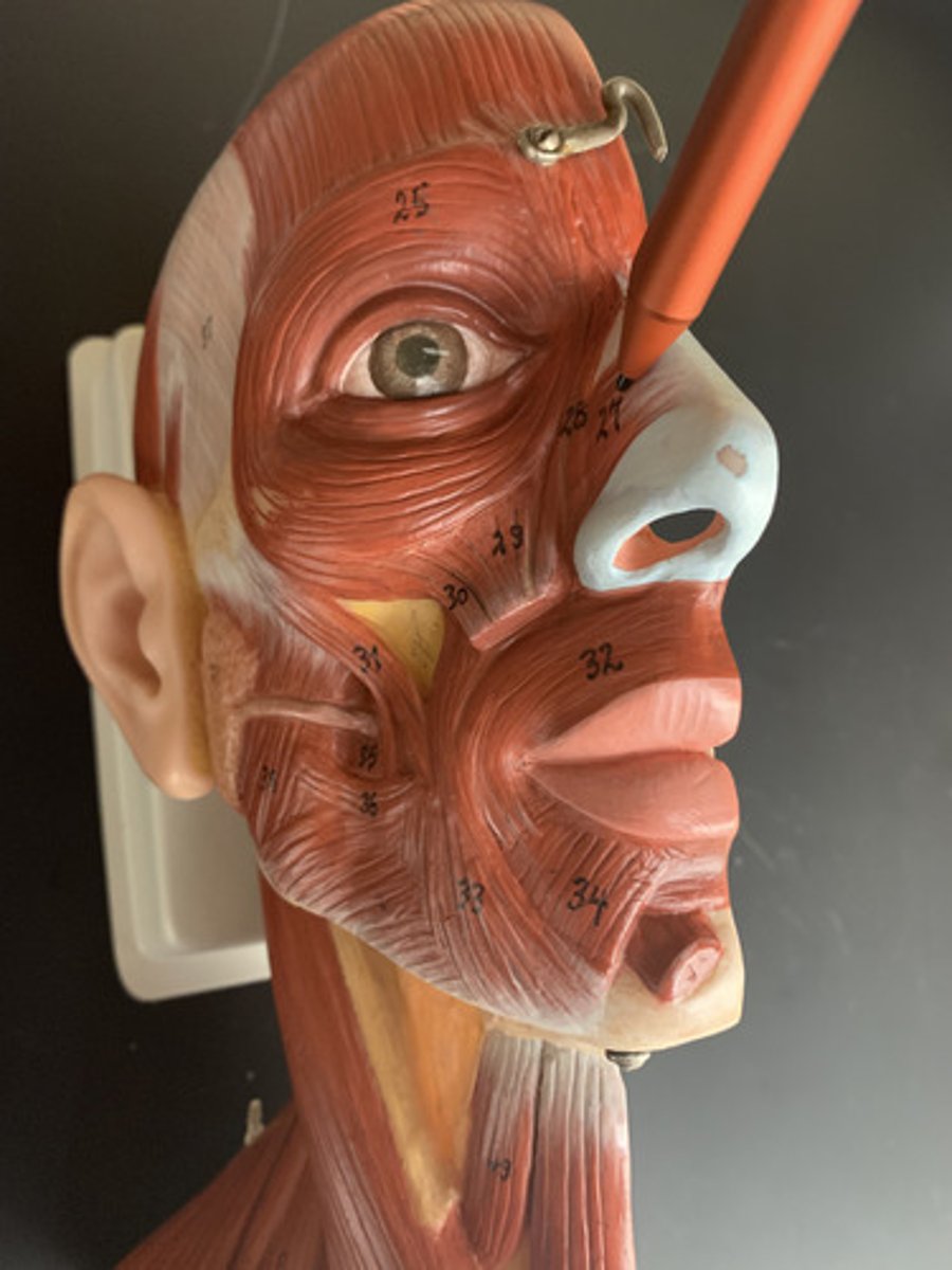
Levator labii superiorus
Origin: Infraorbital margin of maxilla
Insertion: Upper lip
Action: Elevates upper lip
Inn: Facial nerve (CN VII)
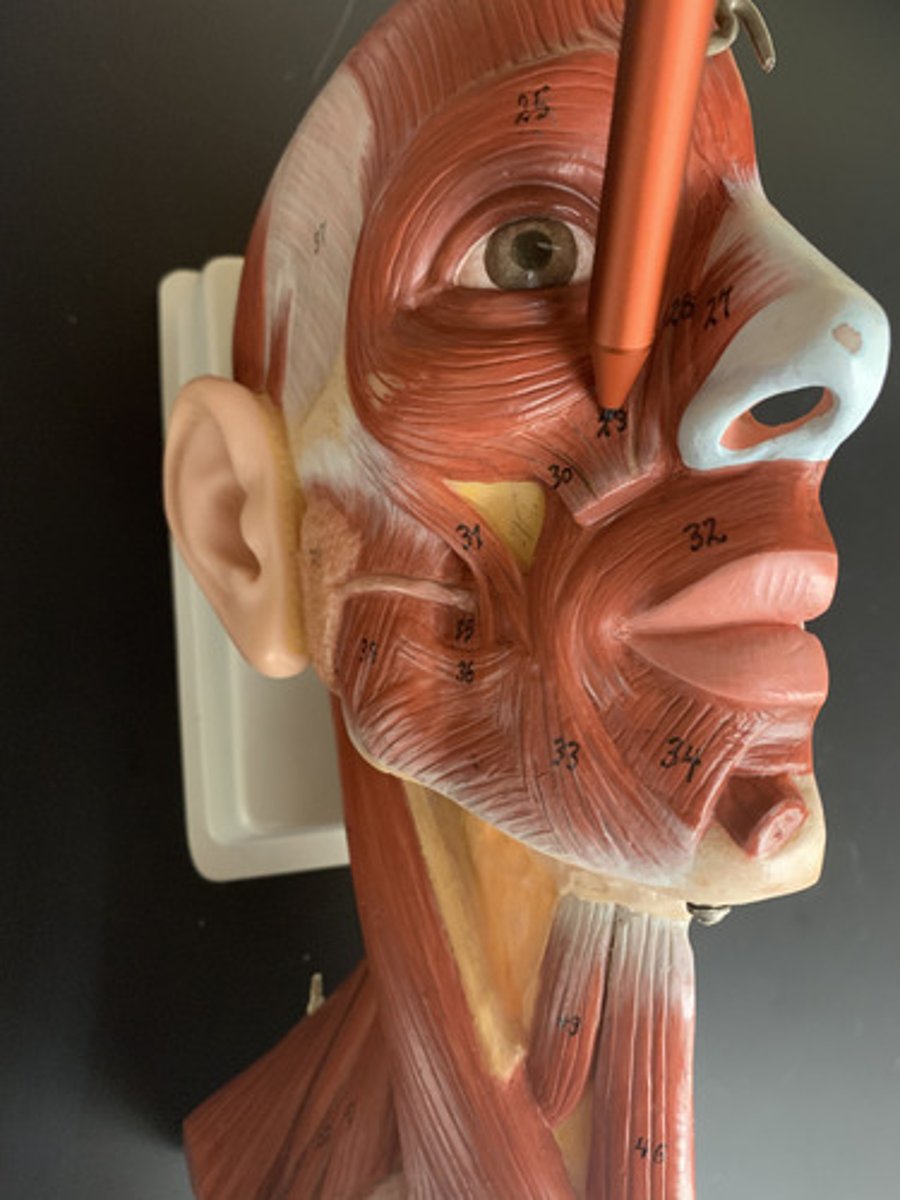
Levator anguli oris
Origin: Canine fossa of maxilla
Insertion: Corners of upper and lower lips
Action: Draws corner of mouth up and medially
Inn: Facial nerve (CN VII)
Zygomaticus major
O: zygomatic bone
I: angle of mouth
A: raises angle of mouth
Inn: Facial nerve (CN VII)
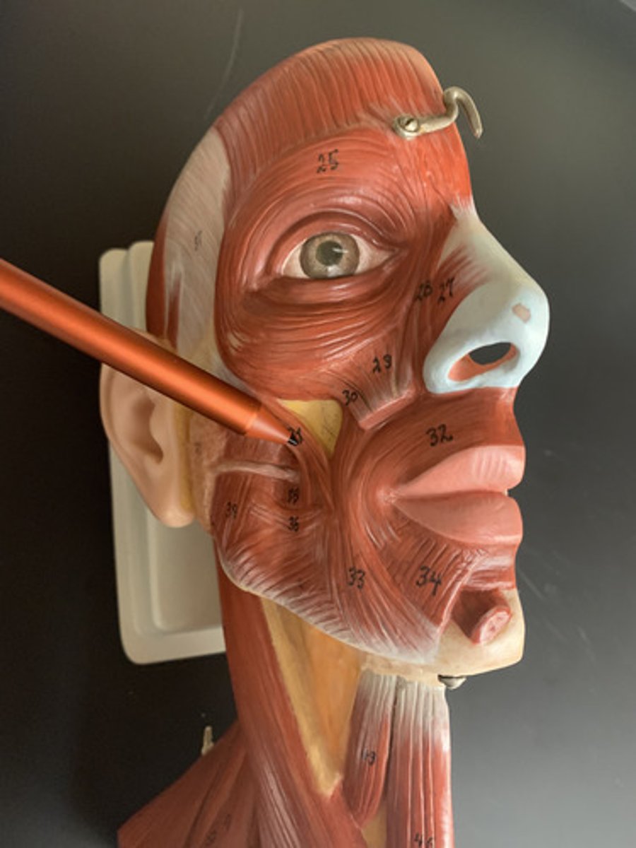
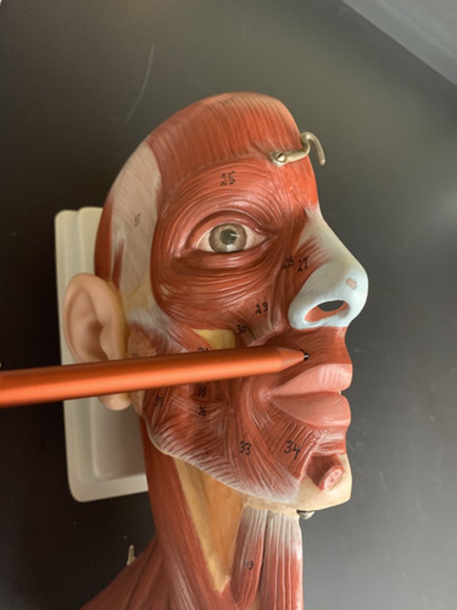
Orbicularis oris
O: Maxilla and mandible
I: Lips
A: Compresses, purses lips
Inn: Facial nerve (CN VII)
Depressor anguli oris
O: mandible
I: angle of mouth
A: depresses angle of mouth
Inn: Facial nerve (CN VII)
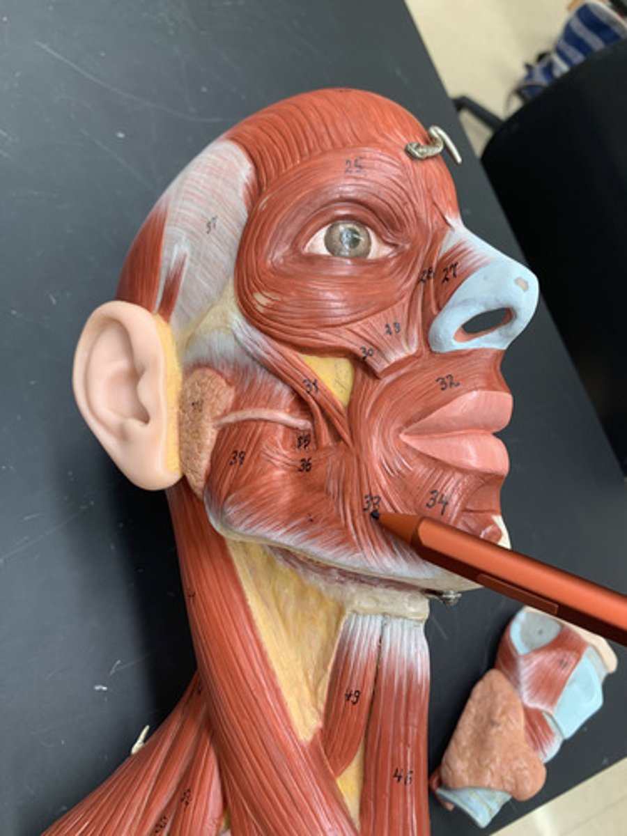
Depressor labii inferioris
O: mandible
I: skin of lower lip
A: depresses lower lip
Inn: Facial nerve (CN VII)
Mentalis
O: body of the mandible
I: skin of the chin
A: elevates, protracts, and depresses lower lip
Inn: Facial nerve (CN VII)
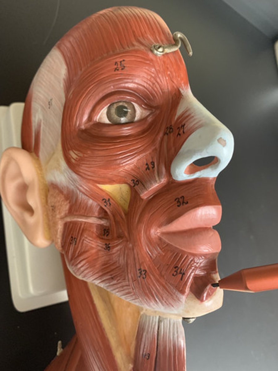
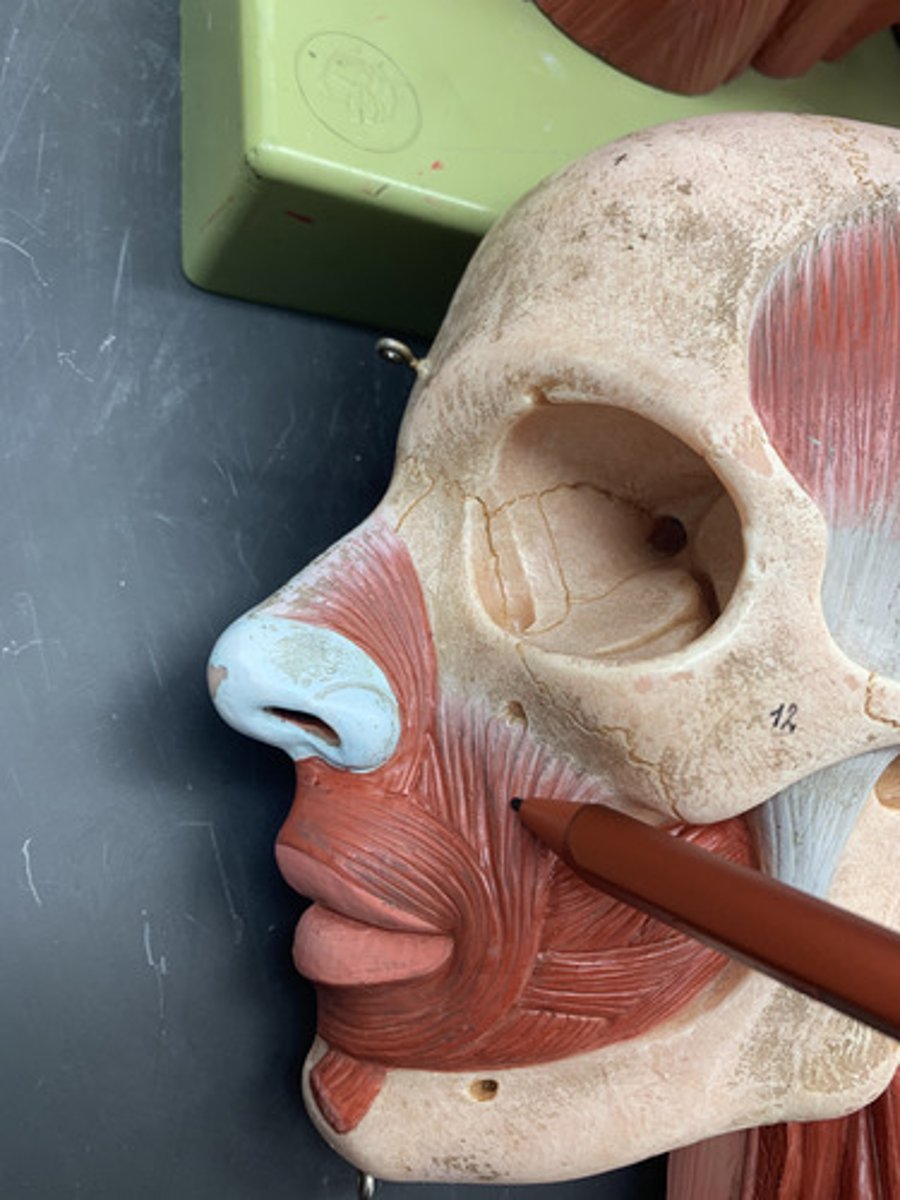
Buccinator
O: aveolar processes of the maxillae and mandible
I: corner of the mouth
A: compress cheeks
Inn: Facial nerve (CN VII)
Risorius
O: fascia associated with masseter
I: angle of mouth
A: draws angle of mouth to side
Inn: Facial nerve (CN VII)
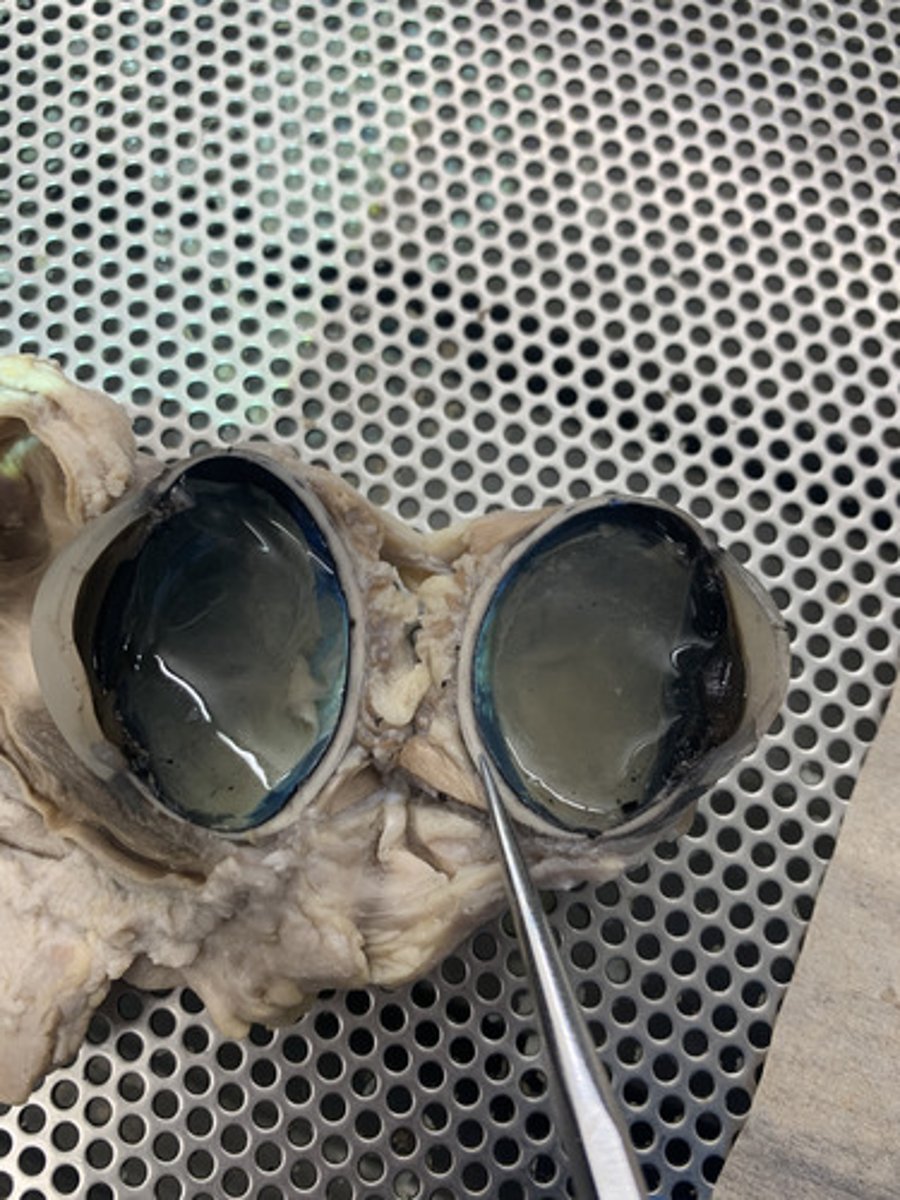
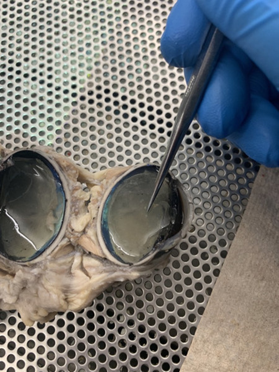
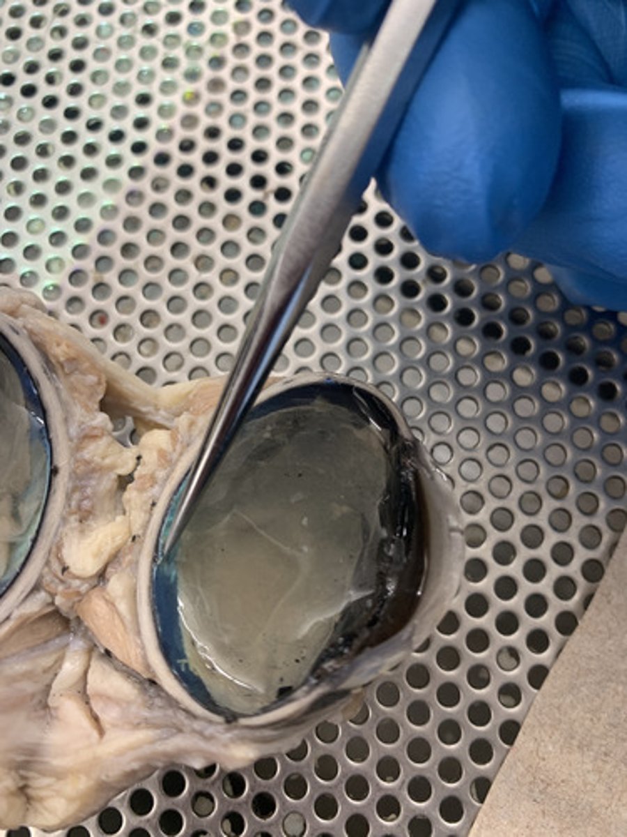
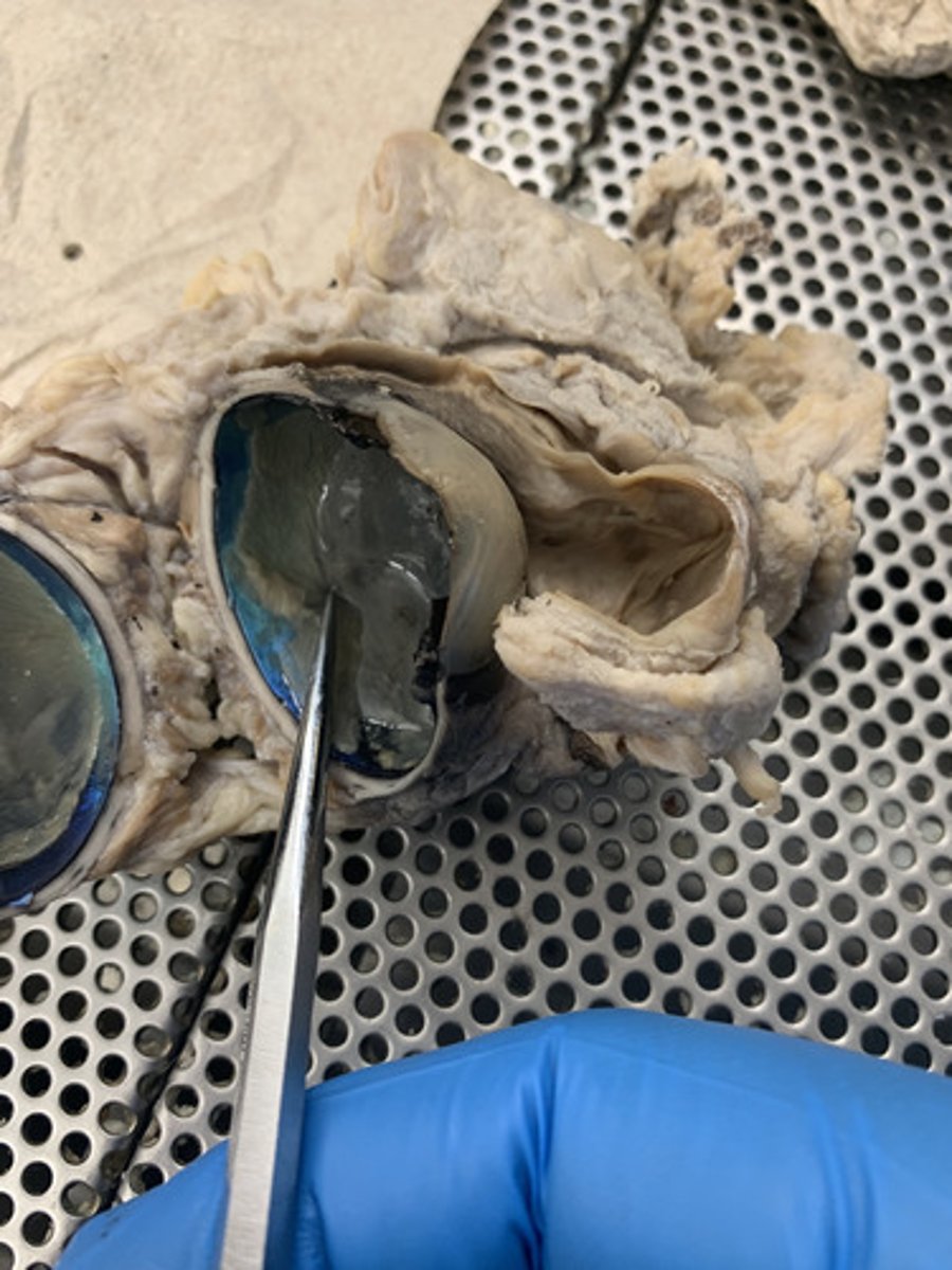
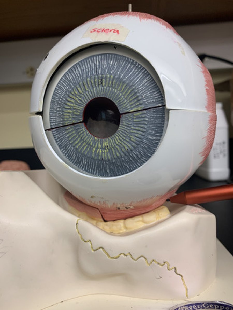
Cornea
The clear tissue of the fibrous tunic that covers the front of the eye
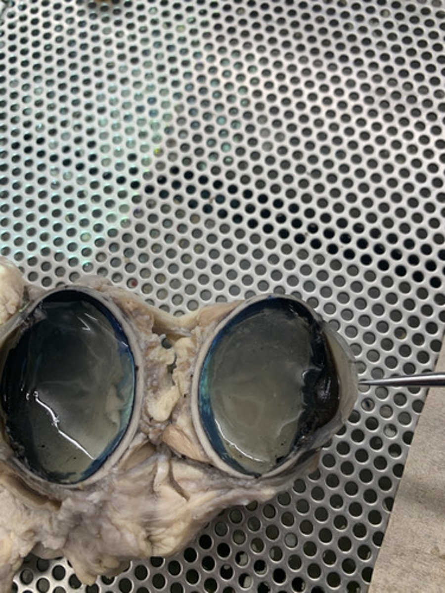
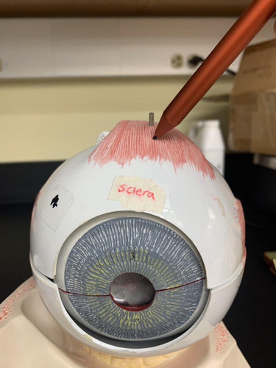
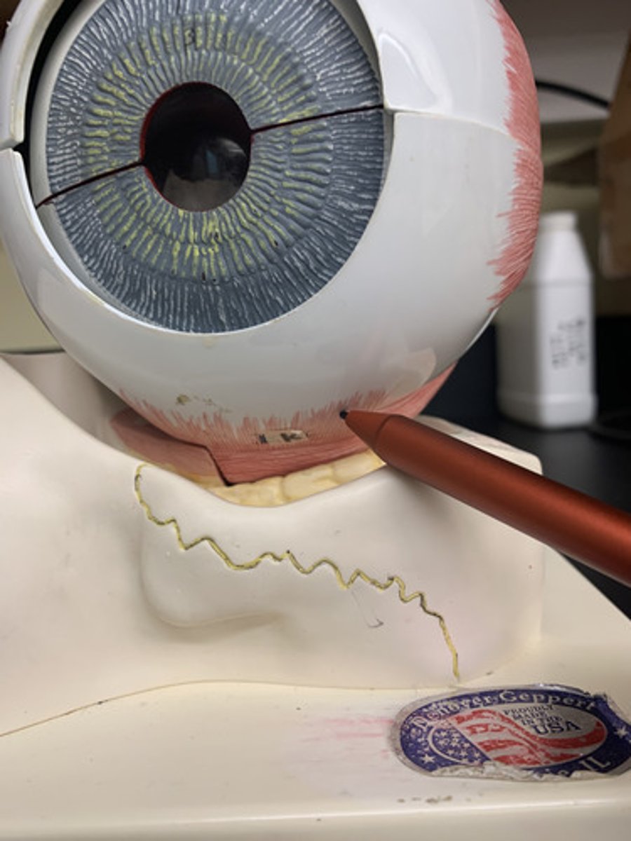
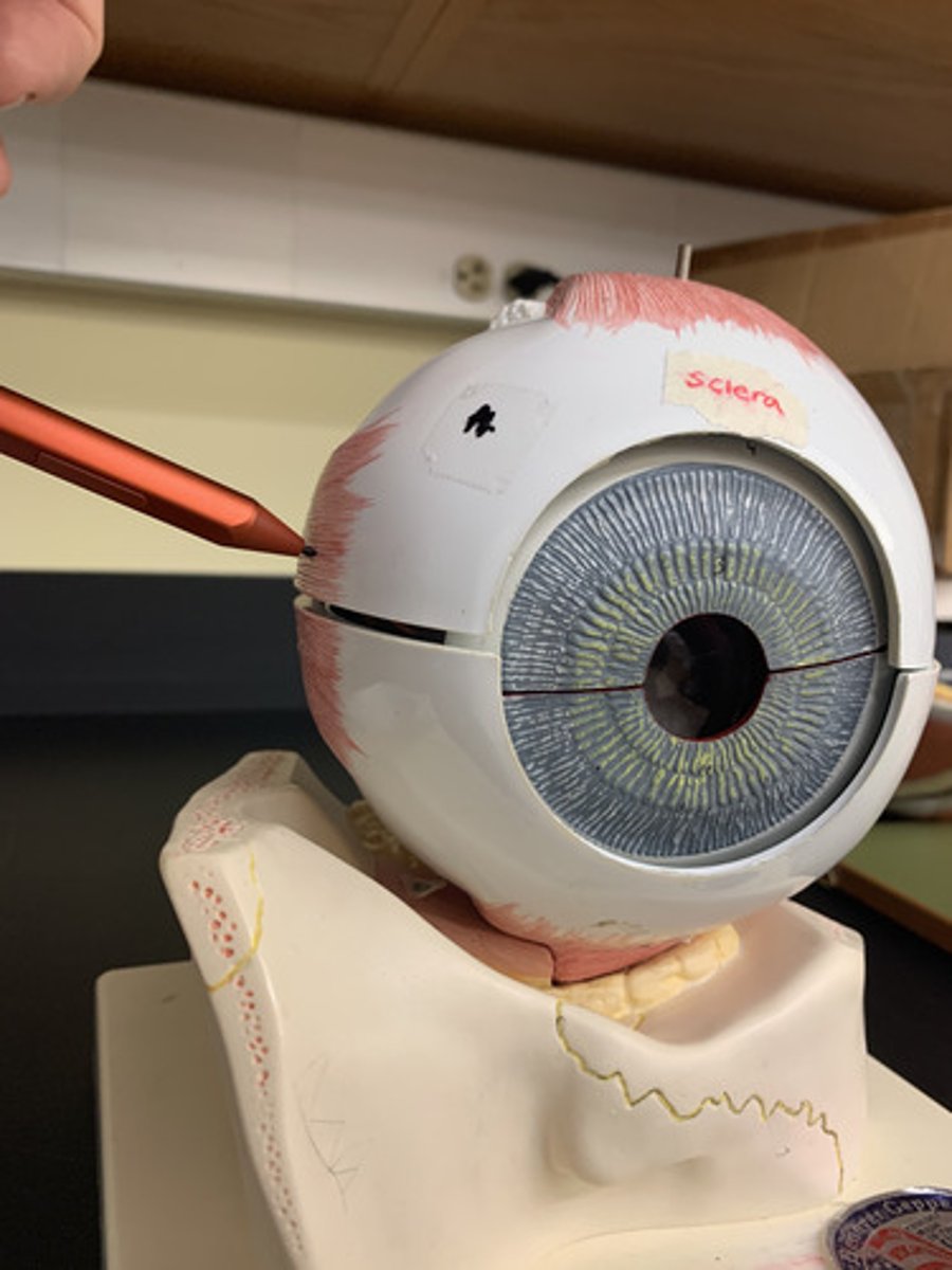
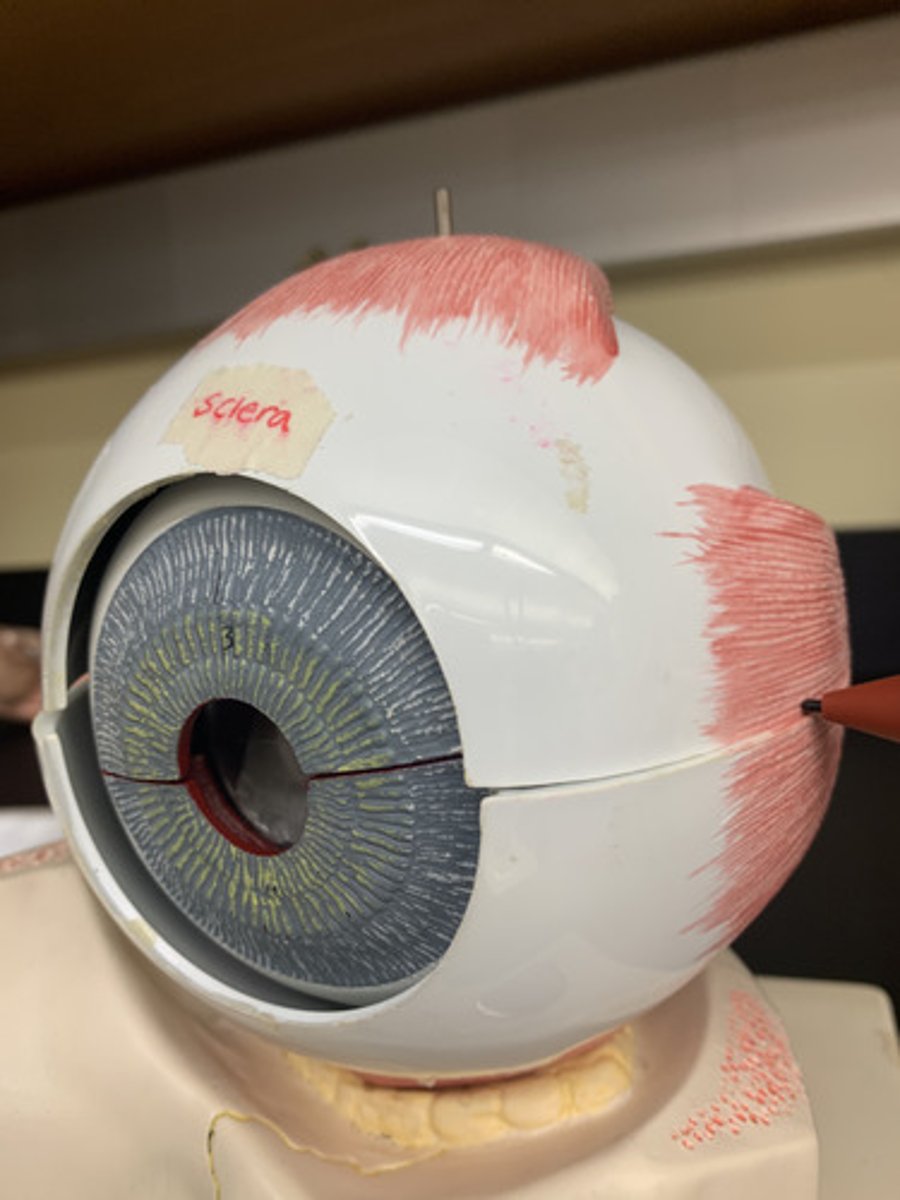
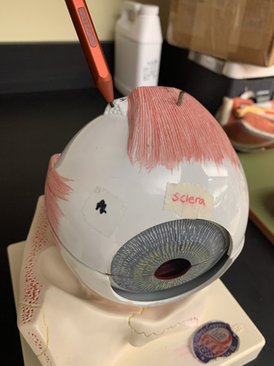
Sclera
white part of the fibrous tunic
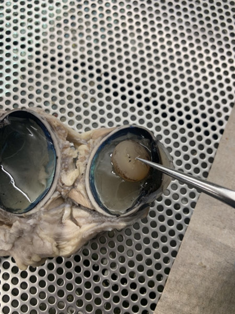
Lens
the transparent structure behind the pupil that changes shape to help focus images on the retina
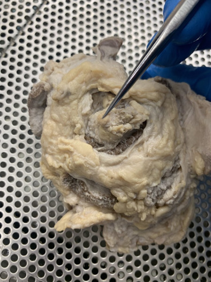
Retina
the light-sensitive inner surface of the eye, containing the receptor rods and cones plus layers of neurons that begin the processing of visual information
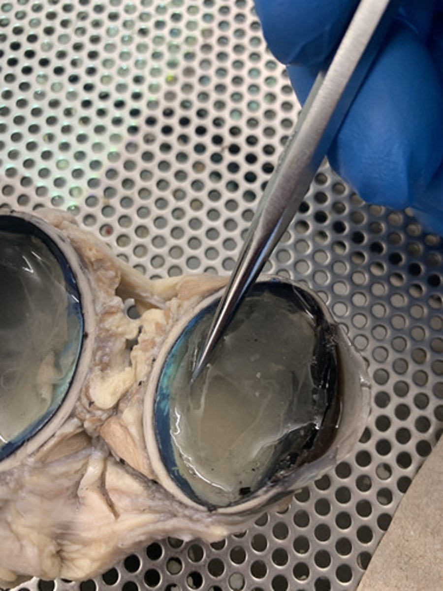
Vitreous humor
jellylike substance found behind the lens in the posterior cavity of the eye that maintains its shape
Ciliary body
ring of tissue behind the peripheral iris that is composed of ciliary muscle and ciliary processes
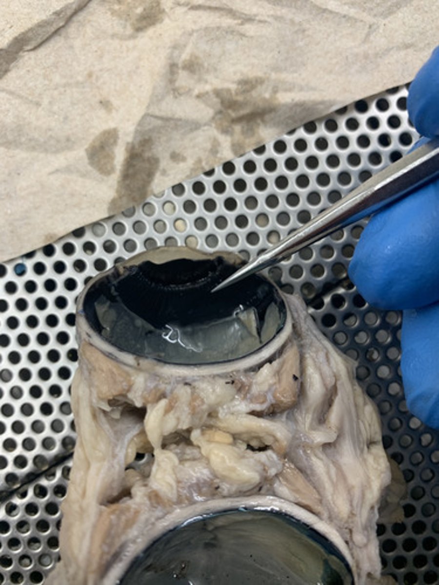
Choroid
middle, vascular layer of the eye, between the retina and the sclera
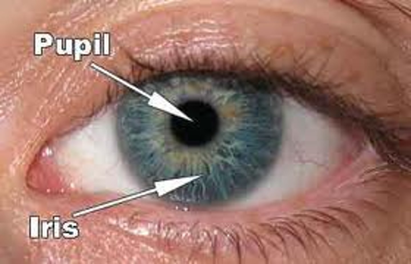
Pupil
the adjustable opening in the center of the eye through which light enters
Optic disc
Region at the back of the eye where the optic nerve meets the retina. It is the blind spot of the eye because it contains only nerve fibers, no rods or cones, and is thus insensitive to light.
Optic nerve (CN II)
the nerve that carries neural impulses from the eye to the brain
Superior rectus
Origin: common tendinous ring
Insertion: superior sclera of eye
Action:elevates front of eye
Inn: Oculomotor nerve (CN III)
Inferior rectus
Origin: Common tendinous ring
Insertion: inferior sclera of eye
Action: depresses front of eye
Inn: Oculomotor nerve (CN III)
Medial rectus
Origin: Common tendinous ring
Insertion: medial sclera of eye
Action: moves front of eye medially
Inn: Oculomotor nerve (CN III)
Lateral rectus
Origin: Common tendinous ring
Insertion: lateral sclera of eye
Action: moves front of eye laterally
Inn: Abducens nerve (CN VI)
Superior oblique
Origin: Common tendinous ring
Insertion: Superolateral sclera of eye
Action: depresses, abducts and medially rotates eye
Inn: Trochlear nerve (CN IV)
Inferior oblique
Origin: Common tendinous ring
Insertion: Inferolateral sclera of eye
Action: Elevates, abducts and laterally rotates eye
Inn: Oculomotor nerve (CN III)
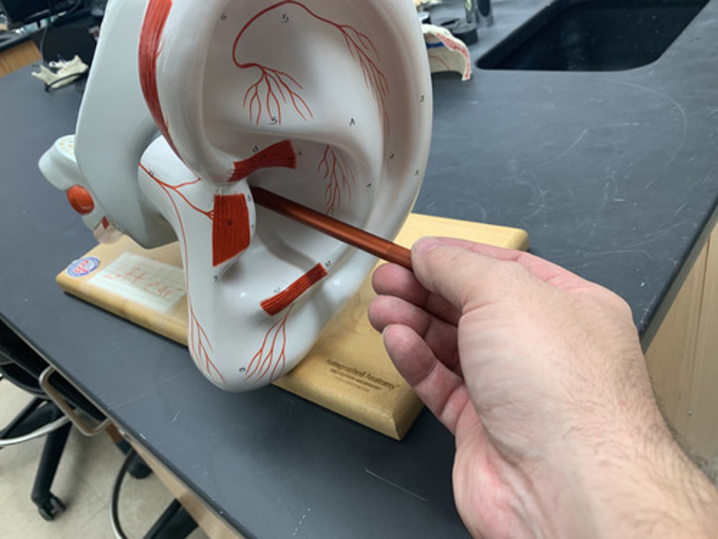
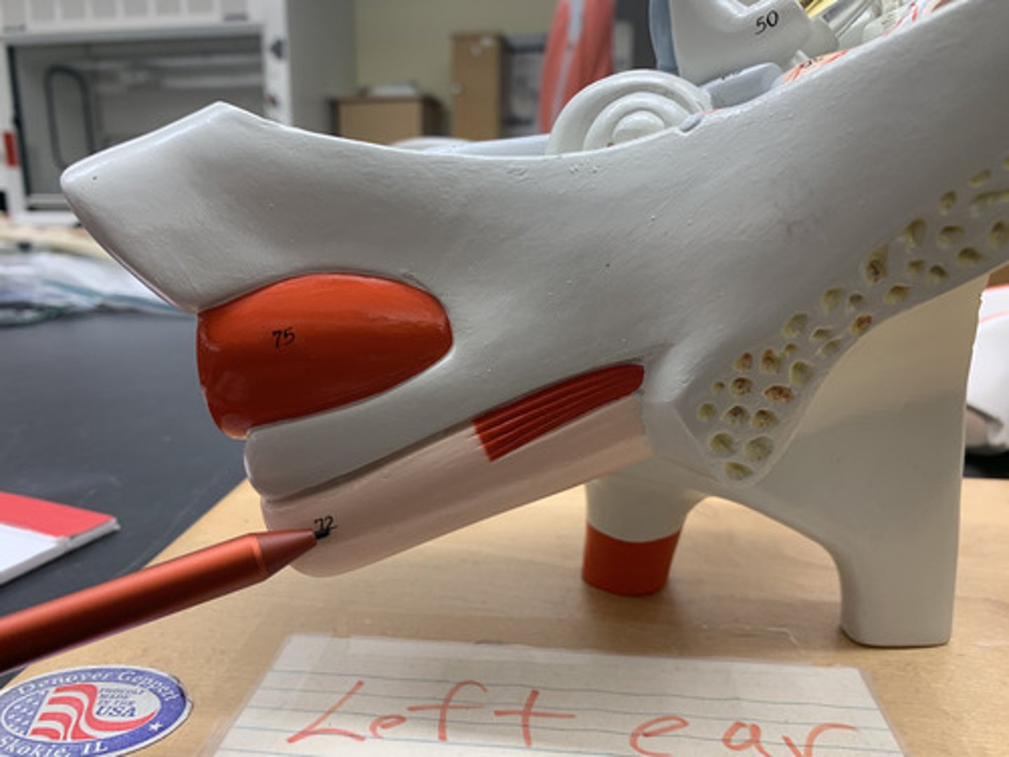
Auricle
external portion of the ear
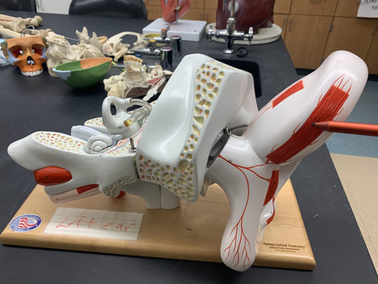
External acoustic meatus
Canal leading to eardrum and middle ear
Tympanic membrane
The eardrum. A structure that separates the outer ear from the middle ear and vibrates in response to sound waves.
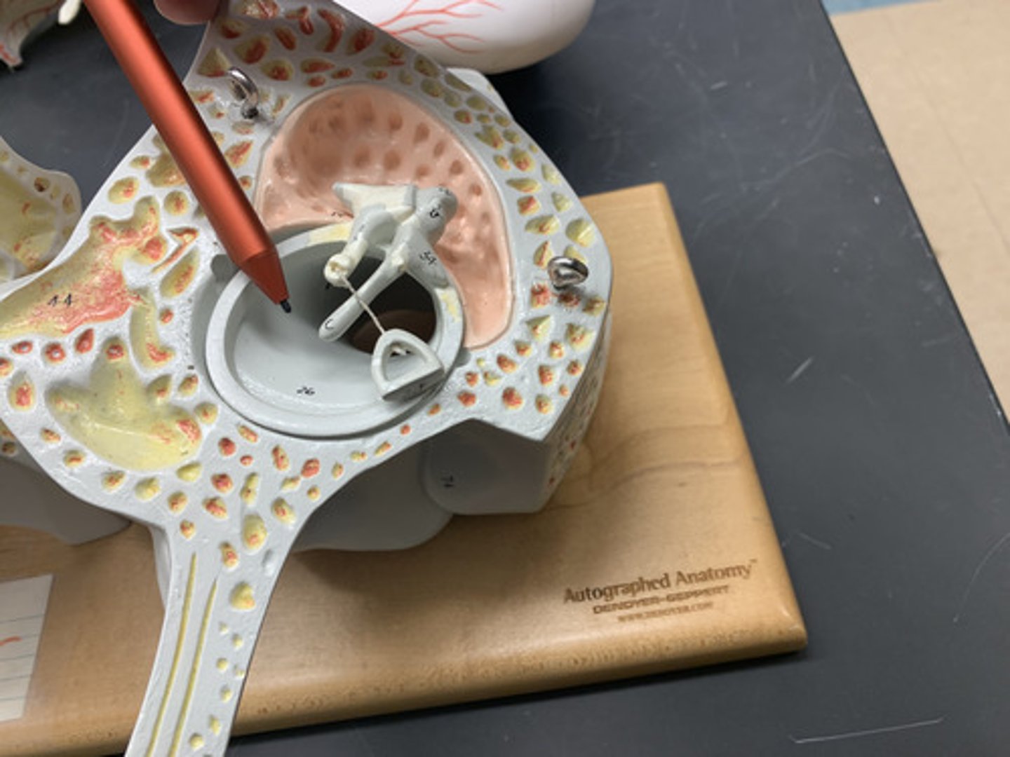
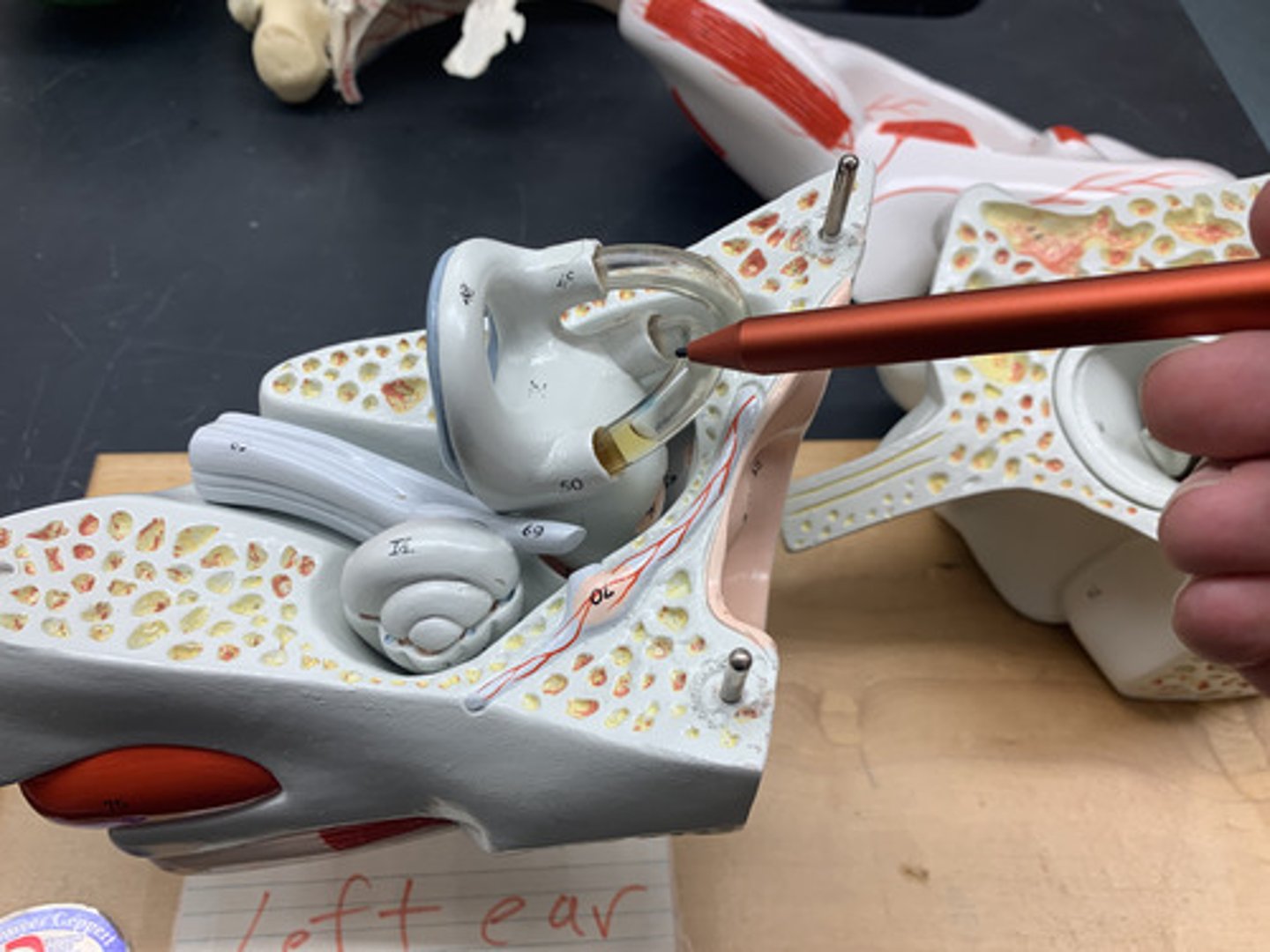
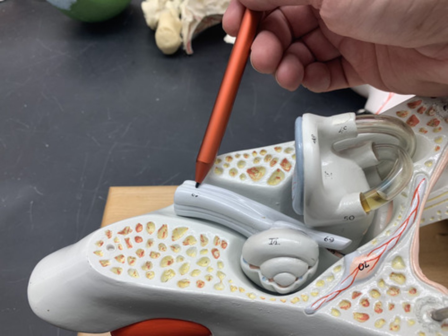
Malleus
hammer; first of the three auditory ossicles of the middle ear
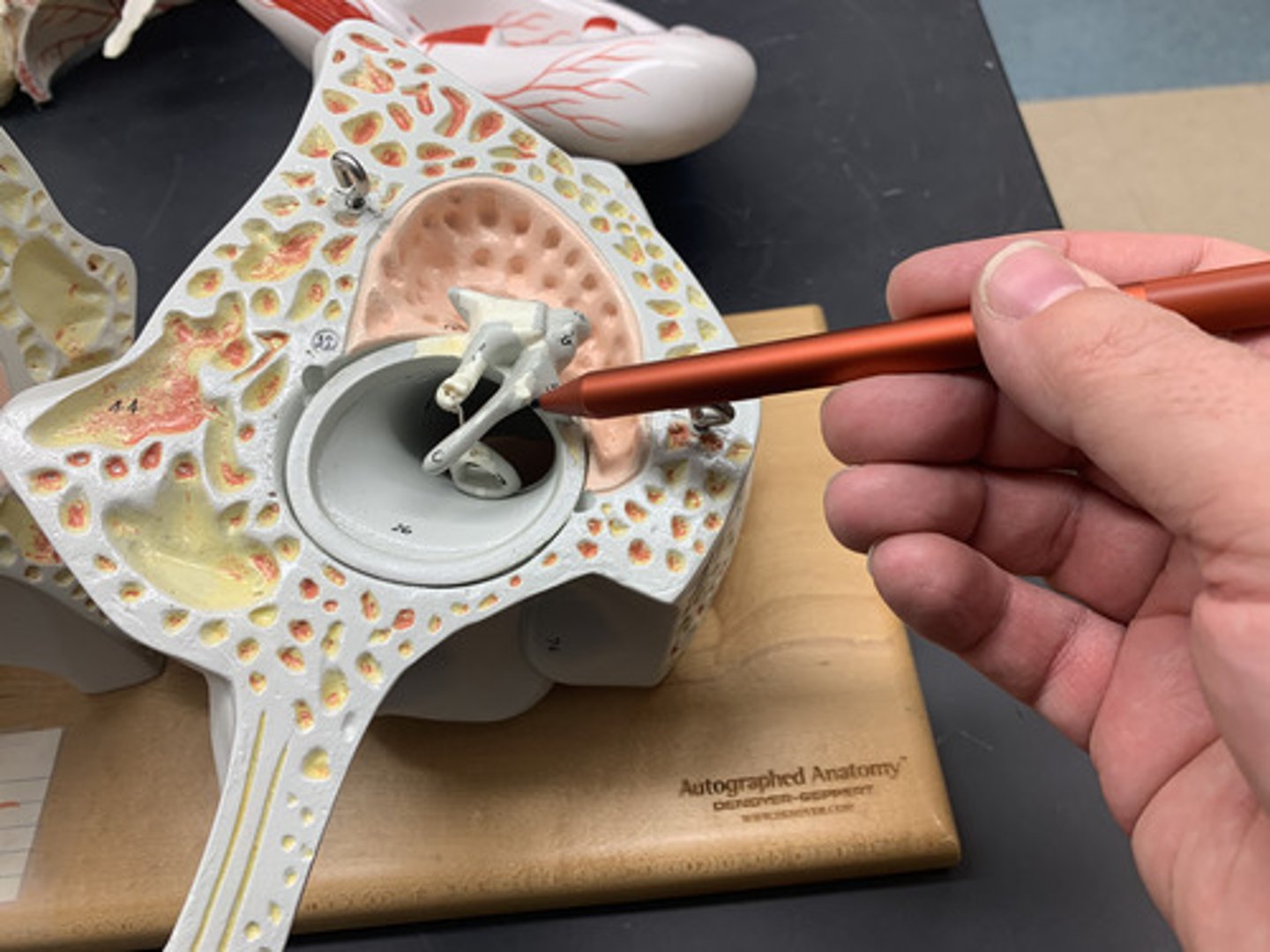
Incus
anvil; middle of the three auditory ossicles of the middle ear
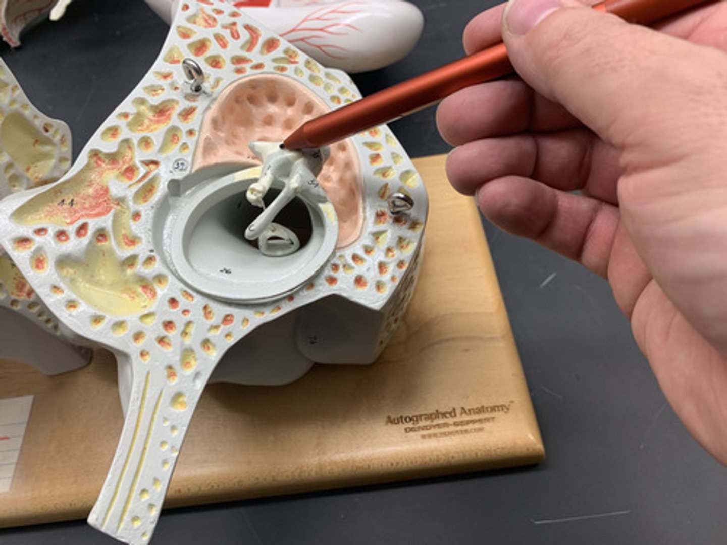
Stapes
stirrup; last of the three auditory ossicles of the middle ear
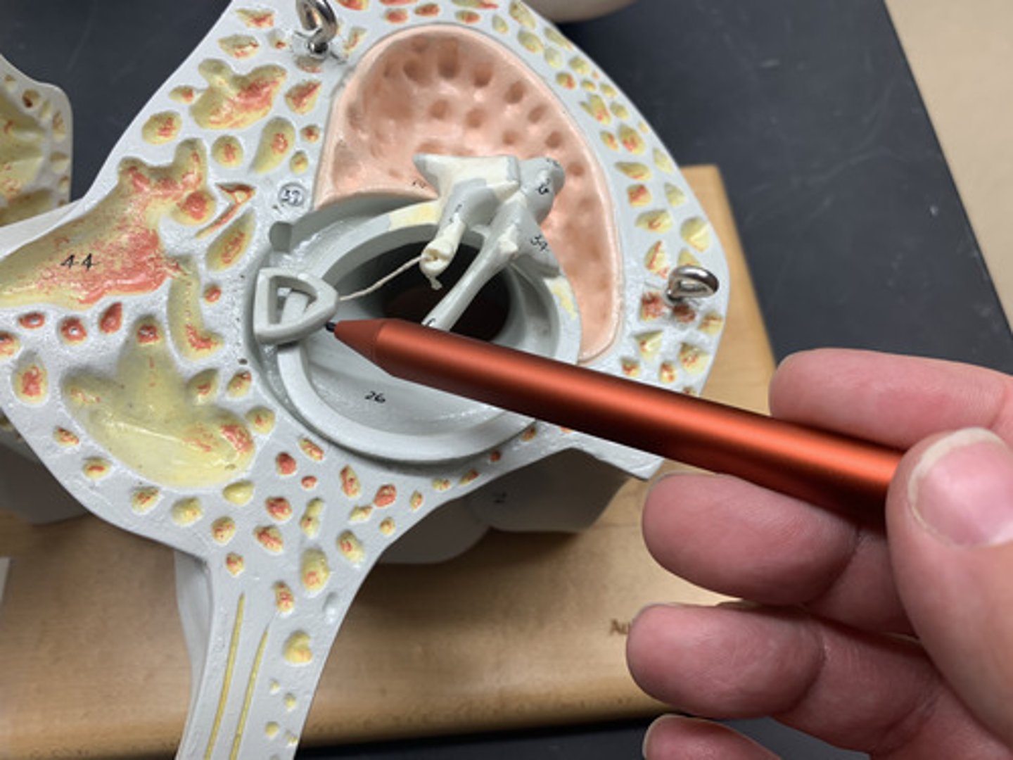
Round window
located just below the oval window; equalize pressure in the inner ear
Oval window
membrane that covers the opening between the middle ear and inner ear
Cochlea
a coiled, bony, fluid-filled tube in the inner ear; sound waves traveling through the cochlear fluid trigger nerve impulses
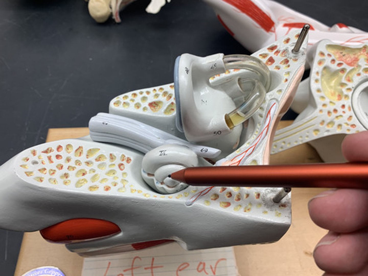
Semicircular canals
three fluid-filled canals in the inner ear responsible for our sense of rotational balance
Vestibule
Fluid filled spaces responsible for our sense of balance
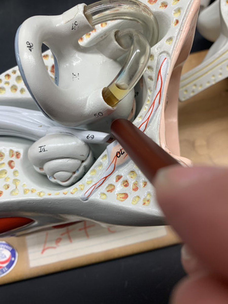
Vestibulocochlear nerve (CN VIII)
cranial nerve that collects sensory information about hearing and balance
Eustachian tube
A narrow tube between the middle ear and the throat that serves to equalize pressure on both sides of the eardrum
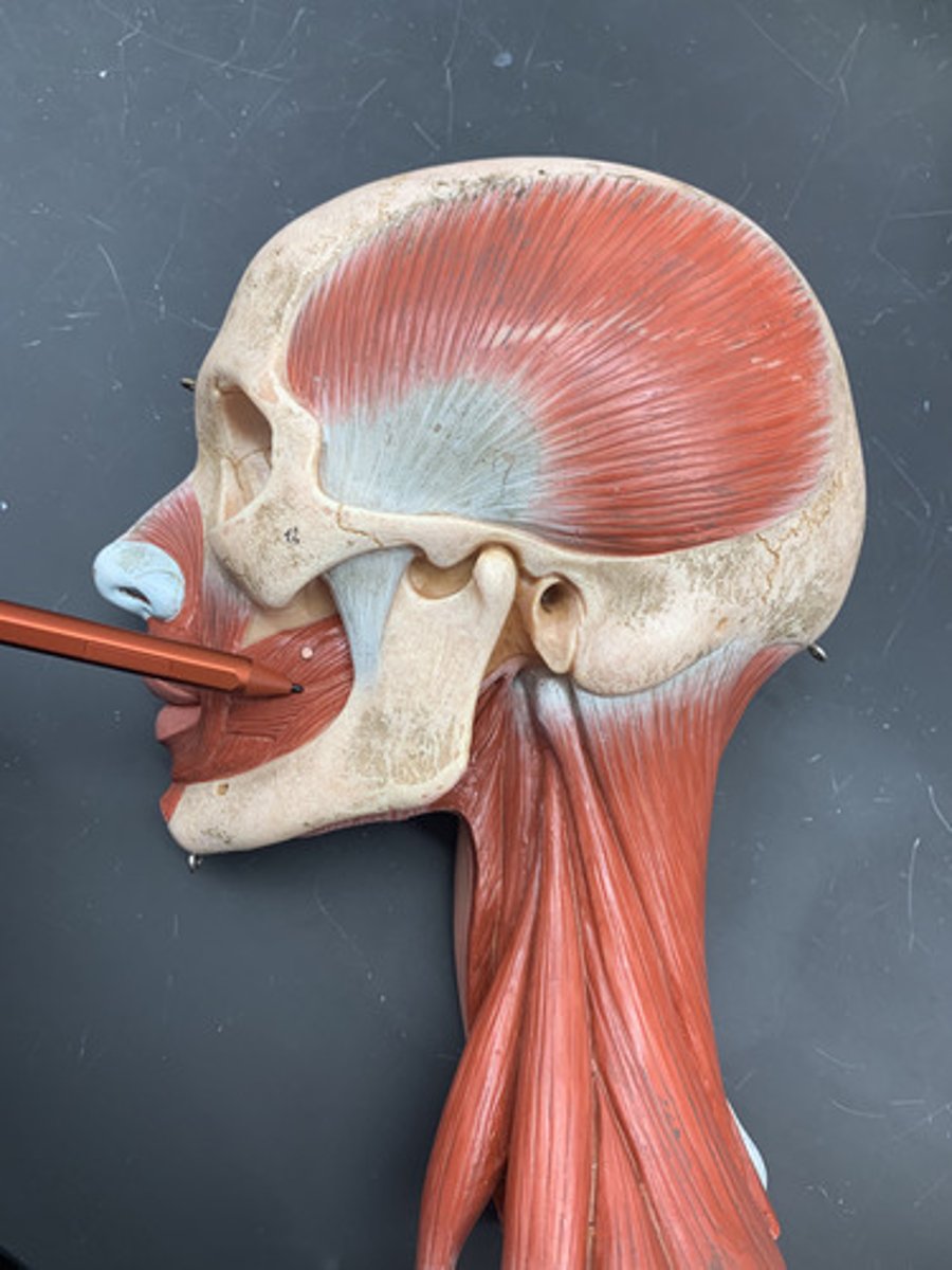
Masseter
Origin: zygomatic arch
Insertion: angle of mandible
Action: elevates & protracts mandible
Inn: Mandibular nerve (CN V3)
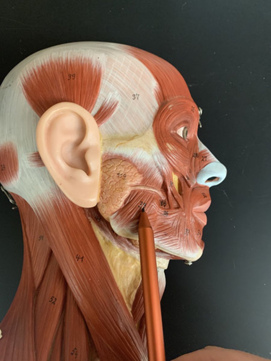
Temporalis
O: temporal fossa
I: coronoid process of mandible
A: Elevates & retracts mandible
Inn: Mandibular nerve (CN V3)
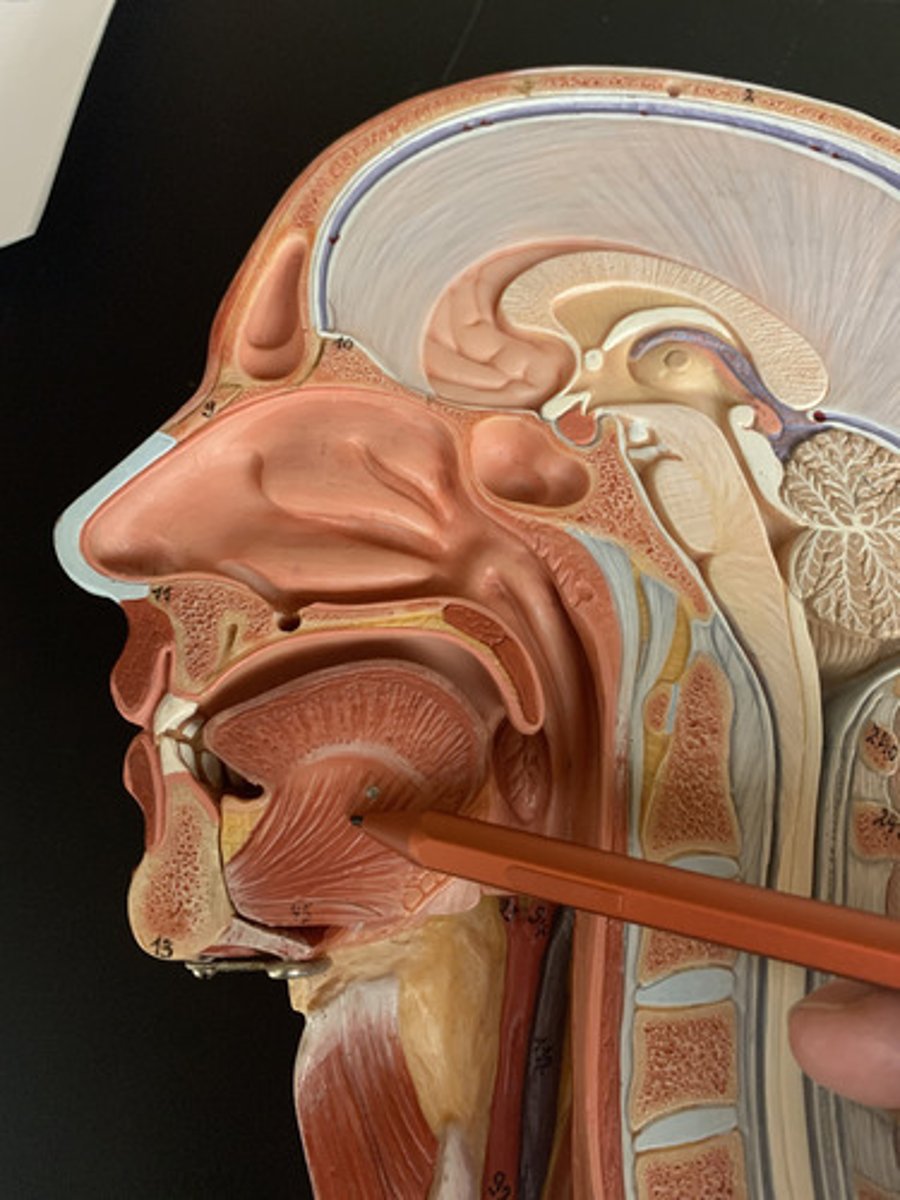
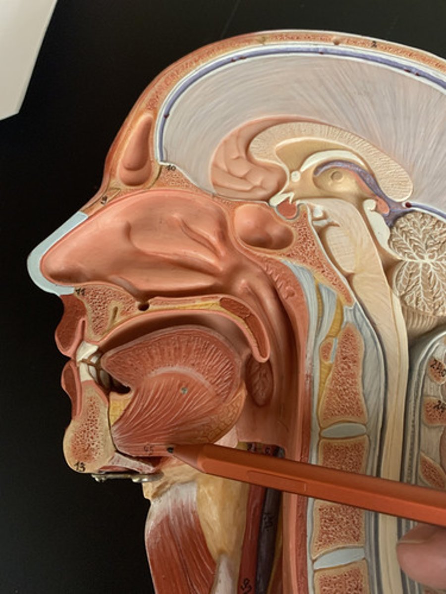
Genioglossus
O: Mandible, internal surface
I: Bottom of tongue, body of hyoid
A: Protrudes tongue
Inn: Hypoglossal nerve (CN XII)
Geniohyoid
O: mandible
I: body of hyoid
A: protracts hyoid
Inn: Hypoglossal nerve (CN XII), C1
Hyoglossus
Origin: Hyoid bone
Insertion: Tongue
Action: Depresses tongue
Inn: Hypoglossal nerve (CN XII)
Palatoglossus
O: Anterolateral palatal aponeurosis
I: Sides of posterior tongue
A: Elevates the tongue or depresses the soft palate
Inn: Pharyngeal plexus (vagus nerve (CN X))
Styloglossus
O: Styloid process of temporal bone
I: Bottom and lateral aspects of tongue
A: Retracts tongue
Inn: Hypoglossal nerve (CN XII)
Hyoid
a U-shaped bone in the neck that supports the tongue.
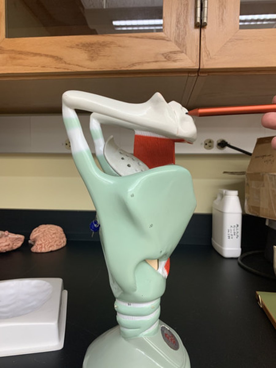
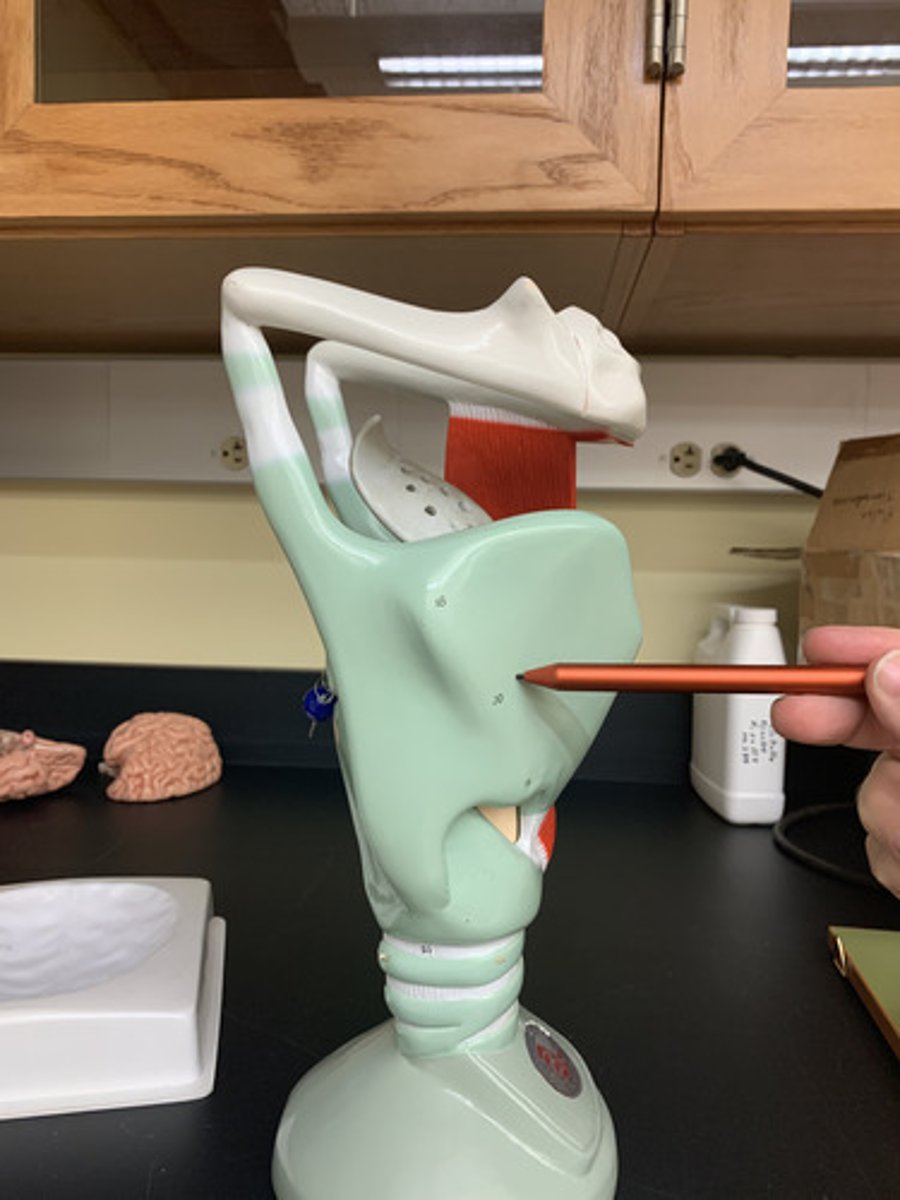
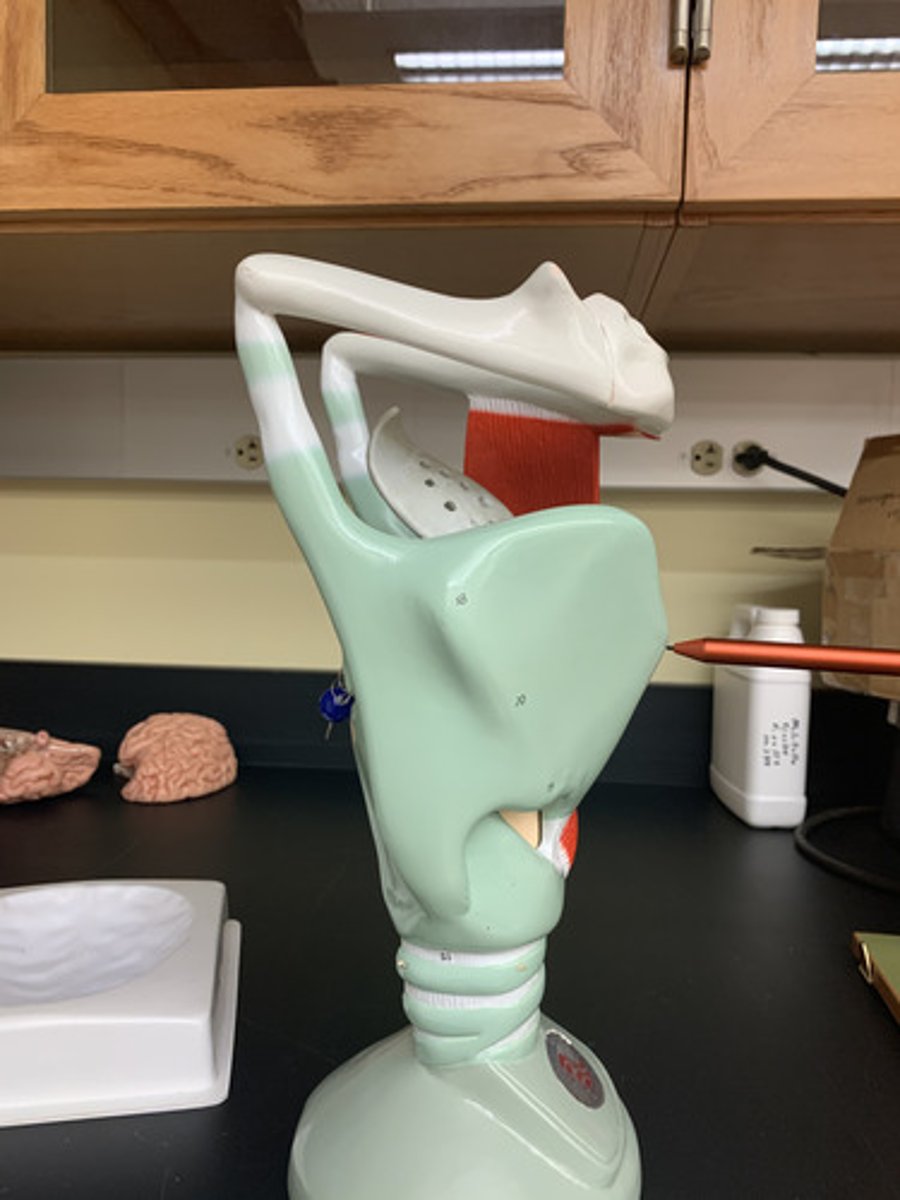
Thyroid cartliage
is the largest piece of cartilage in the larynx & is also known as the Adams Apple
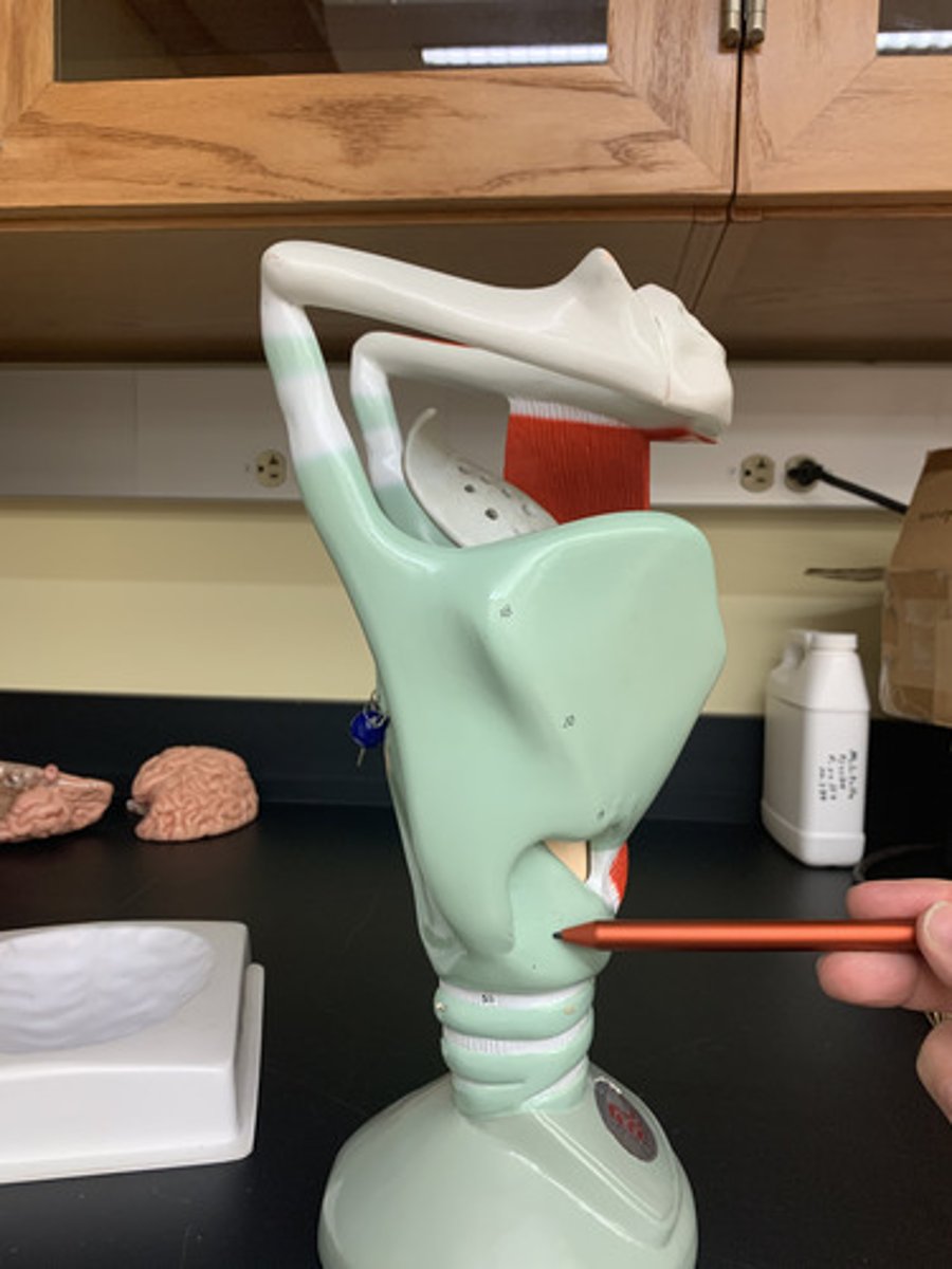
Cricoid cartilage
the ring-shaped structure that forms the lower portion of the larynx
Thyroid prominence
-A protrusion referred to as the "Adam's apple," which is located just below the thyroid notch.
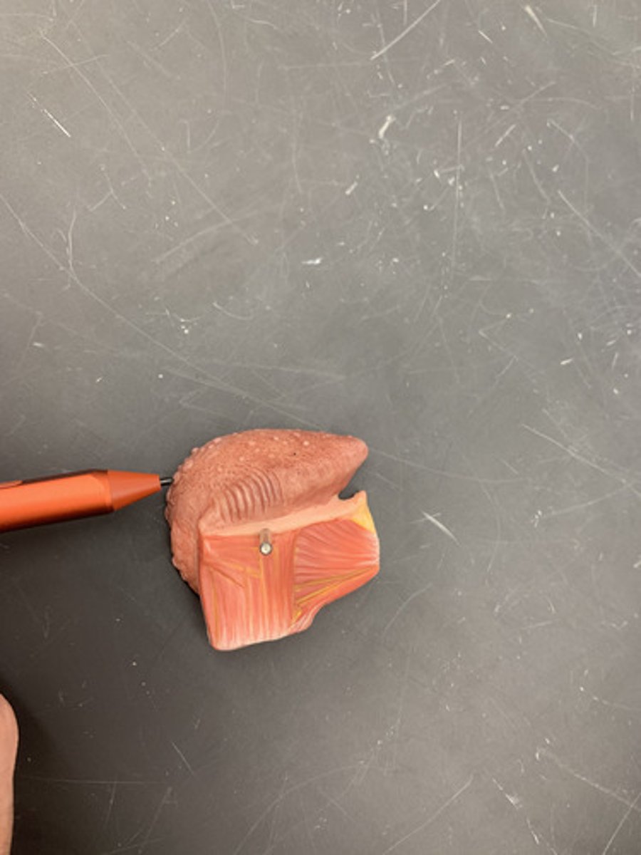
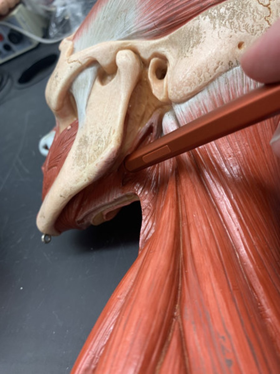
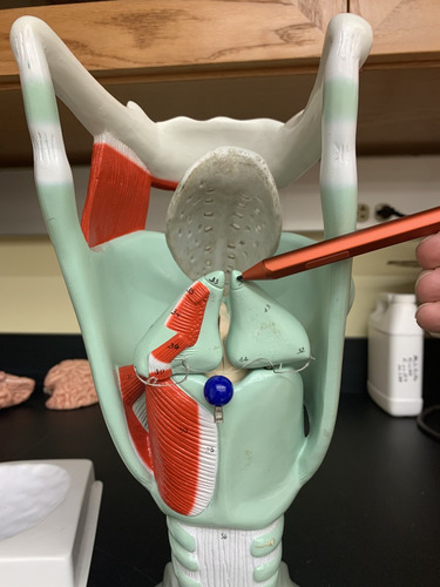
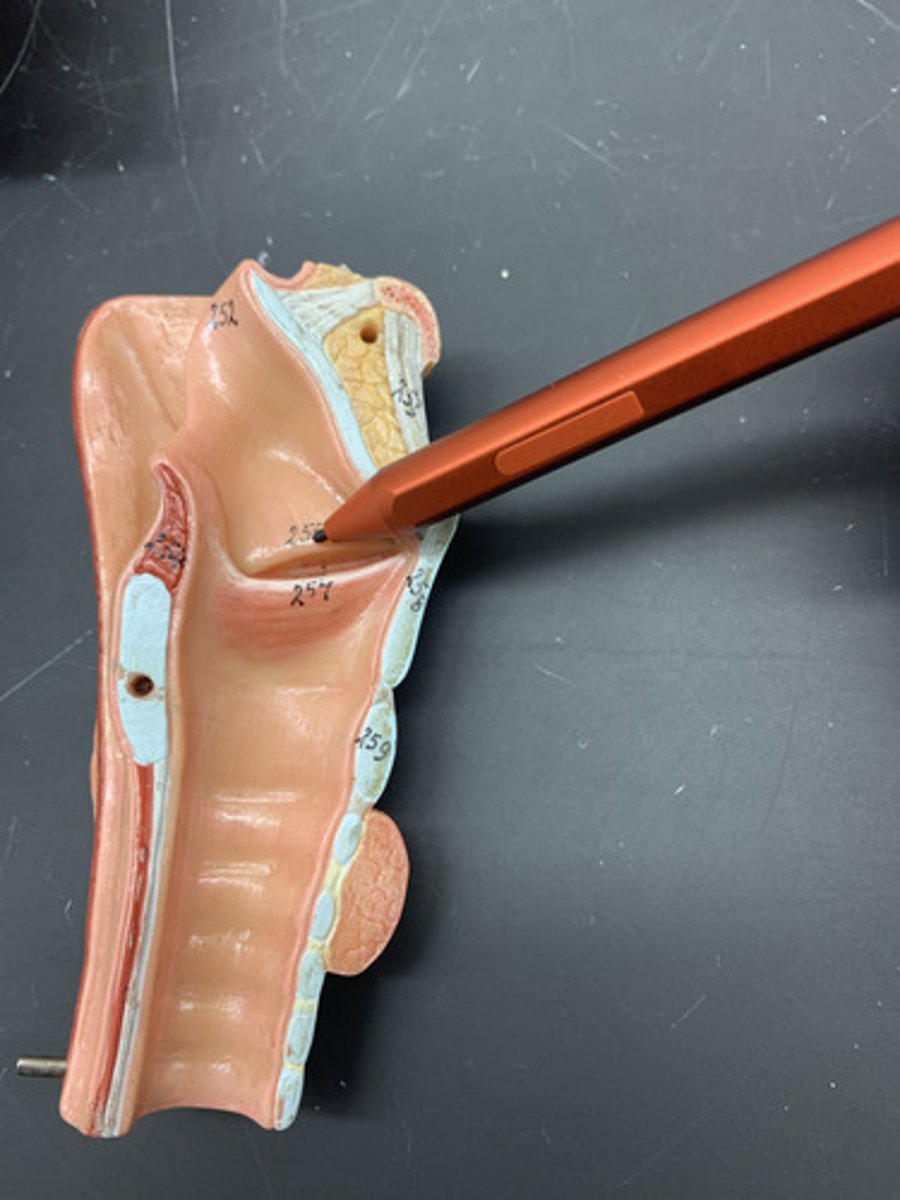
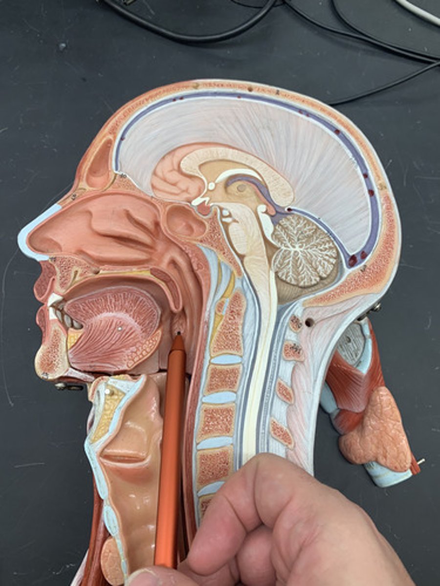
Epiglottis
a flap of cartilage at the root of the tongue, which is depressed during swallowing to cover the opening of the windpipe.
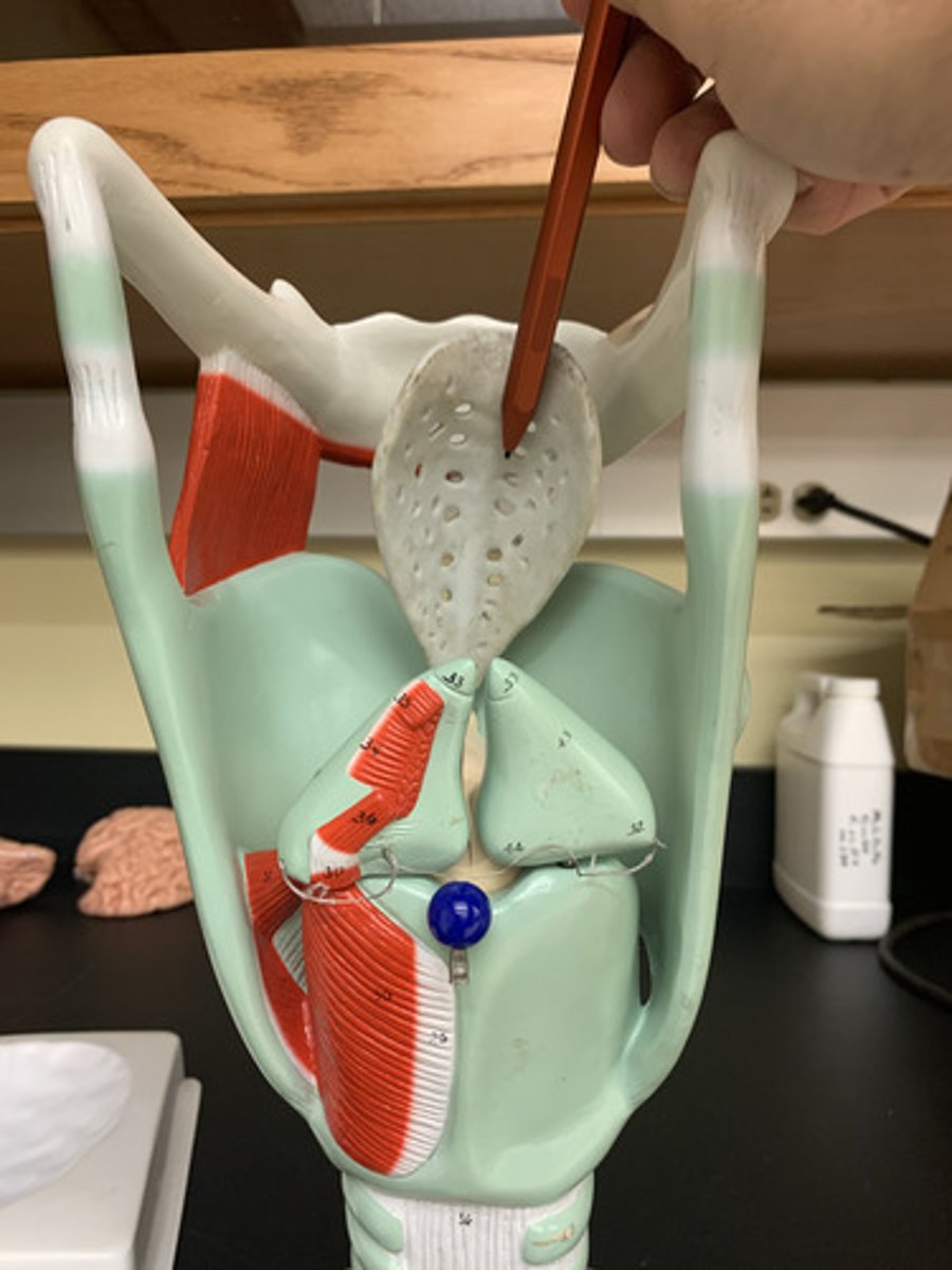
Arytenoid cartilage
Two small cartilages in the larynx, the movements of which abduct and adduct the vocal folds.
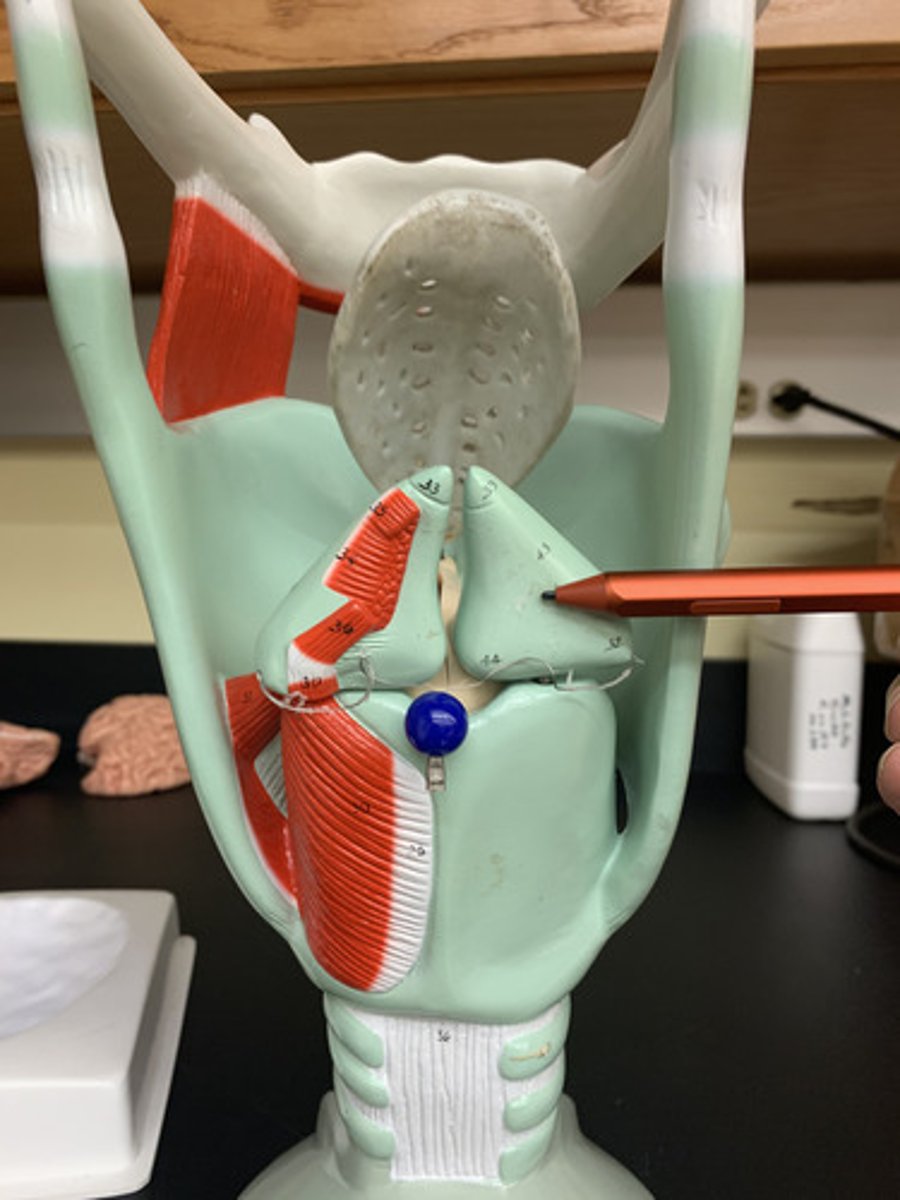
Corniculate cartilage
a pair of horn-like pieces of elastic cartilage located at the apex of each arytenoid cartilage
Cricothyroid ligament
soft piece of connective tissue between the thyroid cartilage and cricoid cartilage
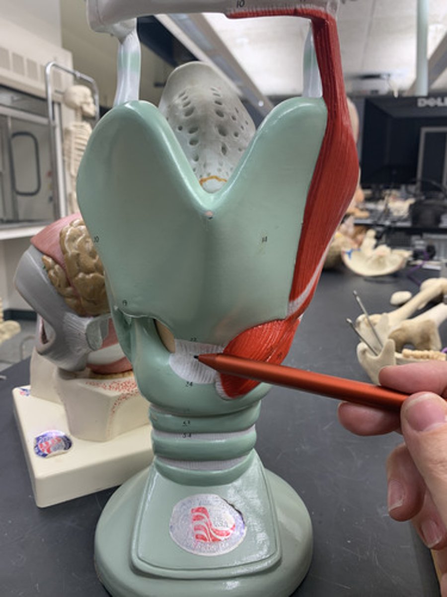
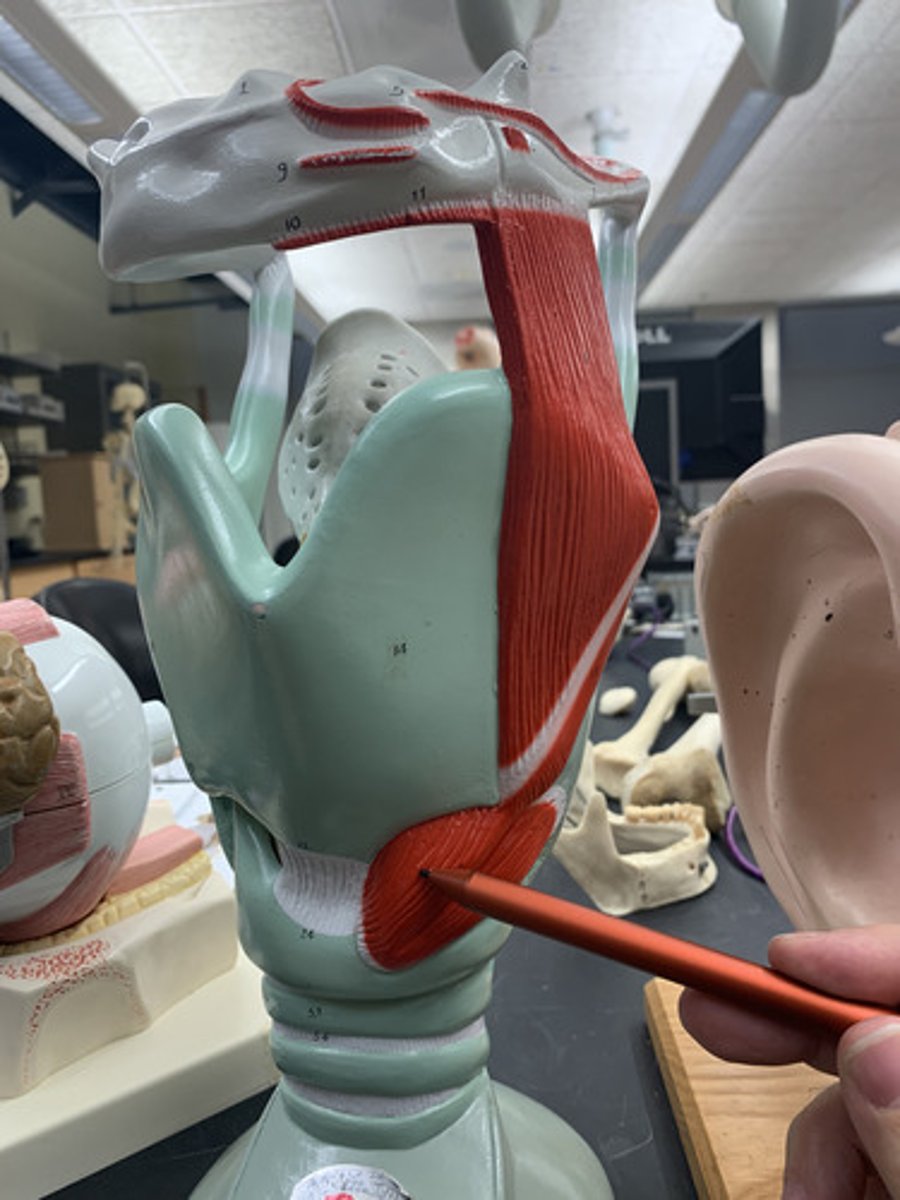
Cricothyroid muscle
O: arch of the cricoid cartilage
I: inferior border of the thyroid cartilage
A: draws the thyroid cartilage forward, lengthening the vocal ligaments
Inn: superior laryngeal nerve, a branch of the vagus nerve (X)
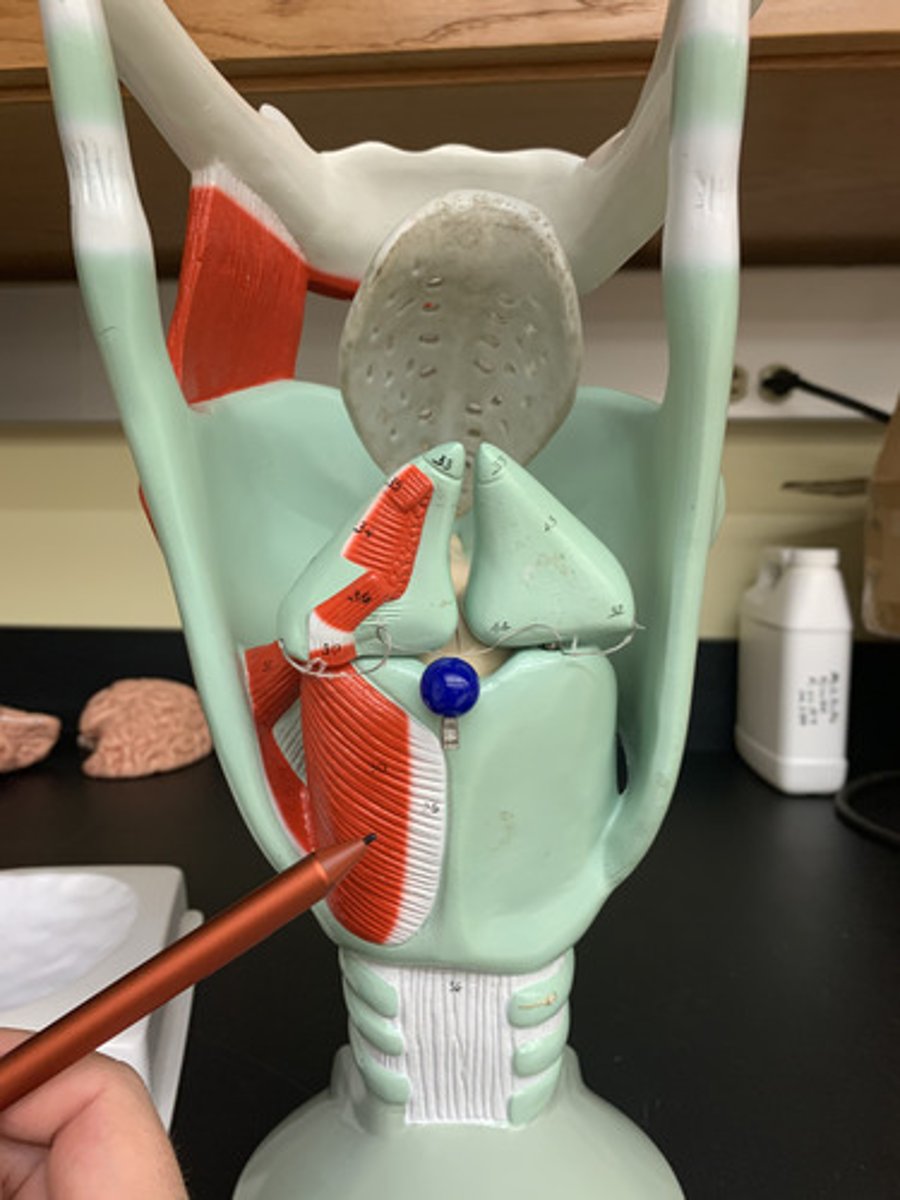
Posterior cricoarytenoid muscle
O: Cricoid cartilage
I: Arytenoid cartilage
A: Abduct vocal folds
Inn: Recurrent laryngeal nerve (CN X)
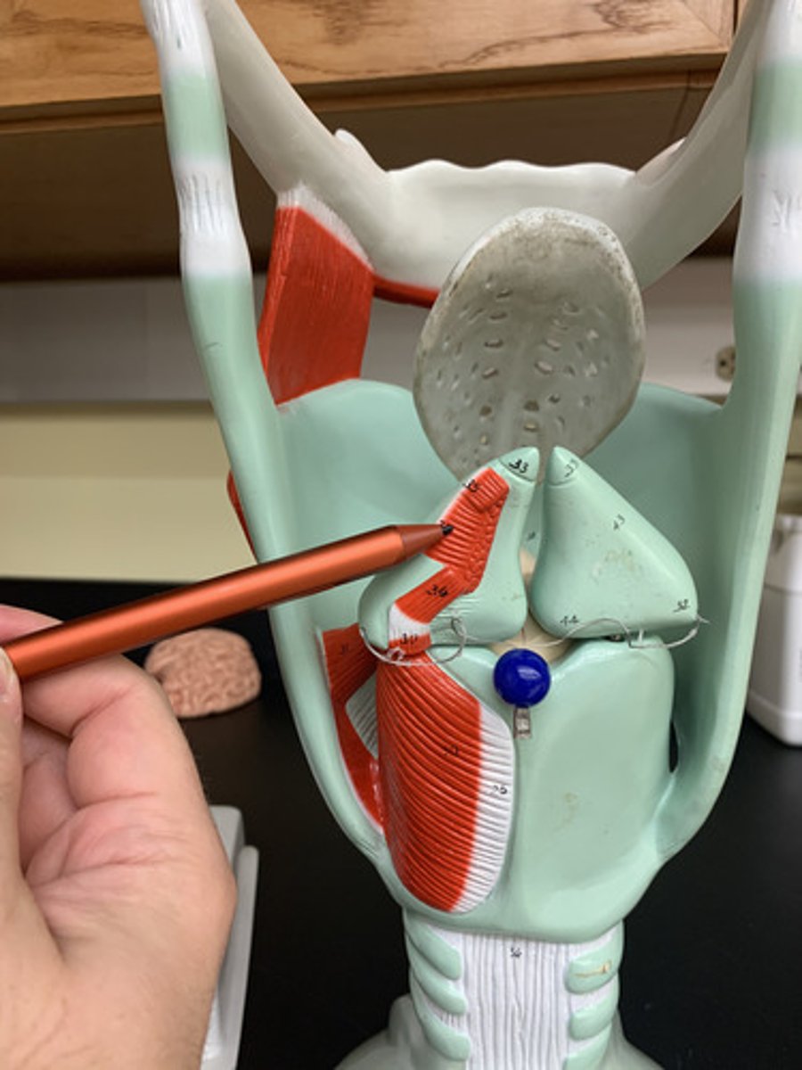
Arytenoideus muscle
O & I: Contralateral sides of arytenoid cartilage
A: Adduct vocal folds
Inn: Recurrent laryngeal nerve (CN X)
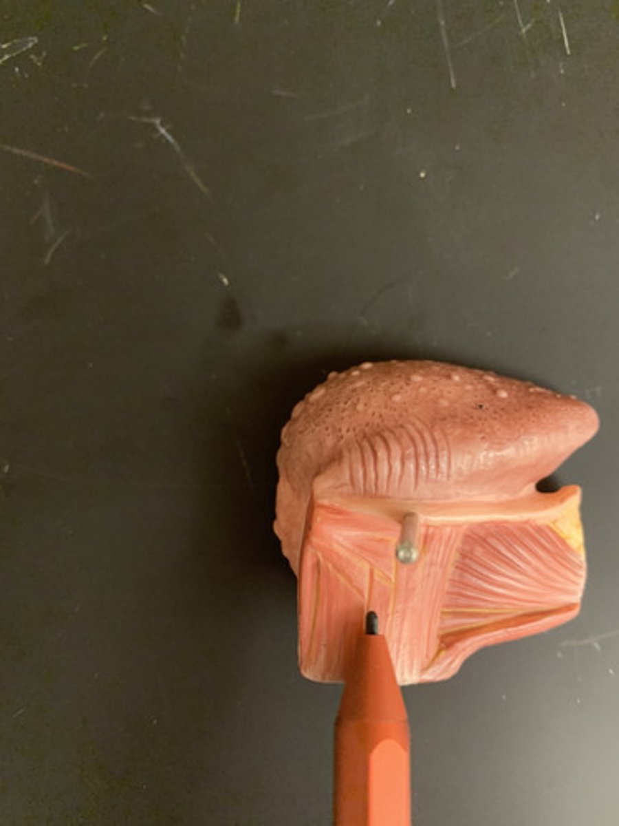
Vocal fold
Mucosal folds that function in voice production (speech); also called the true vocal cords.
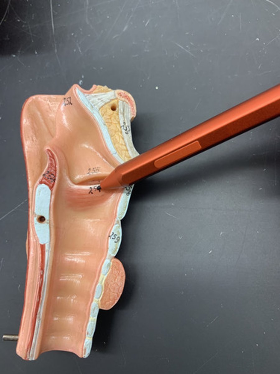
Vestibular fold
close larynx during swallowing; false vocal cords
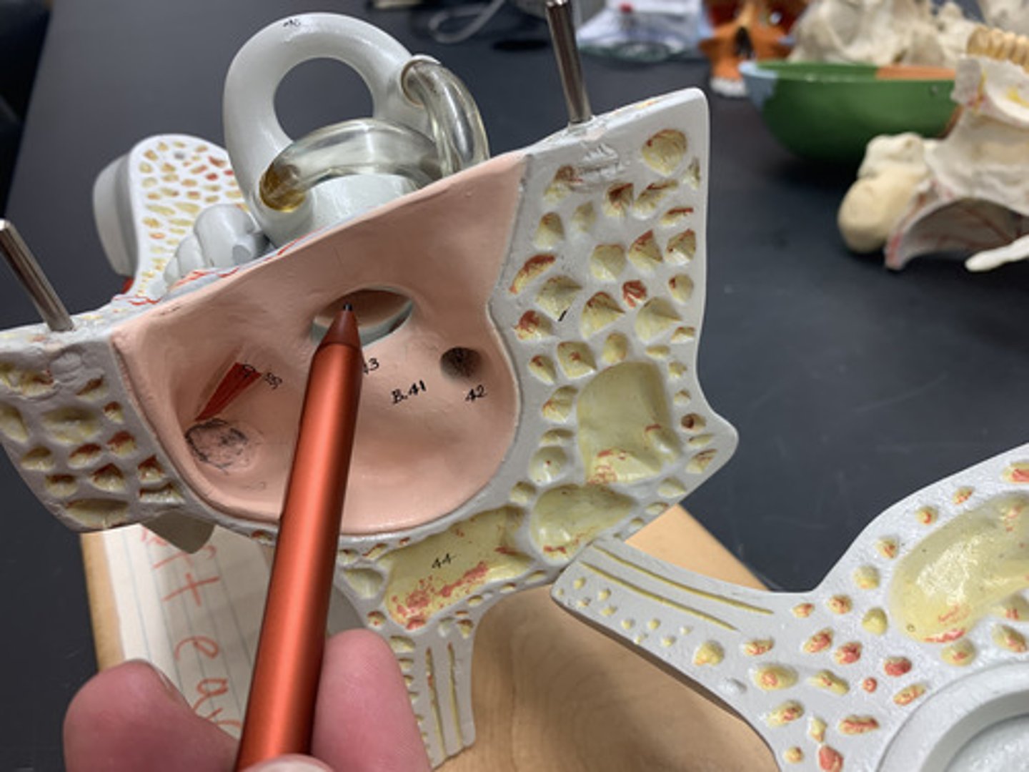
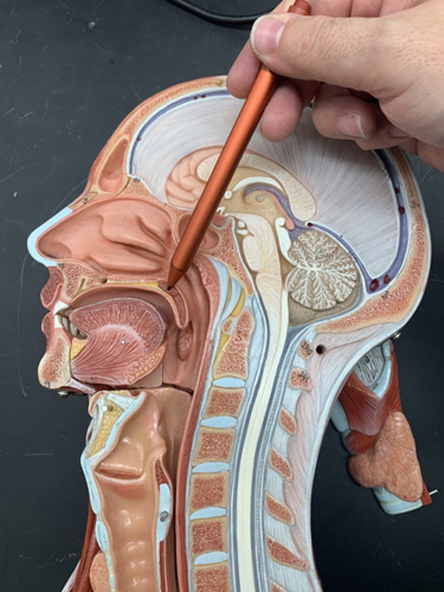
Frontal sinus
cavity within the frontal bone
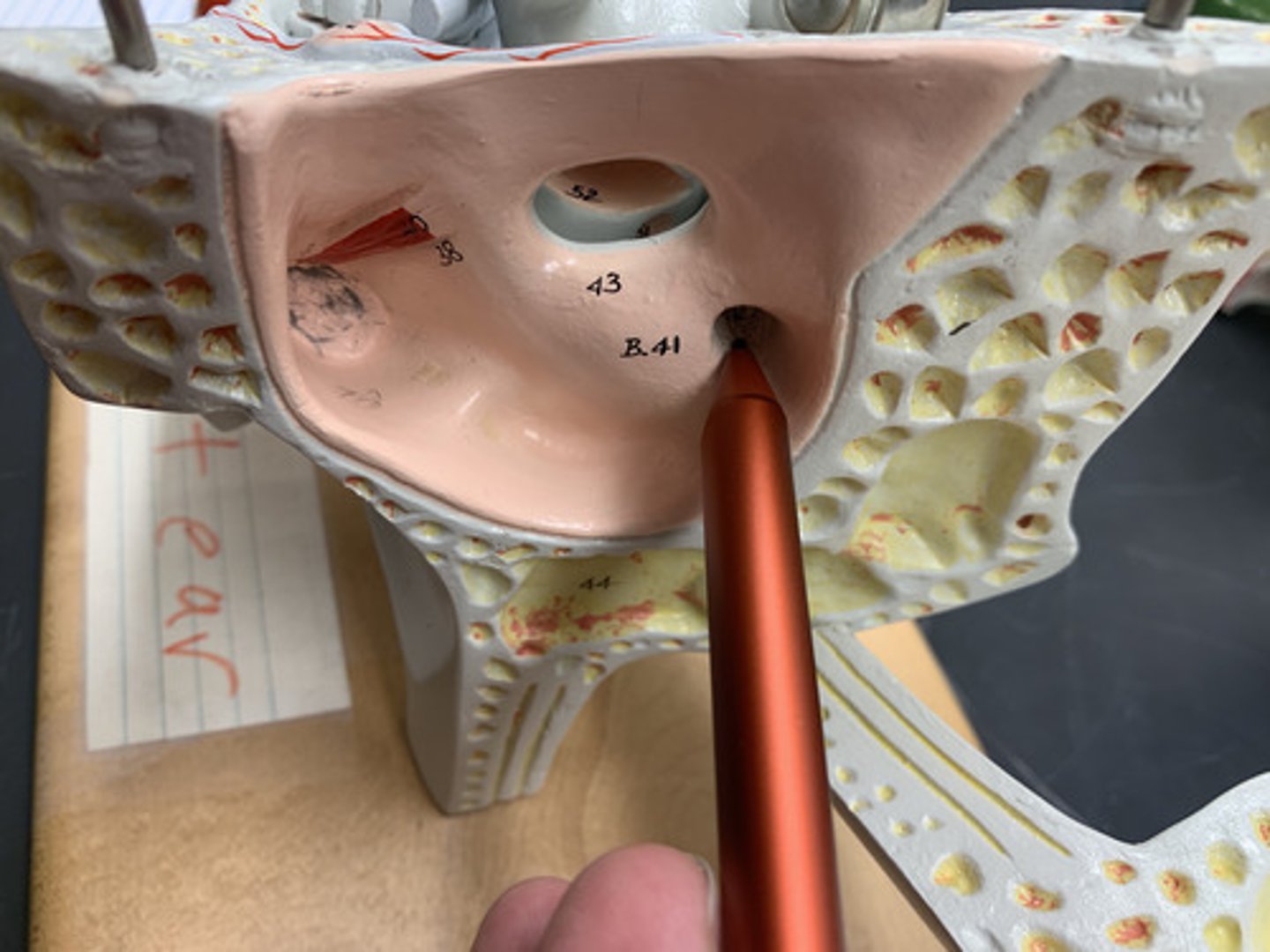
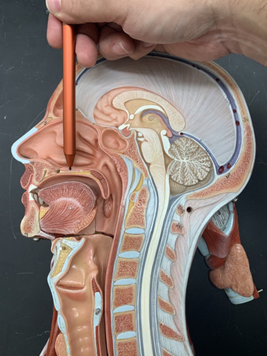
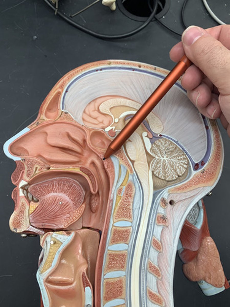
Sphenoid sinus
sinus above and behind the nose
Sella turcica
depression in the sphenoid bone where the pituitary gland is located
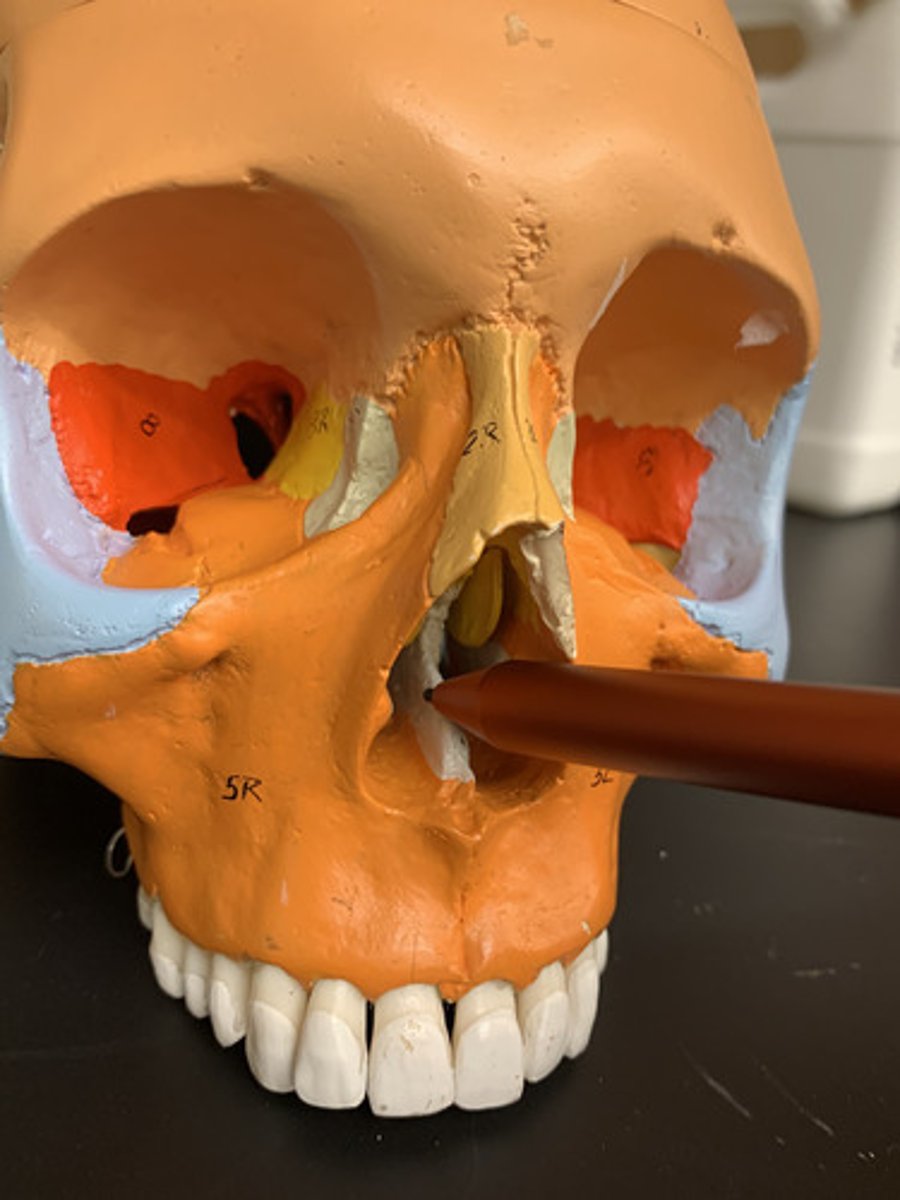
Nasal septum
partition separating the right and left nasal cavities; formed from septal cartilage, perpendicular plate of the ethmoid bone and the vomer
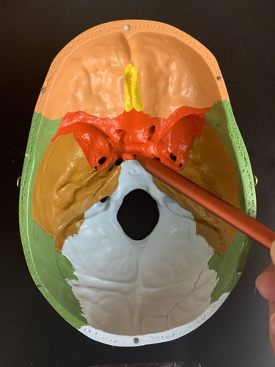
perpendicular plate of ethmoid bone
forms superior part of nasal septum
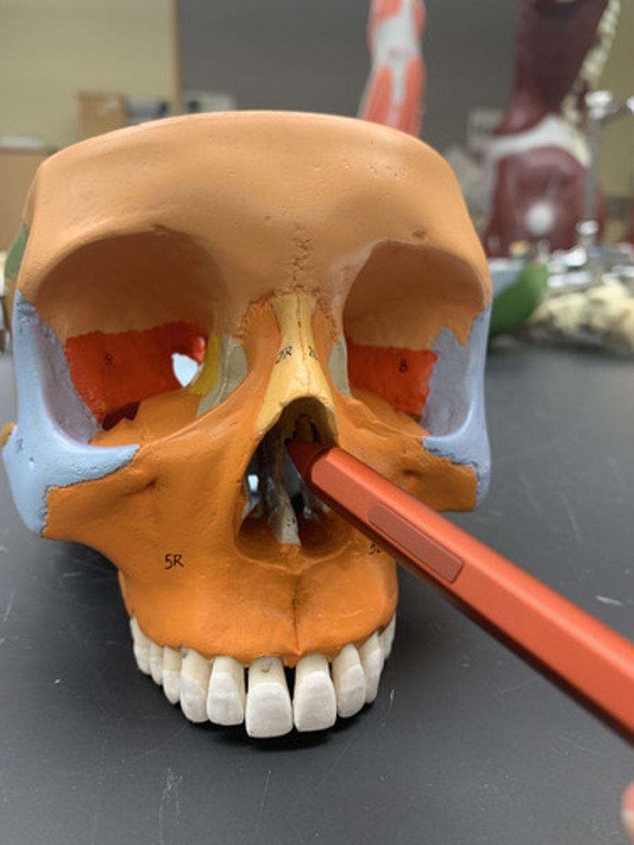
Vomer
forms the inferior portion of the nasal septum
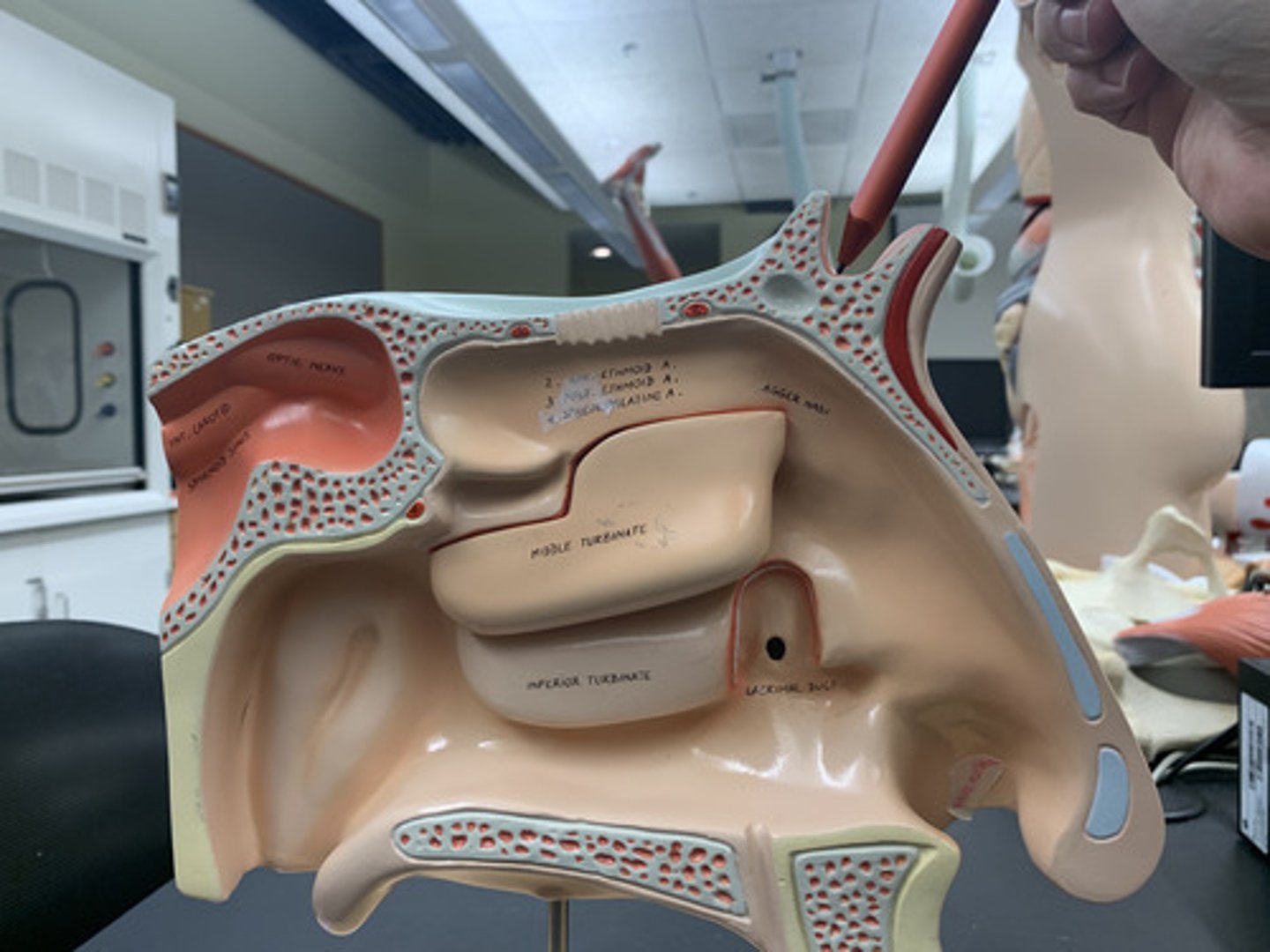
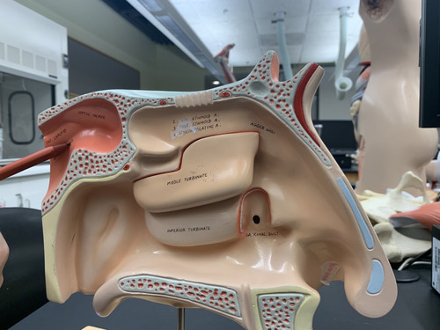
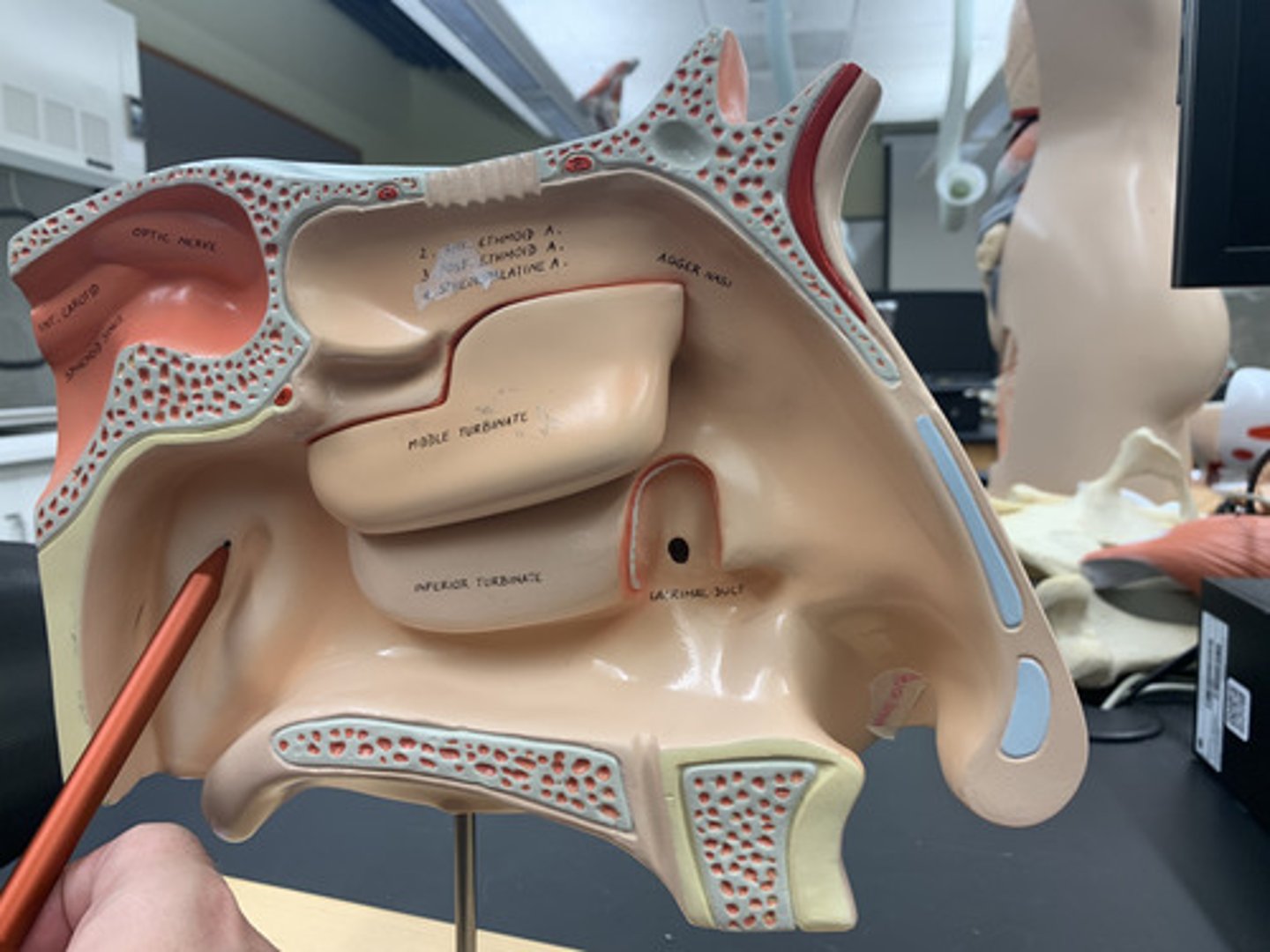
Superior nasal concha
Most superior bony ridge of ethmoid bone that extends from lateral wall of nasal cavity
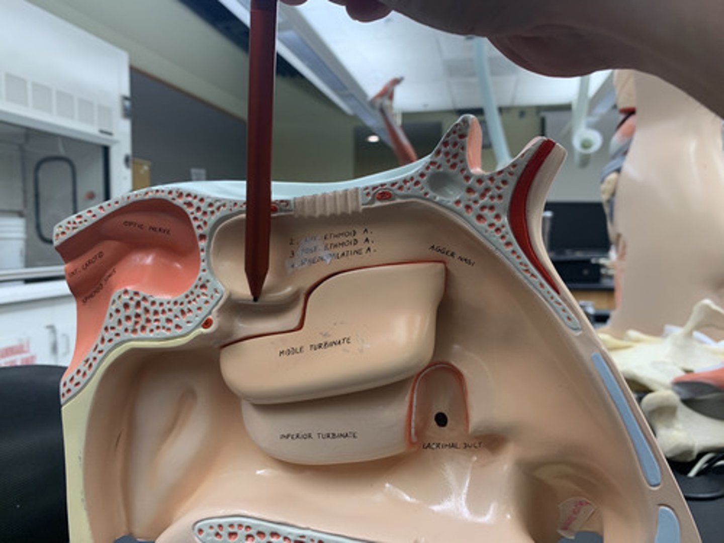
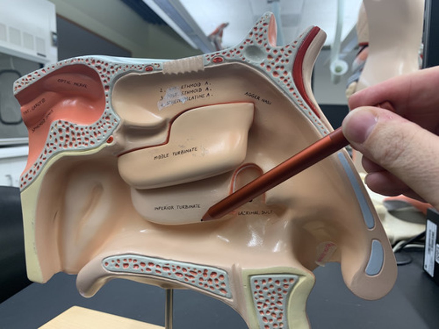
Middle nasal concha
Lower bony ridge of ethmoid bone that extends from lateral wall of nasal cavity
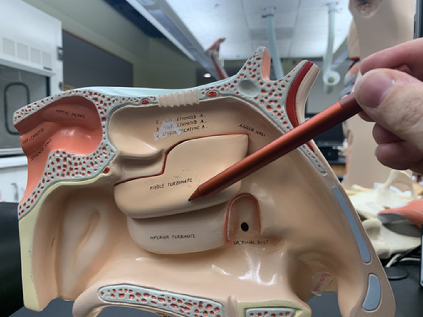
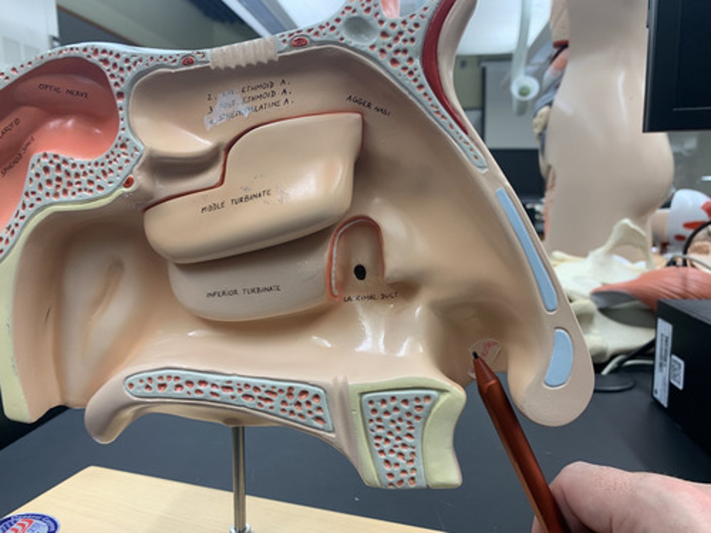
Inferior nasal concha
Lowest bony ridge extending from lateral nasal cavity wall; its own complete bone
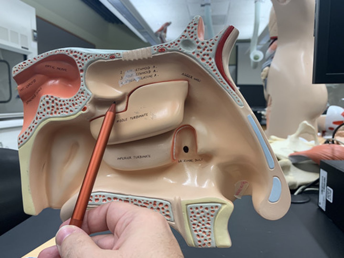
Superior nasal meatus
"valley" between upper two conchae
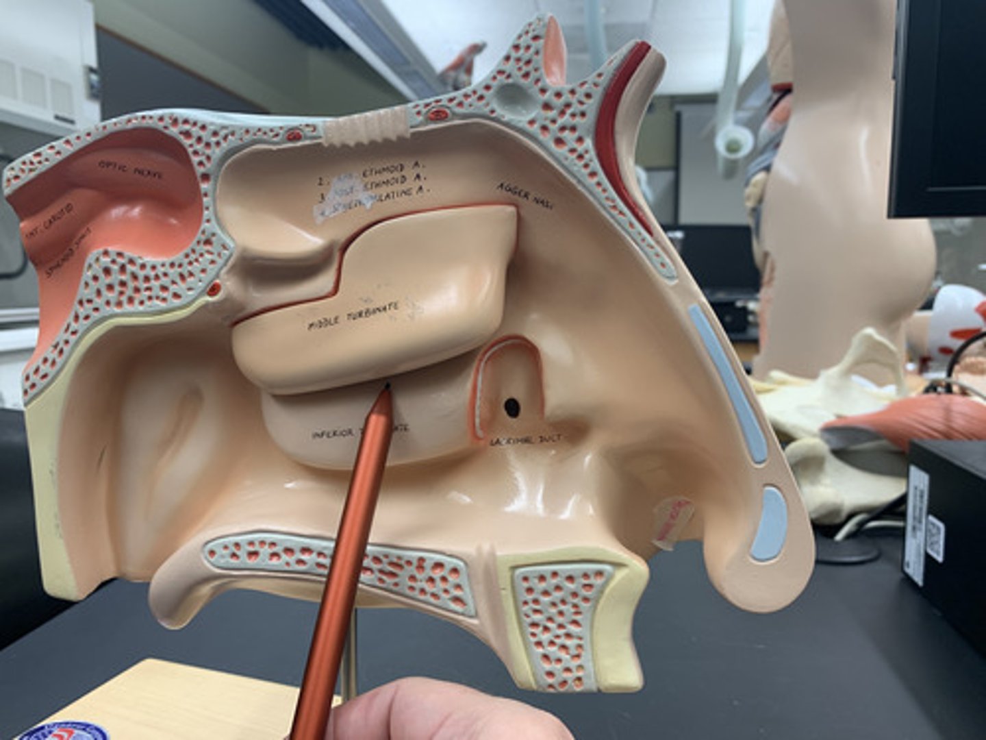
Middle nasal meatus
"valley" between lower two conchae
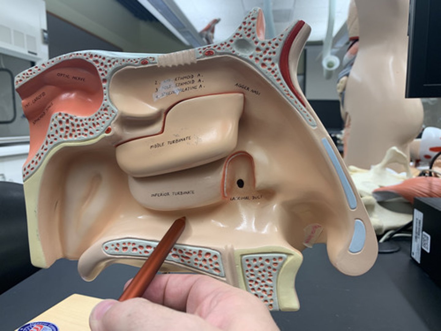
Inferior nasal meatus
"valley" under the inferior nasal concha
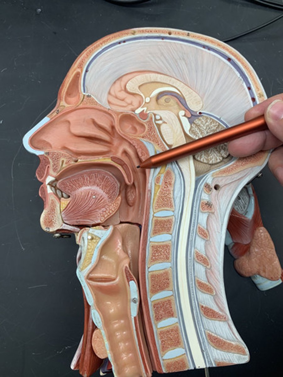
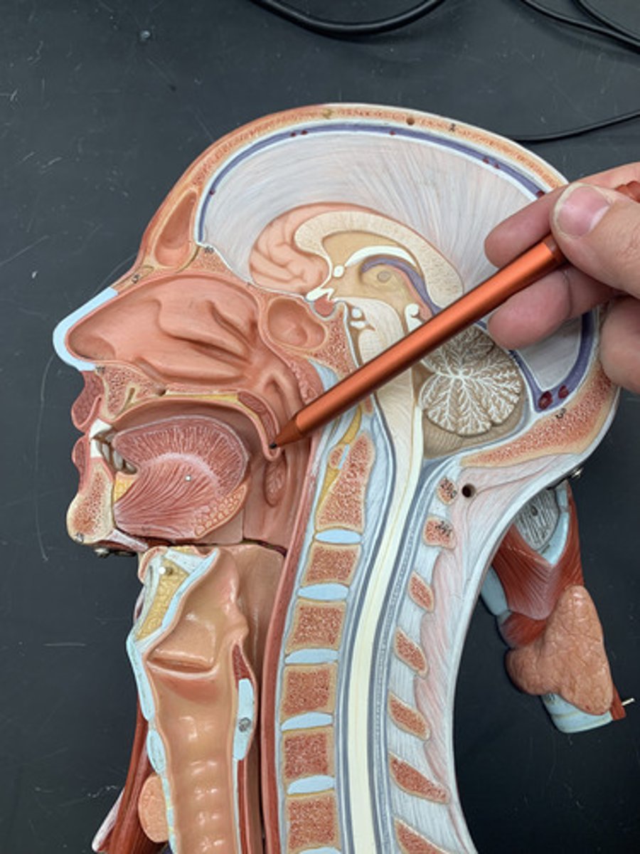
Nasopharynx
region of the pharynx at the back of the nose and above the soft palate
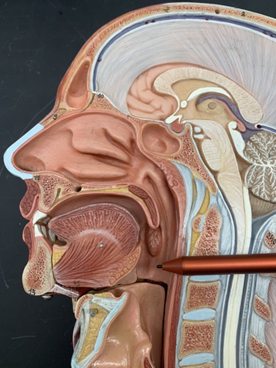
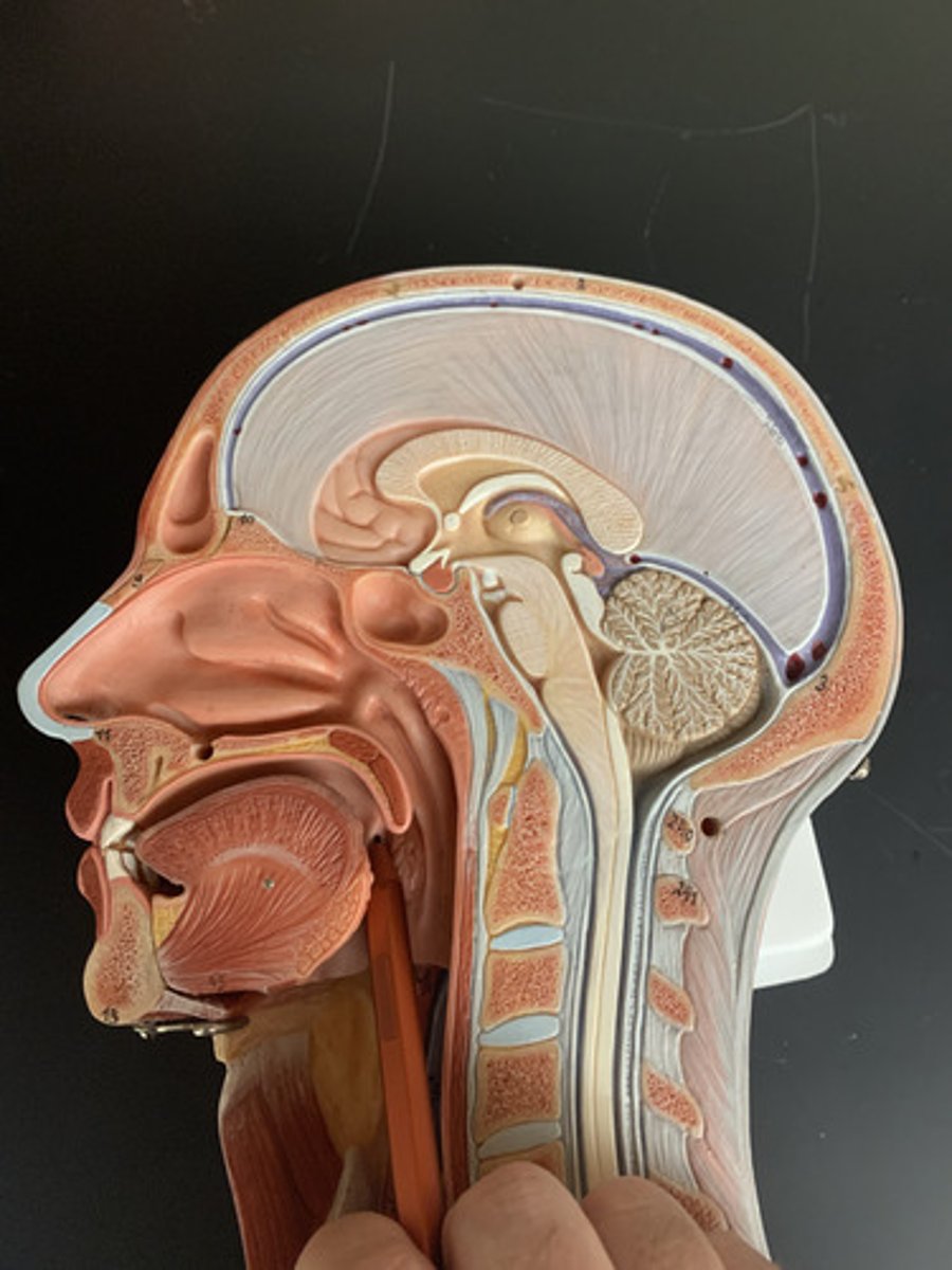
Oropharynx
central portion of the pharynx between the roof of the mouth and the upper edge of the epiglottis
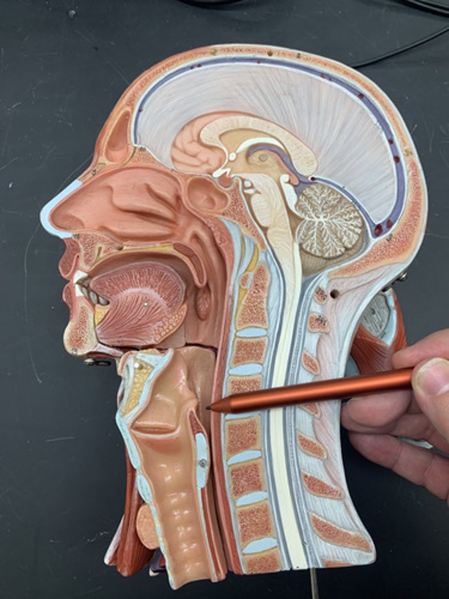
Laryngopharynx
lower part of the pharynx, just below the oropharyngeal opening into the larynx and esophagus
Nasal vestibule
space contained within the flexible tissues of the nose
Torus tubarius
elevation of cartilage caused by the auditory tube
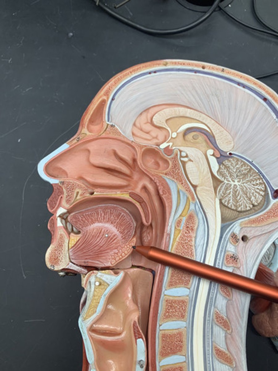
Soft palate
a muscular flap that closes off the nasopharynx during swallowing or speaking
Hard palate
bony anterior (front) portion of the palate
Uvula
free projection hanging down from the middle of the soft palate
Pharyngeal tonsil
also called adenoids; located in posterior wall of nasopharynx
Palatine tonsil
one of a pair of almond-shaped masses of lymphatic tissue in the oropharynx
Lingual tonsil
mass of lymphoid tissue located at the base of the tongue
Palatoglossal fold
1st arch, formed by palatoglossal muscle covered w/mucosa
Palatopharyngeal fold
formed by palatopharyngeus muscle covered with mucosa
Vallate papillae
largest taste buds with 8-12 forming "V" at back of tongue