Lower Limb
1/273
There's no tags or description
Looks like no tags are added yet.
Name | Mastery | Learn | Test | Matching | Spaced | Call with Kai |
|---|
No analytics yet
Send a link to your students to track their progress
274 Terms
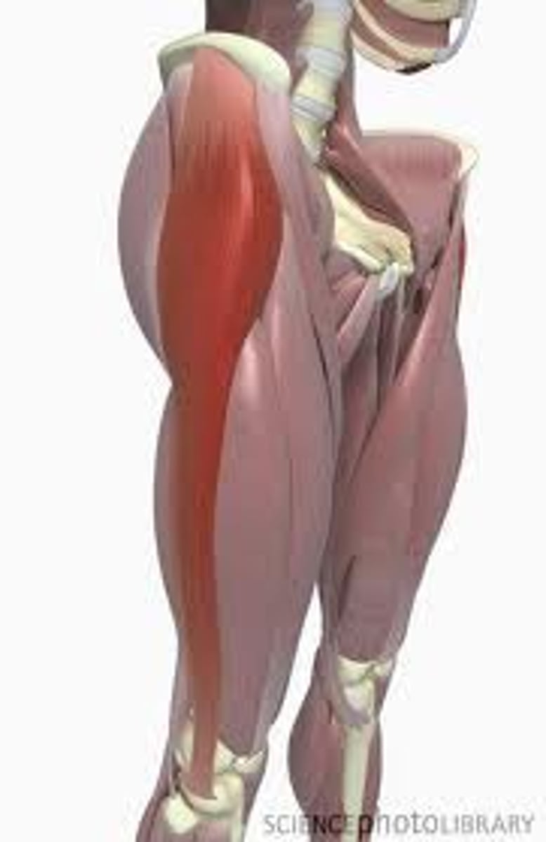
Gluteus Maximus Proximal
Ilium posterior to gluteal line; dorsal surface of sacrum and coccyx; sacrotuberous ligament

Gluteus Maximus Distal
most fibers end in iliotibial tract, which inserts into lateral condyle of tibia; some fibers insert on gluteal tuberosity

Gluteus Maximus Innervation
Inferior gluteal nerve (L5, S1, S2)

Gluteus Maximus Action
extends hip joint (especially from flexed position) and assists in lateral rotation; fixes hip joint and assists in rising from sitting position
Gluteus Maximus Blood Supply
inferior gluteal artery

Gluteus Medius proximal
external surface of ilium between anterior and posterior gluteal lines

Gluteus Medius distal
lateral surface of greater trochanter of femur
Gluteus Medius innervation
superior gluteal nerve (L5, S1)

Gluteus Medius action
abduct and medially rotate hip joint; keep pelvis level when ipsilateral limb is weight bearing and advance opposite (unsupported) side during its swing phase

Gluteus Medius blood supply
superior gluteal artery

Gluteus Minimus proximal
external surface of ilium between anterior and inferior gluteal lines
Gluteus Minimus distal
anterior surface of greater trochanter of femur

Gluteus Minimus innervation
superior gluteal nerve (L5, S1)

Gluteus Minimus action
abduct and medially rotate hip joint; keep pelvis level when ipsilateral limb is weight bearing and advance opposite (unsupported) side during its swing phase

Gluteus Minimus blood supply
superior gluteal artery

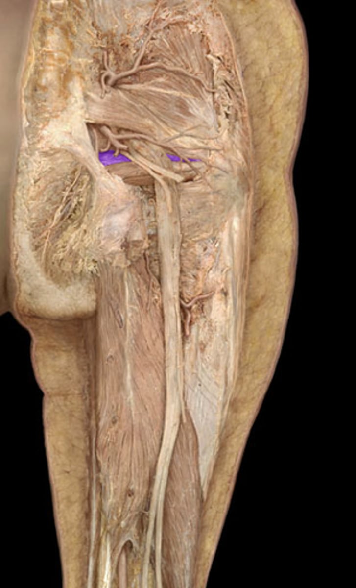
Piriformis proximal
anterior surface of sacrum; sacrotuberous ligament

Piriformis distal
superior border of greater trochanter of femur

Piriformis innervation
branches of anterior rami of S1 and S2

Piriformis action
laterally rotate extended hip joint and abduct hip joint when flexed; stabilize hip joint
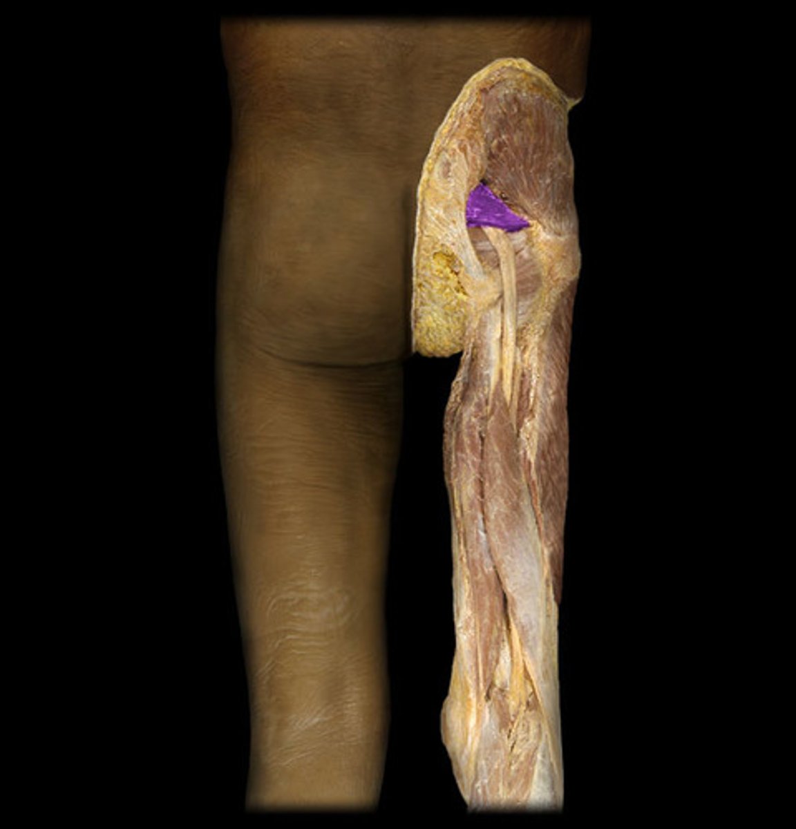
Piriformis blood supply
superior gluteal artery and inferior gluteal artery

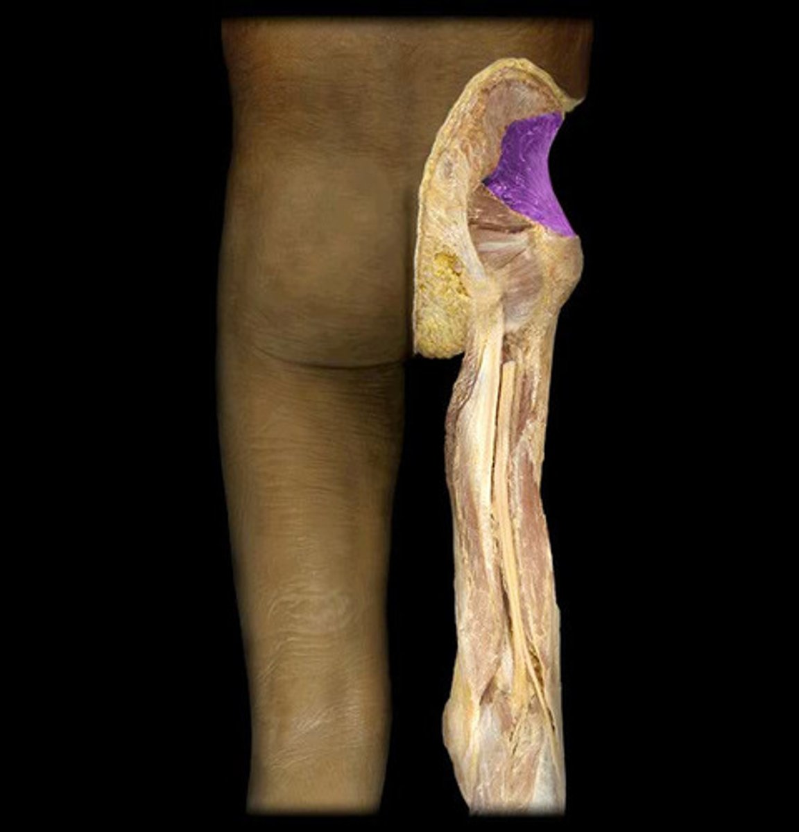
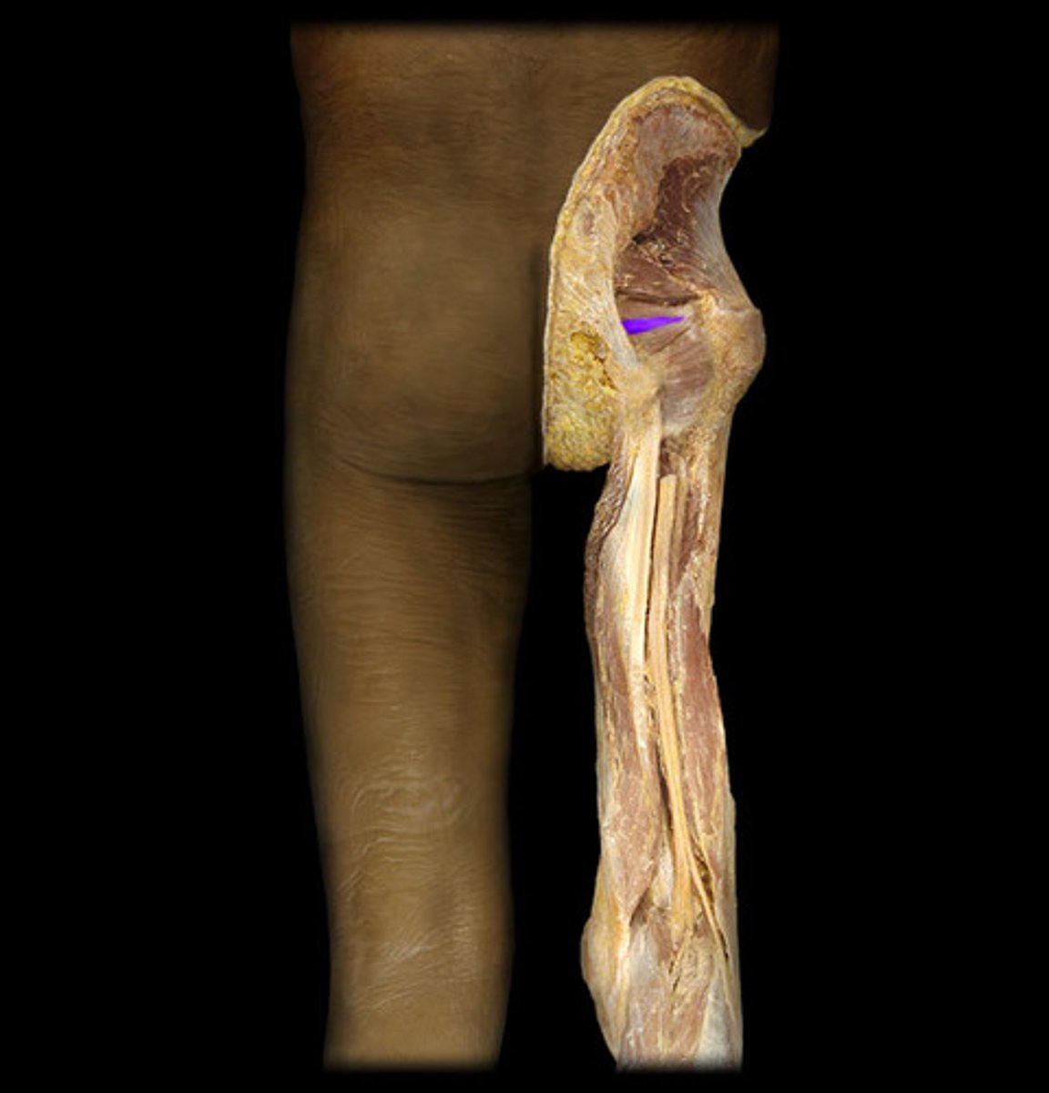
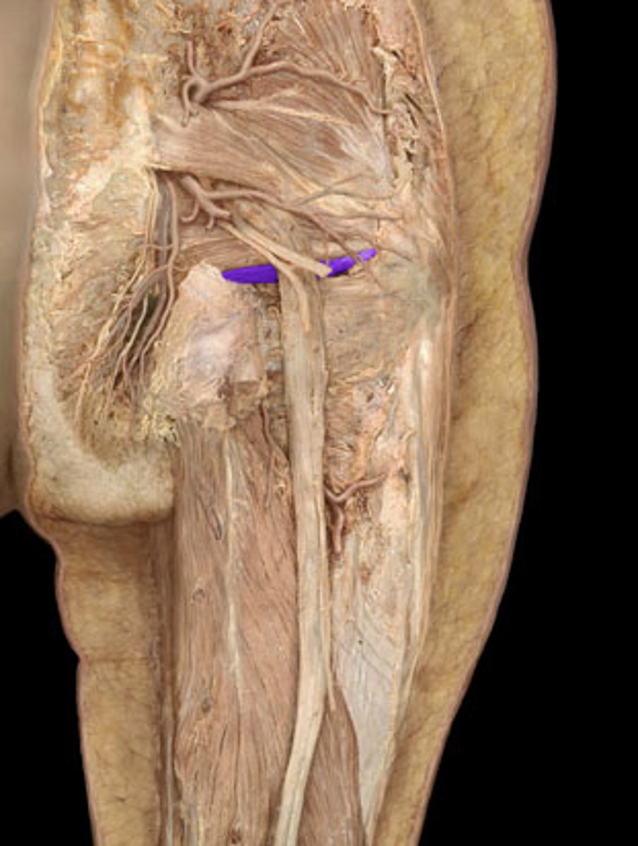
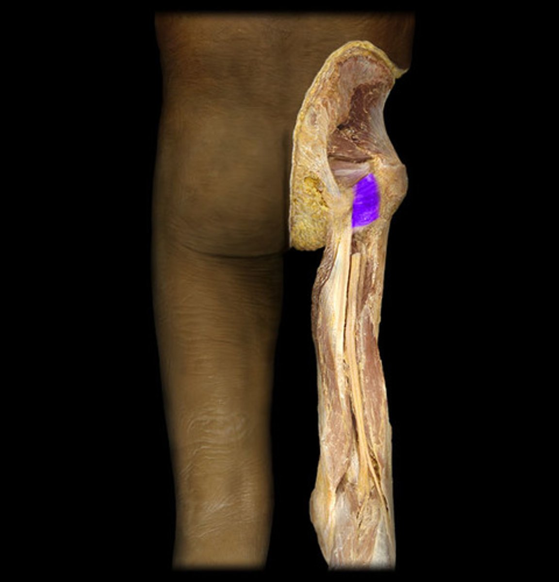
Gemellus superior proximal
ischial spine

Gemellus superior distal
medial surface of greater trochanter of femur (trochanteric fossa)
Gemellus superior innervation
nerve to obturator internus (L5, S1)

Gemellus superior action
laterally rotate extended hip joint and abduct hip joint when flexed; stabilize hip joint

Gemellus superior blood supply
inferior gluteal artery

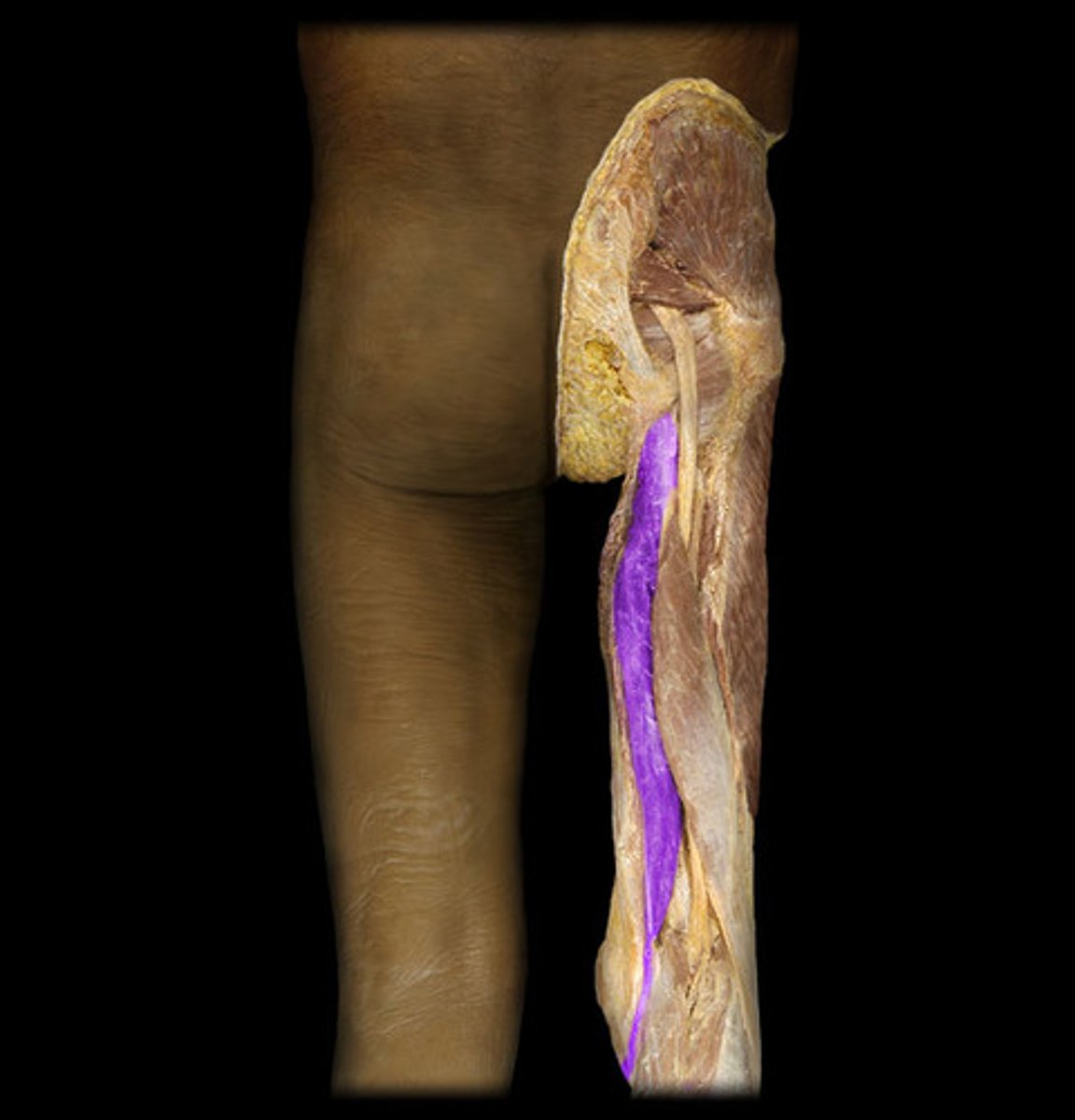
Obturator Internus proximal
pelvic surface of obturator membrane and surrounding bones
Obturator Internus distal
medial surface of greater trochanter of femur

Obturator Internus innervation
nerve to obturator internus (L5, S1)

Obturator Internus action
laterally rotate extended hip joint and abduct hip joint when flexed; stabilize hip joint

Obturator Internus blood supply
internal pudendal and obturator artery

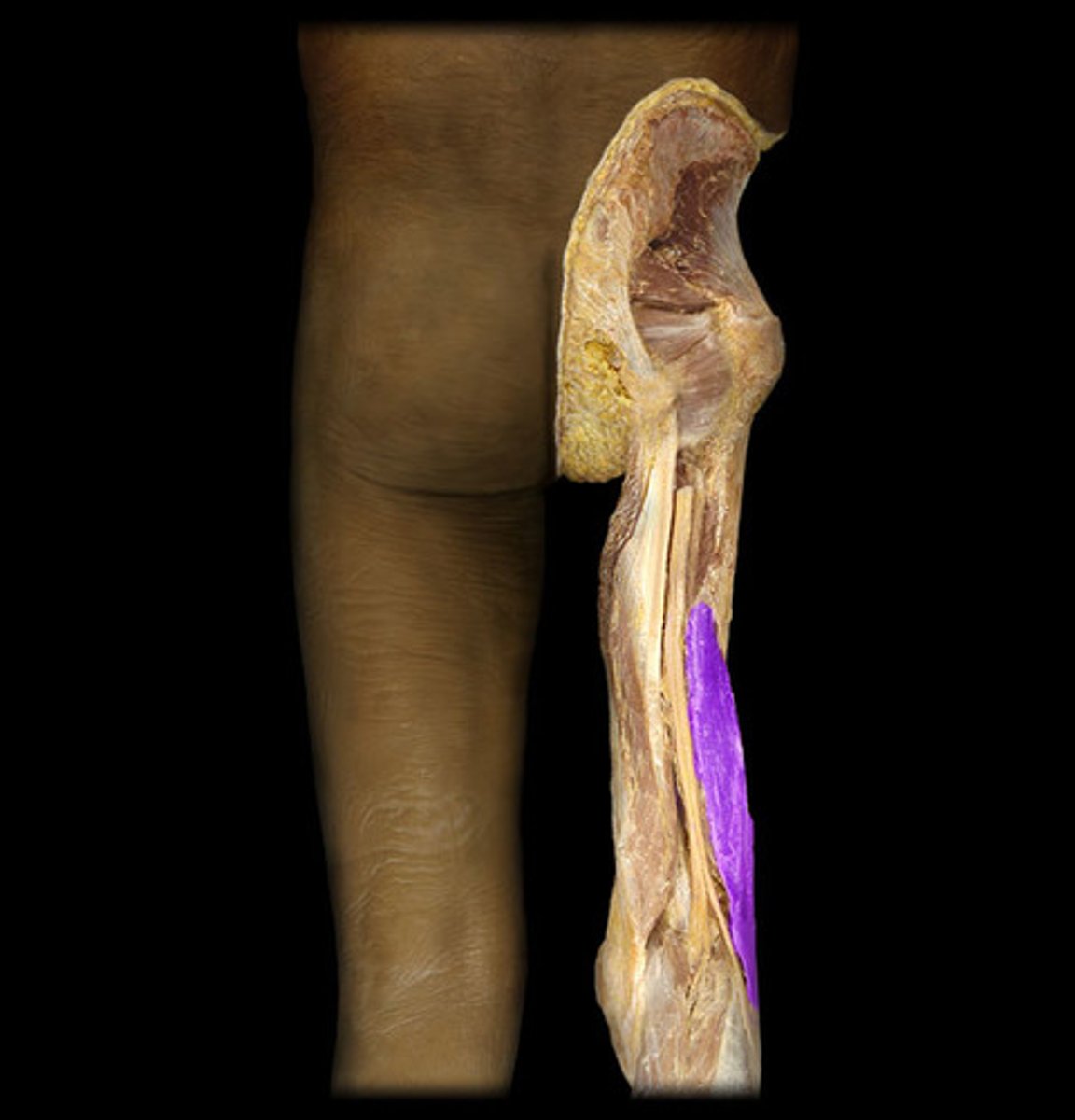
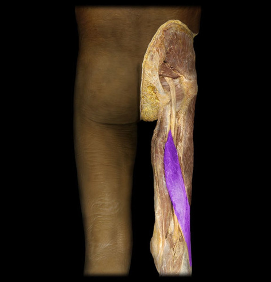
Gemellus inferior proximal
ischial tuberosity

Gemellus inferior distal
medial surface of greater trochanter of femur

Gemellus inferior innervation
nerve to quadratus femoris (L5, S1)
Gemellus inferior action
laterally rotate extended hip joint and abduct hip joint when flexed; stabilize hip joint

Gemellus inferior blood supply
medial circumflex femoral artery

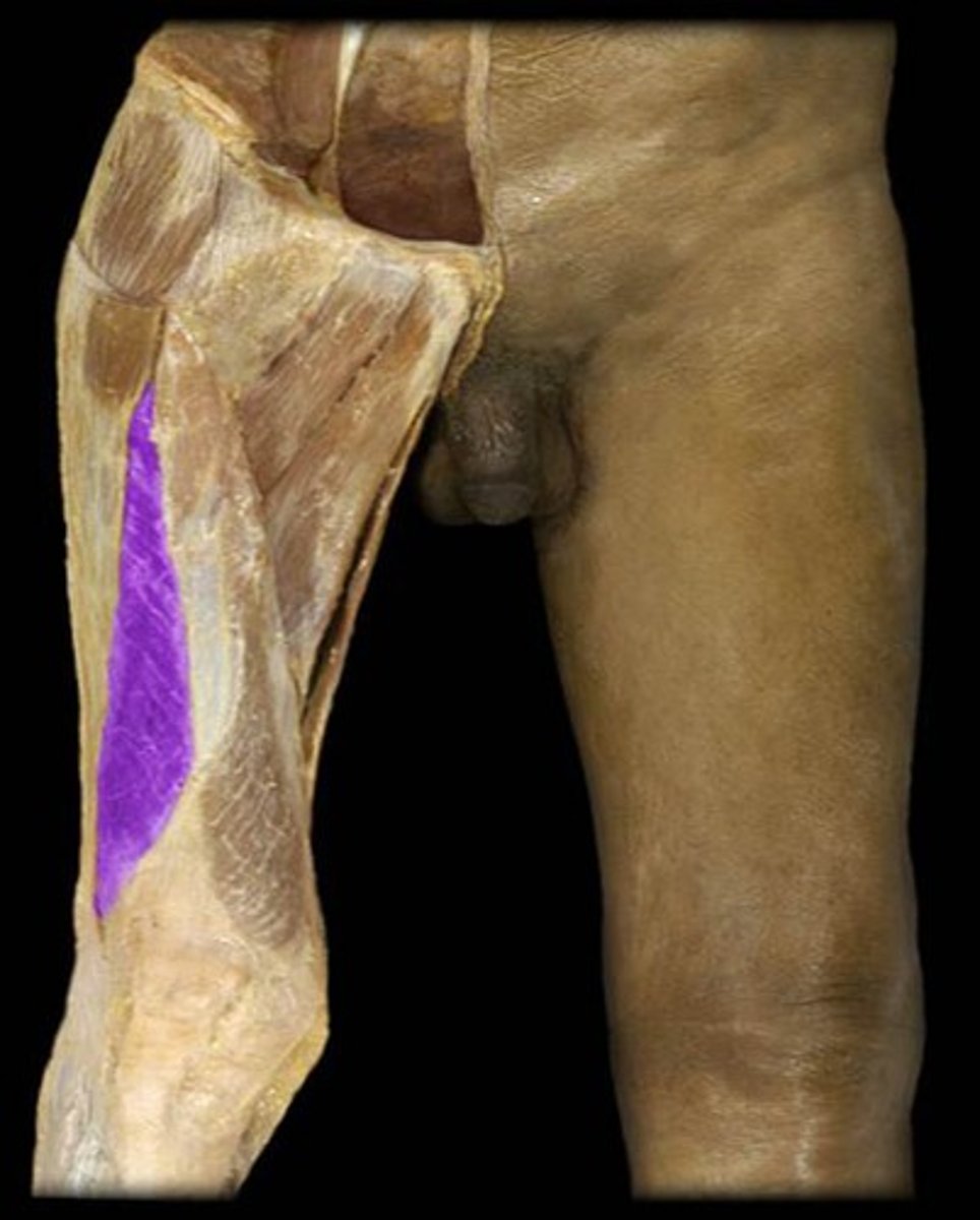
Tensor Fascia Lata proximal
anterior superior iliac spine; anterior part of iliac crest
Tensor Fascia Lata distal
Iliotibial tract, which attaches to lateral condyle of tibia

Tensor Fascia Lata innervation
superior gluteal nerve (L5, S1)

Tensor Fascia Lata action
abduct and medially rotate hip joint; keep pelvis level when ipsilateral limb is weight bearing and advance opposite (unsupported side) during its swing phase

Tensor Fascia Lata blood supply
lateral circumflex femoral artery

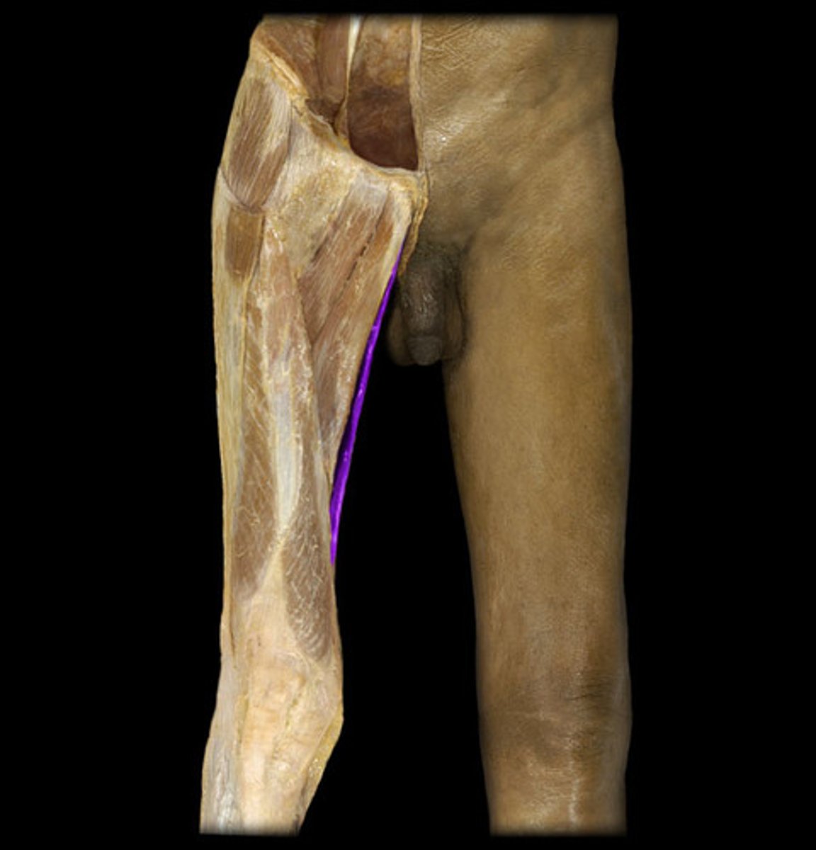
Obturator Externus proximal
margins of obturator foramen and obturator membrane
Obturator Externus distal
trochanteric fossa of femur
Obturator Externus innervation
obturator nerve (L3, L4)
Obturator Externus action
laterally rotates hip joint; stabilizes hip joint
Obturator Externus blood supply
medial circumflex femoral artery and obturator artery
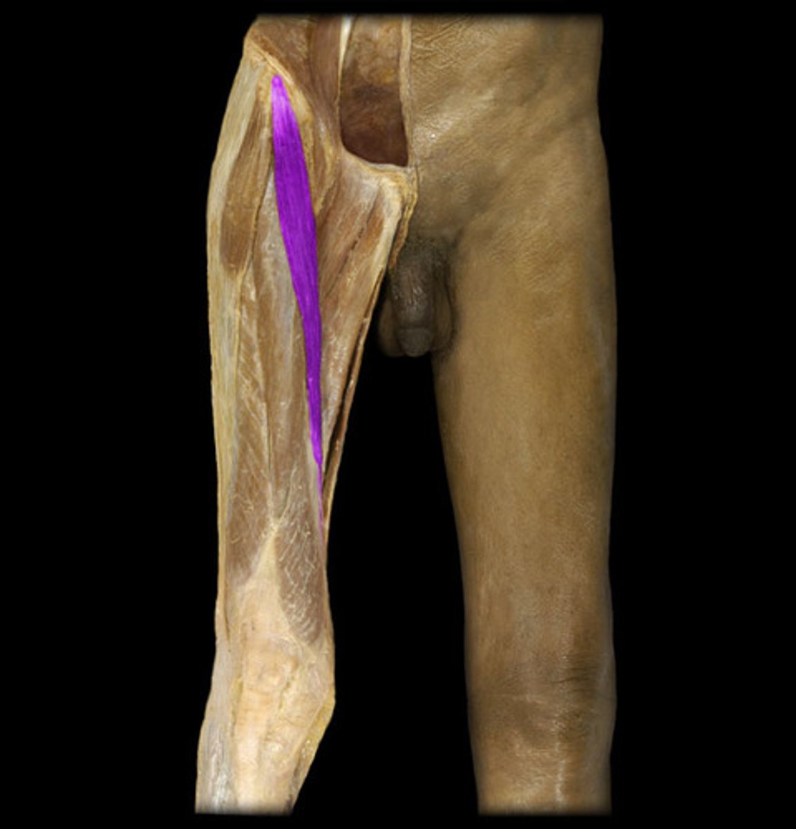
Quadratus femoris proximal
lateral border of ischial tuberosity

Quadratus femoris distal
quadrate tubercle on intertrochanteric crest of femur and area inferior to it

Quadratus femoris innervation
nerve to quadratus femoris (L5, S1)
Quadratus femoris action
laterally rotates hip joint; stabilizes hip joint

Quadratus femoris blood supply
medial circumflex femoral artery

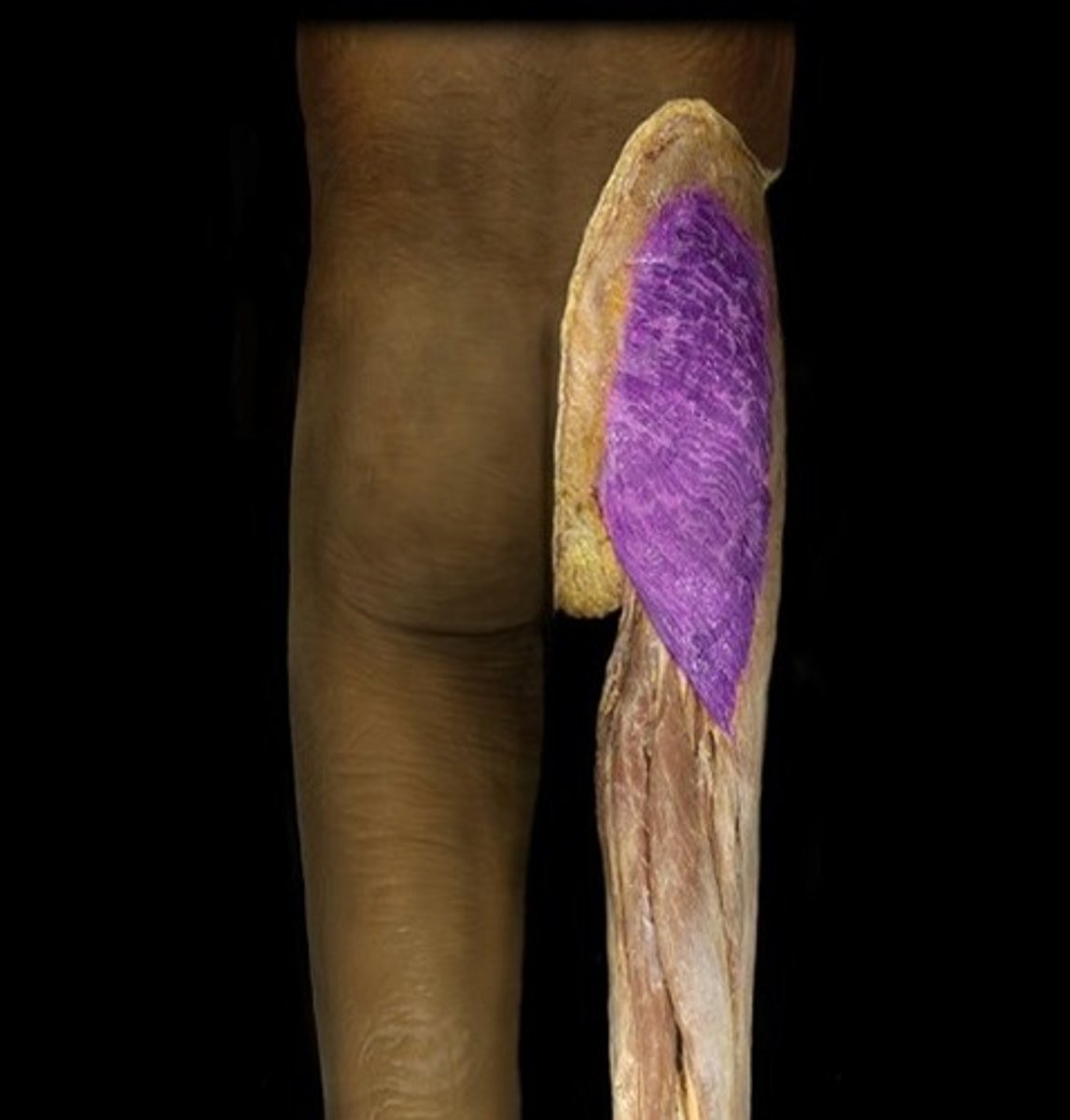
Vastus lateralis proximal
greater trochanter and lateral lip of linea aspera of femur

Vastus lateralis distal
via common tendinous (quadriceps tendon) and independent attachments to base of patella; indirectly via patellar ligament to tibial tuberosity; medial and lateral vasti also attach to tibial tuberosity; medial and lateral vast also attach to tibia and patella via aponeuroses (medial and lateral patellar retinacula)

Vastus lateralis innervation
Femoral nerve (L2, L3, L4)
Vastus lateralis action
extends knee joint

Vastus lateralis blood supply
lateral circumflex femoral artery and profunda femoris artery

Biceps Femoris Short Head proximal
linea aspera and lateral supracondylar line of femur

Biceps Femoris Short Head distal
lateral side of head of fibula. tendon is split at this stie by fibular collateral ligament of knee

Biceps Femoris Short Head innervation
common fibular division of sciatic nerve (L5, S1, S2)
Biceps Femoris Short Head action
flexes knee joint and laterally rotates it when flexed

Biceps Femoris Short Head blood supply
perforating branches of profunda femoris, inferior gluteal, and medial circumflex femoral arteries

Biceps Femoris Long Head proximal
ischial tuberosity

Biceps Femoris Long Head distal
lateral side of head of fibula, tendon is split at this stie by fibular collateral ligament of knee

Biceps Femoris Long Head innervation
tibial division of sciatic nerve (L5-S2)

Biceps Femoris Long Head action
flexes knee joint and laterally rotates it when flexed; extends hip joint

Biceps Femoris Long Head blood supply
perforating branches of profunda femoris, inferior gluteal, and medial circumflex femoral arteries
Sartorius proximal
anterior superior iliac spine and superior part of notch inferior to it

Sartorius distal
superior part of medial surface of tibia (pes anserinus)

Sartorius innervation
femoral nerve (L2, L3)

Sartorius action
flexes, abducts, ad lateral rotates hip joint; flexes knee joint (medially rotates leg when knee joint is flexed)
Sartorius blood supply
femoral artery

Gracilis proximal
body and inferior ramus of pubis

Gracilis distal
superior part of medial surface of tibia (as part of pes anserinus)

Gracilis innervation
obturator nerve (L2, L3)

Gracilis action
Adducts hip joint; flexes knee joint, medially rotating it when flexed
Gracilis blood supply
profunda femoris artery, medial circumflex femoral artery

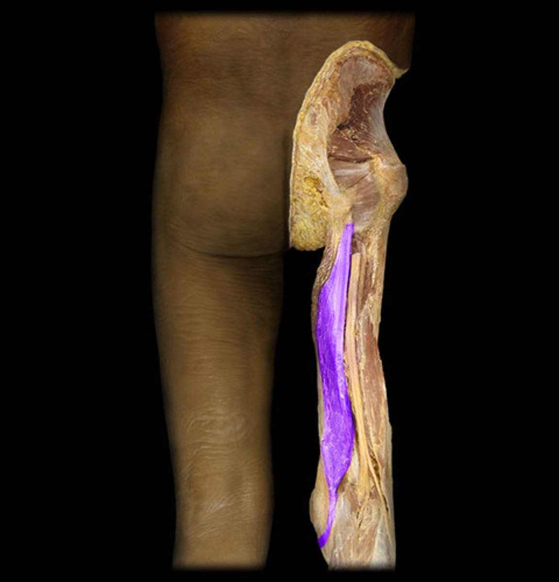
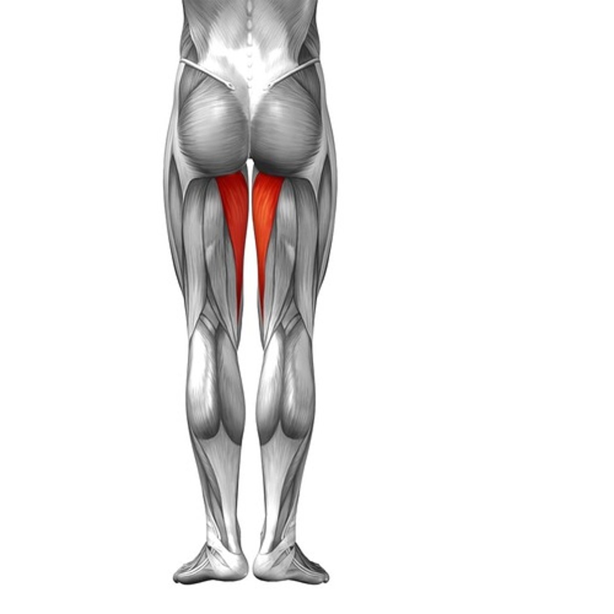
Semitendinosus proximal
ischial tuberosity

Semitendinosus distal
superior part of medial surface of tibia (pes anserinus)

Semitendinosus innervation
tibial division of sciatic nerve part of tibia (L5, S1, S2)

Semitendinosus action
extend hip joint; flex knee joint and medially rotate it when flexed. when hip and knee joints are flexed (sitting) these muscles can extend trunk at hip joint (to rise)
Semitendinosus blood supply
perforating branch of profunda femoris and medial circumflex femoral arteries

Semimembranosus proximal
ischial tuberosity

Semimembranosus distal
posterior part of medial condyle of tibia. reflected attachment forms oblique popliteal ligament (to lateral femoral condyle)

Semimembranosus innervation
tibial division of sciatic nerve part of tibia (L5, S1, S2)
Semimembranosus action
extend hip joint; flex knee joint and medially rotate it when flexed. when hip and knee joints are flexed (sitting) these muscles can extend trunk at hip joint (to rise)

Semimembranosus blood supply
perforating branch of profunda femoris and medial circumflex femoral arteries

Gastrocnemius proximal
lateral head: lateral aspect of lateral condyle of femur
medial head: popliteal surface of femur, superior to medial condyle

Gastrocnemius distal
posterior surface of calcaneus via calcaneal tendon
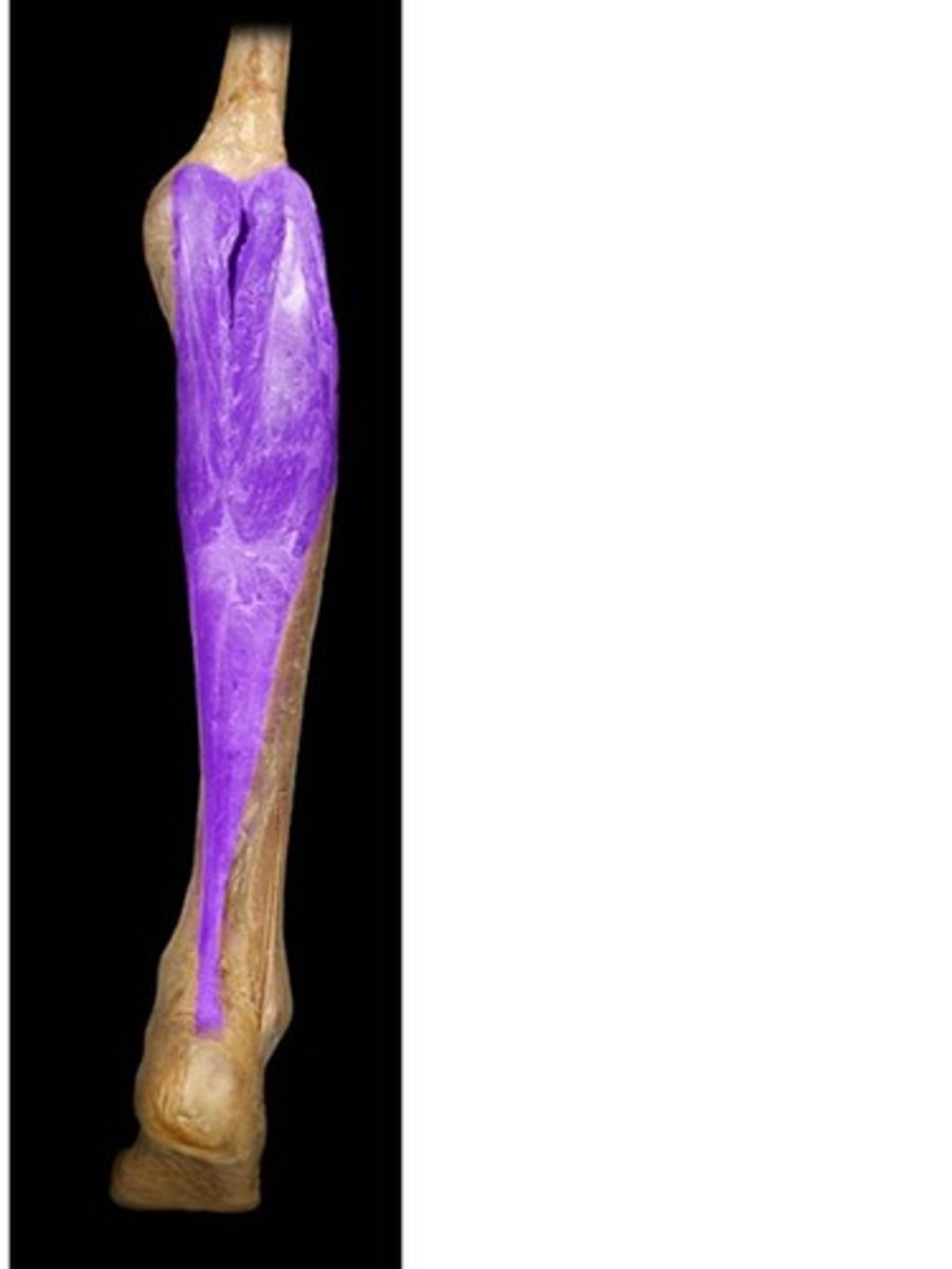
Gastrocnemius innervation
tibial nerve (S1, S2)

Gastrocnemius action
plantarflexes ankle joint when knee joint is extended; raises heel during walking; flexes knee joint

Gastrocnemius blood supply
popliteal and posterior tibial arteries

Adductor magnus proximal
adductor part: inferior ramus of pubis, ramus of ischium
hamstring part: ischial tuberosity

Adductor magnus distal
adductor part: gluteal tuberosity, linea aspera, medial supracondylar line
hamstring part: adductor tubercle of femur

Adductor magnus innervation
adductor part: obturator nerve (L2, L3, L4) and branches of posterior division
hamstring part: tibial part of sciatic nerve (L4)
Adductor magnus action
adducts hip joint
adductor part: flexes hip joint
hamstring part: extends hip joint

Adductor magnus blood supply
femoral, profunda femoris, and obturator arteries

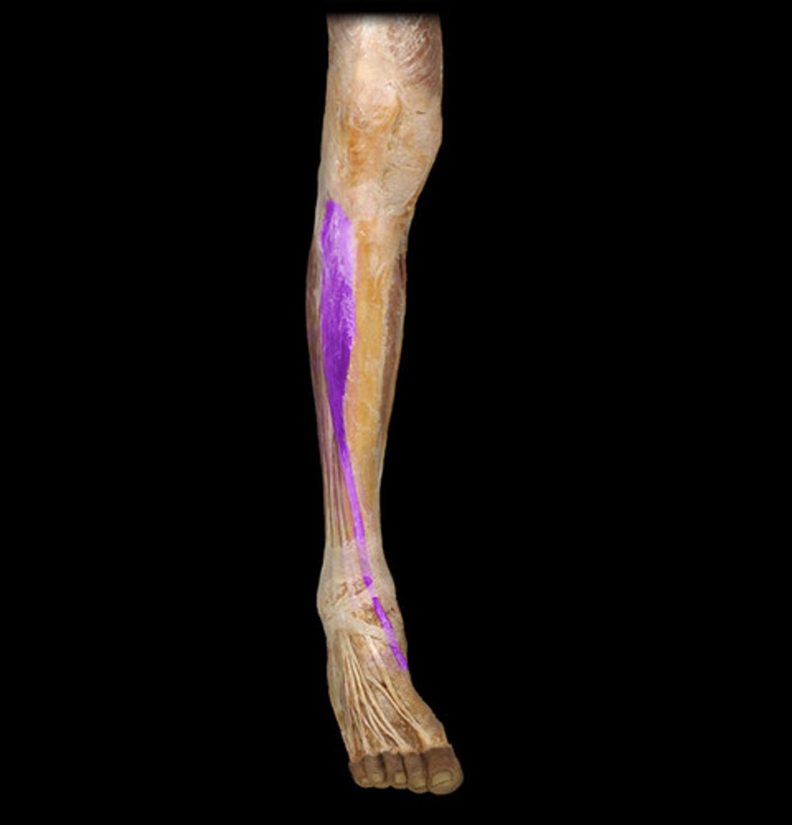
Tibialis Anterior proximal
lateral condyle and superior half of lateral surface of tibia and interosseous membrane

Tibialis Anterior distal
medial and inferior surfaces of medial cuneiform and base of 1st metatarsal

Tibialis Anterior innervation
deep fibular nerve (L4, L5)

Tibialis Anterior action
dorsiflexes ankle joint and inverts subtalar joint

Tibialis Anterior blood supply
anterior tibial artery