Lesson 18 Visual System
1/59
There's no tags or description
Looks like no tags are added yet.
Name | Mastery | Learn | Test | Matching | Spaced | Call with Kai |
|---|
No analytics yet
Send a link to your students to track their progress
60 Terms
Retina
thin layer of cells that transduces light energy into neuronal activity
Layers of the retina
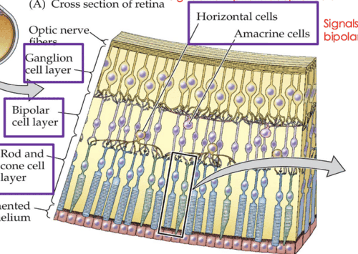
Path of visual information in the retina:
Photoreceptors → (horizontal cells)
bipolar cells → (amacrine cells)
ganglion cells
how does light pass through the retina?
the light passes through the ganglion cells and bipolar cells and hits the rods and cones first (photoreceptors), then it is processed and moved back through the bipolar cell layer and then to the ganglion cell layer
photoreceptors
are sensory neurons that detect light
types of photoreceptors
rods and cones
Bipolar cells receive input from photoreceptors and
synapse onto ganglion cells whose axons from the ...
optic nerve which carries information to the brain
outline the pathway starting at the photoreceptor
Photoreceptor → bipolar cell → ganglion cell → optic nerve → optic chiasm → optic tract → LGN → V1
Rods
-Long, cylindrical Outer Segment with
many discs
➢ Higher photopigment concentration
➢ 1000 times more sensitive to light
➢ 92 million of these in each human
retina
➢ Contribute to vision in scotopic
conditions
➢ Bulk of contribution in nighttime
lighting
➢ Absent in the fovea
➢ Rhodopsin - pigment
Cones
-Shorter, tapering outer segment with
fewer disks
➢ Lower photopigment concentration
➢ 5 million of these in each human retina
➢ Bulk of contribution in photopic (light)
conditions
➢ Bulk of contribution in daytime lighting
➢ Concentrated in the fovea
➢ Perception of Color
➢ Contain one of three opsins (short,
medium, and long wavelength
activated) pigment
Both rods and cones
-Outer segment
➢ Inner segment
➢ Synaptic terminal
➢ Cell body
➢ Mesopic conditions (Intermediate light levels)
visual acuity is a measure of how much
detail we see and is sharper in the center of the visual field
fovea
The center region of the retina has a high density of
cones
- This region receives direct light input that does not pass through other cells or blood vessels
-Rods are more numerous in the periphery and are absent in the fovea
-Rods have high sensitivity to dim light but low acuity
In photopic conditions...
our visual acuity is much higher in our central retina because of the sole presence of cones there
why do we have a blindspot?
a lack of photoreceptors, so no visual information is gathered
Transduction
the process by which an environmental stimulus (light) causes an electrical response (receptor potential) in a sensory receptor cell
steps of phototransduction
1. Light activates (bleaches) the
photopigment (opsin or rhodopsin)
- The G-protein is stimulated
2. The effector enzyme is activated
3. PDE is activated and cGMP
levels are reduced
4. Na+ channels close, and the
cell membrane hyperpolarizes
Phototransduction in cones is virtually the same as in rods, but the difference is ...
the type of opsins in the membranous disks of the cone outer segments.
Receptive field
area of retina or visual space that when light is applied
the firing rate of the neuron changes
Ganglion cells have what kind of receptive fields?
concentric receptive fields (a circular central area with a ring around it, the surround)
Bipolar and ganglion cells have two types of receptive fields, what are they?
-On-center / Off-surround
- Off-center / On-Surround
The center and surround are always...
opposites
how does membrane potential of a cell change in response to light?
the membrane potential will hyperpolarize because photons strike the discs inside the photoreceptors are captured by special photopigment receptor molecules causing Na+ channels to close.
when light shine on an off area what happens?
hyper polarization
when light shines on an on area what happens?
depolarization
P-type ganglion cells
-Parvocellular
-90% of the ganglion cell
population
-Smaller Receptive fields
-Smaller cells
-Some are sensitive to differences in wavelength of light (color detection!)
-Color opponent cells (red versus green and blue versus yellow)
M-type ganglion cells
-Magnocellular
-5% of the ganglion cell population
-Larger Receptive fields
-Larger Cells
-Conduct AP more rapidly
-MORE sensitive to low-contrast stimuli
which light stimulus would produce the most action potential?
the one with the most depolarization
Ganglion cells carry information from the retina to the
Thalamus (LGN)
The structures in the retinofugal
pathway (neural pathway leaving the
retina, in order they receive visual
information, are...
(1)Optic nerve
(2)Optic Chiasm
(3)Optic Tract
Trace the pathway of visual information from retina to the cortex:
Photorceptor → (horizontal cells) bipolar cell → (amacrine cells) ganglion cells → optic nerve → optic
chiasm → LGN → V1
Visual field
entire region of space (measured in degrees of visual angle) that can be seen with both eyes looking straight ahead
Binocular visual field
central portion of visual field viewed by both retinas
Describe why humans have a binocular visual field
Proportionally more axons cross the midline in prey animals that have laterally placed eyes with little
overlap in their fields of vision
-This gives a prey animal an
especially wide field of view at the cost of poor depth perception
In contrast, predators have greater overlap in their visual fields due to their front-facing eyes, and much
better depth perception
label the visual fields and the major components of the visual system.
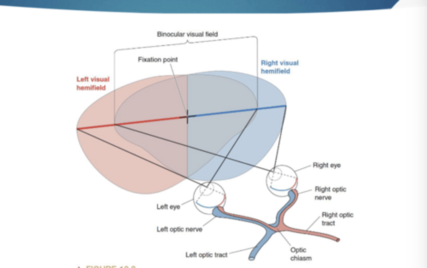
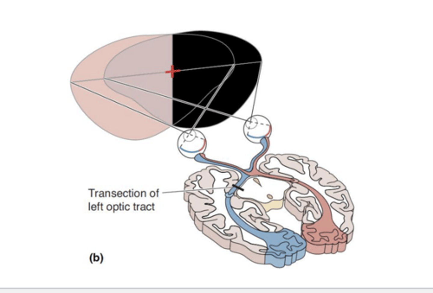
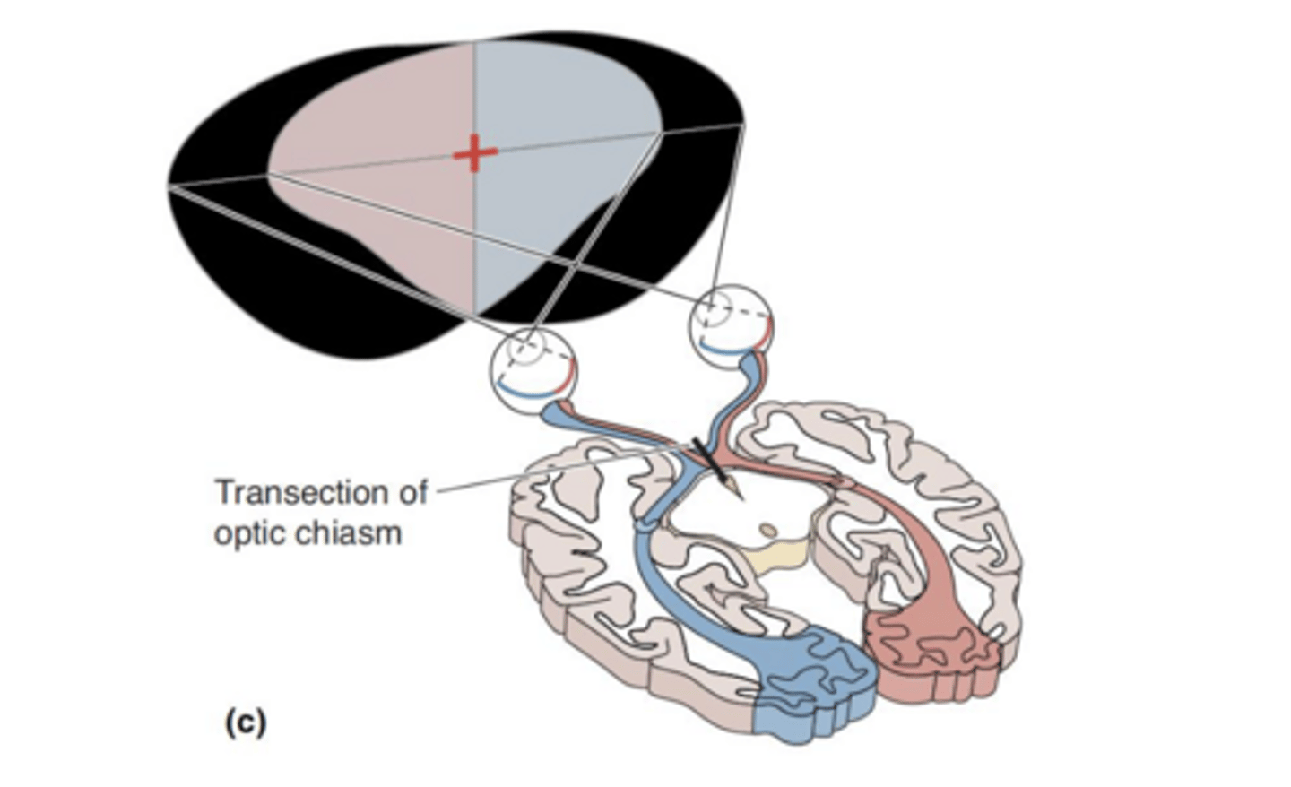
What you see in one visual field is processed...
by the opposite tract
- ex: something seen in your left visual field is processed by the right tract
outline what is seen by the nasal retina and temporal retina
Temporal retina - does cross the optic chasm
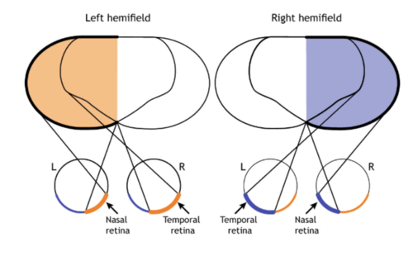
to see your left visual hemifield, you use your
1. nasal retina of the left eye
2. temporal retina of the right eye
lesions
areas of tissue that have been pathologically altered by injury, wound, or infection
lesion in the left optic nerve
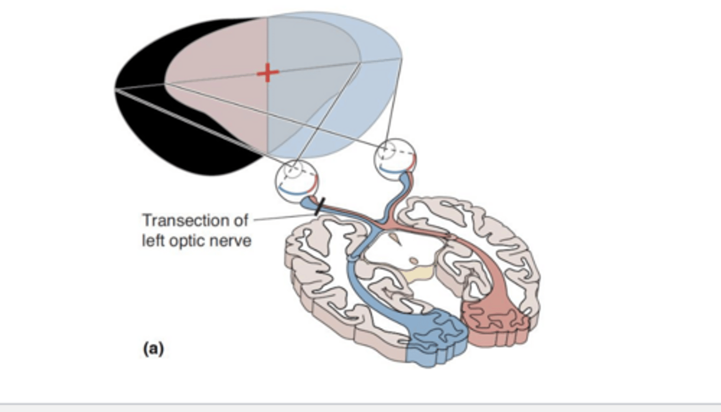
lesion in the left optic tract
lesion at optic chiasm
Ganglion cells send action potentials...
down their axons (the optic nerve)
In vertebrates, some or all of each optic nerve crosses the midline at the ...
optic chiasm; after crossing, it is known as the optic tract.
Most axons of the optic tract terminate on cells in the...
lateral geniculate nucleus (LGN) of the thalamus.
Axons of LGN neurons terminate in the
primary visual cortex (V1), also called the striate cortex.
The cortical visual areas are organized into two major streams...
Dorsal stream: spatial location and movement of objects
Ventral stream: identification and recognition of objects
ventral stream
-Projects toward temporal lobe
• Perception of visual world and object recognition
• Shape and color perception
• Fusiform Face Area (FFA)
• Some cells in this stream respond nearly exclusively to faces
• Colors and abstracts shapes good stimuli for cells in this stream
• prospagnosia
Dorsal stream
-Projects toward parietal lobe
-Visual motion analysis and visual
control of action
-Cells selective for motion (linear, radial,
& circular)
- Proposed roles: navigation, directing
eye movements, motion perception
In order to get a sharply focused image to fall
onto the retina...
-The cornea bends light entering the eye, & light then travels through the pupil
• The lens changes shape to focus the light onto the retina
In those with myopia
the lens focuses the image in front of the retina.
In those with farsightedness (hyperopia)...
the lens focuses the image behind the retina.
What happened during lassie/ wearing contacts
-In LASIK eye surgery, the cornea is reshaped.
-Contacts and LASIK alter the light refraction prior to it hitting the lens, so the image will focus on the retina.
Color is perceived by ...
The visual system as we detect differences in the wavelength of photons within
Color perception is influenced by
light intensity, prior exposure to different stimulus, and surrounding colors
trichromatic hypothesis of color perception
Three different types of cones, each with peak responding to a specific part of the spectrum
-Short (S): 420 nm
- Medium (M): 530 nm
- Long (L): 560 nm
-Each has a separate pathway to the brain, color recognized based on which receptors are activated
Colorblindness
a recessive x-linked disorder in which an individual cannot distinguish between certain colors
-color blindness is more common in
males than females (~8% vs. 0.5%)
Genes encoding L & M photopigments are carried on the
X chromosome
Red-Green color blindness:
Absence or abnormal M or L cones
Blue-Yellow color blindness
Absence or abnormal S cones