NASM CPT 7th Edition (Chapter 5)
1/104
There's no tags or description
Looks like no tags are added yet.
Name | Mastery | Learn | Test | Matching | Spaced | Call with Kai |
|---|
No analytics yet
Send a link to your students to track their progress
105 Terms
Human Movement System (HMS)
The collective components and structures that work together to move the body: muscular, skeletal, and nervous systems
Kinetic Chain
A concept that describes the human body as a chain of interdependent links that work together to perform movement
Nervous System
A network of specialized cells called neurons that transmit and coordinate signals, providing a communication network within the human body
Neuron
A specialized cell that is the functional unit of the nervous system
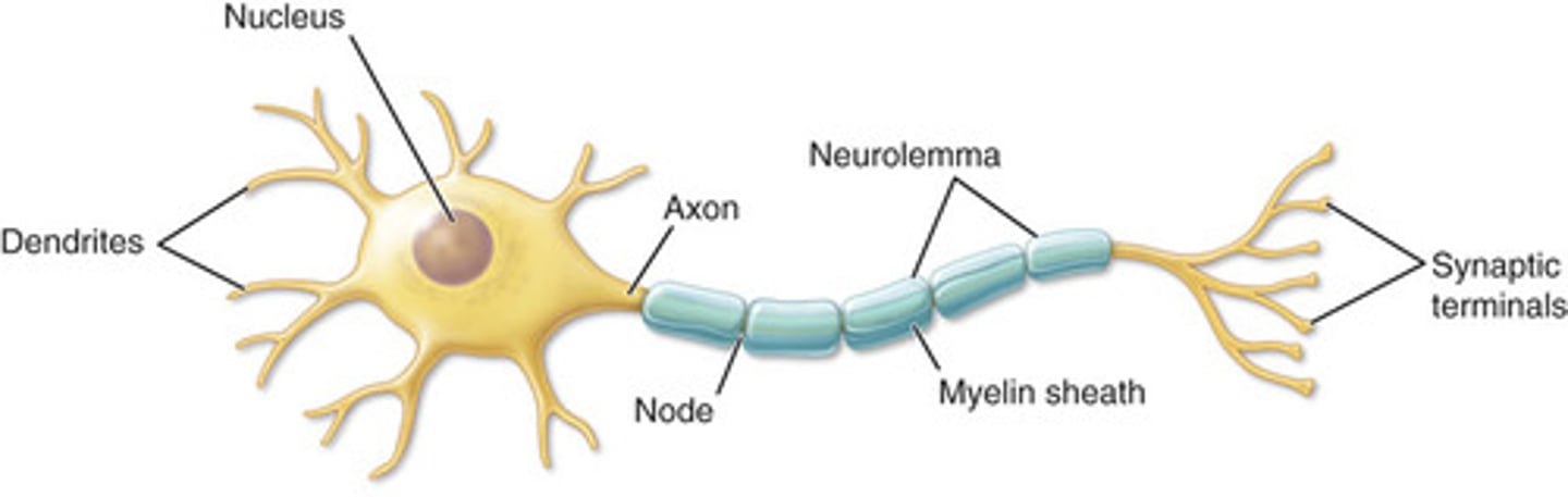
Nucleus
Cellular structure or organelle that contains the majority of the cell's genetic material in the form of chromosomes
Organelles
Tiny cellular structures that perform specific functions within a cell. Examples include nuclei, mitochondria, lysosomes, ribosomes, and the endoplasmic reticulum
Mitochondria
The parts of the cell that use nutrients to create energy for the cell; commonly known as the powerhouses of the cell
Effector Sites
A part of the body, such as a muscle or organ, that receives a signal from a neuron to produce a physiological response
Electrolytes
Minerals that have an electrical charge to help transmit nerve impulses throughout the body, such as sodium, potassium, and magnesium
Central Nervous System (CNS)
A division of the nervous system that includes the brain and spinal cord
Peripheral Nervous System (PNS)
Nerves that connect the rest of the body to the central nervous system
Afferent Pathway
Sensory pathway that relays information to the central nervous system
Efferent Pathway
A motor pathway that relays information from the central nervous system to the rest of the body
Interneurons
Neurons located within the spinal cord and brain that transmits impulses between afferent and efferent neurons
Mechanoreceptors
Specialized structures that respond to mechanical forces (touch and pressure) within tissues and then transmits signals through sensory nerves
Somatic Nervous System
Nerves that serve the outer areas of the body and skeletal muscle and are largely responsible for the voluntary control of movement
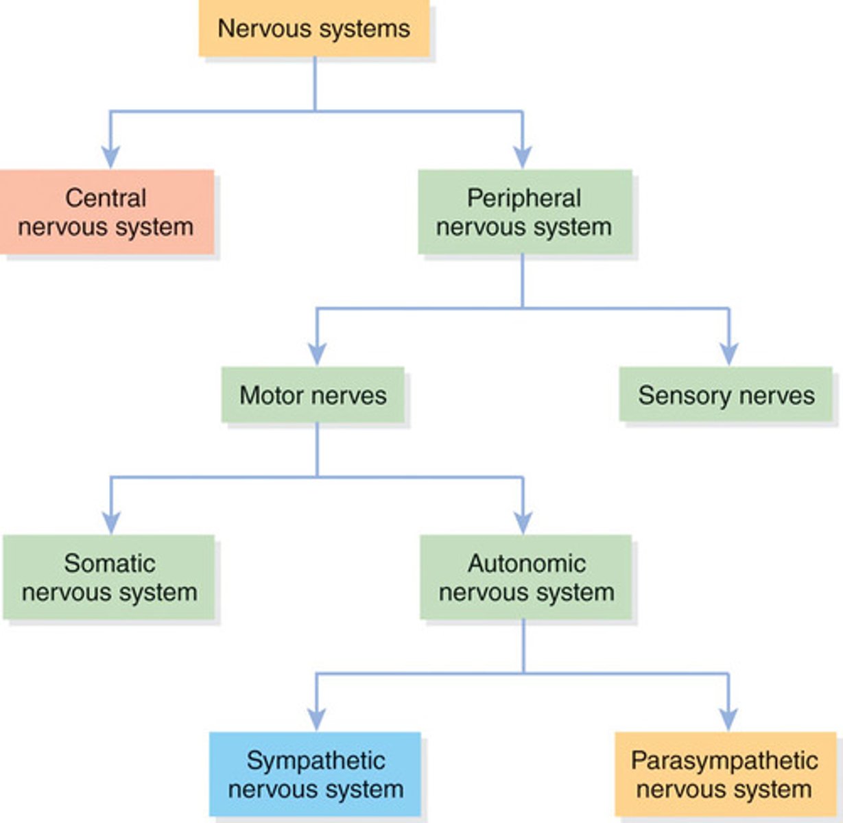
Autonomic Nervous System (ANS)
A division of the peripheral nervous system that supplies neural input to organs that run the involuntary processes of the body (e.g., circulating blood, digesting food, producing hormones)
Sympathetic Nervous System
Subdivision of the autonomic nervous system that works to increase neural activity and put the body in a heightened state
Parasympathetic Nervous System
Subdivision of the autonomic nervous system that works to decrease neural activity and put the body in a more relaxed state
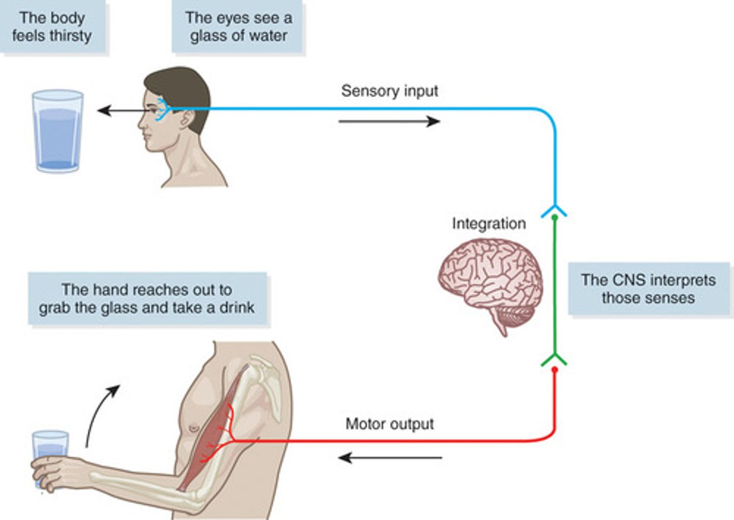
Sensory Function
Ability of the nervous system to sense changes in either the internal or external environment
Proprioception
The body's ability to naturally sense its general orientation and relative position of its parts
Integrative Function
The ability of the nervous system to analyze and interpret the sensory information to allow for proper decision-making, which produces an appropriate response
Motor Function
The neuromuscular (or nervous and muscular systems) response to the integrated sensory information
Muscle Spindles
Sensory receptors sensitive to change in length of the muscle and the rate of that change
Stretch Reflex
Neurological signal from the muscle spindle that causes a muscle to contract to prevent excessive lengthening
Sensory, Integrative, and Motor Function
Golgi Tendon Organ (GTO)
A specialized sensory receptor located at the point where skeletal muscle fibers insert into the tendons of skeletal muscle; sensitive to changes in muscular tension and rate of tension change
Joint Receptors
Receptors located in and around the joint capsule that respond to pressure, acceleration, and deceleration of the joint
Neuroplasticity
The concept that the brain will continually change or grow, reforming neural pathways throughout an individual's entire life span
Neurocircuitry
The interconnection of neurons in the brain and spinal cord
Motor Skills
Specific movements through the coordinated effort of the sensory and motor subsystems
Motor Skills Development Process
Stage 1 (cognitive): The client is just learning a skill. They understand the goals of the skill and develop movement strategies and can perform the skill but with inconsistent performance.
Stage 2 (associative): The client begins to understand the skill. Through practice, they refine the skill and movement strategy and can perform the skill with less error.
Stage 3 (autonomous): The client has mastered the skill. They perform the skill consistently with no error and independently modify the skill without error.
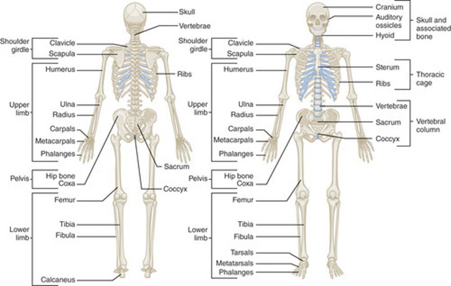
Skeletal System
A description of the bones of the body
Osteoporosis
A condition of reduced bone mineral density, which increases risk of bone fracture
Joints
The sites where two bones meet and movement occurs as a result of muscle contraction
Axial Skeleton
A division of the skeletal system consisting of the skull, the rib cage, and the vertebral column
Appendicular Skeleton
A division of the skeletal system consisting of the arms, legs, and pelvic girdle
Levers
Rigid rods where muscles attach
Remodeling
The process by which bone is constantly renewed by the resorption and formation of the bone structure
Osteoclasts
Special cells that break down and remote old bone tissue
Osteoblasts
Special cells that form and lay down new bone tissue
Wolff's Law
Scientific explanation of how remodeling (new bone growth) occurs along the lines of stress placed on the bone
Long Bones
Characterized by the their long cylindrical body, with irregular or widened bony ends. They are shaped much like a beam and exhibit a slight curvature that is necessary for efficient force distribution. Long bones are composed predominantly of compact bone tissue to ensure strength and stiffness. However, they do have a considerable amounts of spongy bone tissue for shock absorption.
Short Bones
Similar in length and width and appear somewhat cubical in shape. They consist predominantly of spongy bone tissue to maximize shock absorption. The carpals of the wrists and tarsals of the ankles fit this category for bones
Flat Bones
Flat bones are thin bones comprising two layers of compact bone tissue surrounding a layer of spongy bone tissue. These bones protect internal structures and provide broad attachment sites for muscles
Irregular Bones
Bones of unique shape and function that do not fit the characteristics of the other categories
Sesamoid Bones
Small bones embedded in a joint capsule or found in locations where a tendon passes over a joint. Found in the hands and feet. Sesamoid bones develop within particular tendons at a site of considerable friction or tension. They serve to improve leverage and protect the joint from damage
Processes
Projections protruding from the bone where tendons and ligaments can attach
Vertebral Column
Bones that house the spinal cord; consists of the cervical, thoracic, lumbosacral regions
Spinal Cord
Bundle of nerves housed within the vertebrae
The Spine
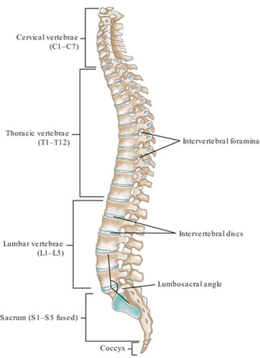
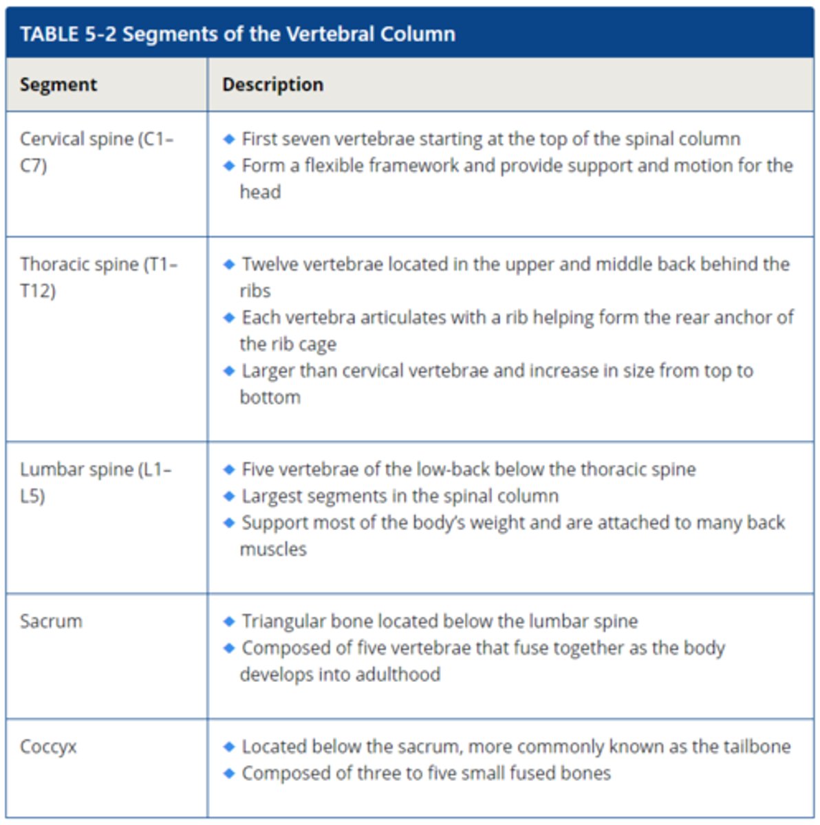
Segments of the Vertebral Column
Intervertebral Discs
Fibrous cartilage structures between vertebrae that act as shock absorbers and assist with movement
Neutral Spine
Represents a position in which the vertebrae and associated structures are under the least amount of load and can most optimally support functional movement
Osteokinematics
Movement of a limb that is visible
Arthrokinematics
The description of joint surface movement; consists of three major types: roll, slide, and spin
Synovial Joints
A joint with a fluid-filled joint capsule
Nonaxial Joints
A gliding joint that moves in only one plane, either back and forth or side to side
Nonsynovial Joints
Joints that have no joint capsule, fibrous connective tissue, or cartilage in the uniting structure
Ligament
A fibrous connective tissue that connects bone to bone
Collagen
A protein found in connective tissue, muscles, and skin that provides strength and structure. It is the most abundant protein in the human body
Elastin
A protein that provides elasticity to skin, tendons, ligaments, and other structures
Growth Plate
A specialized cartilage disc located in the epiphysis that is responsible for longitudinal bone growth
Skeletal Muscle
The type of muscle tissue that connects to bones and generates the forces that create movement
Fascia
Connective tissue that surrounds muscles and bones
Epimysium
Inner layer of fascia that directly surrounds an entire muscle, commonly referred to as the "deep fascia"
Fascicles
Largest bundles of fibers within a muscle. Fascicles are surrounded by perimysium
Perimysium
Connective tissue surrounding a muscle fascicle
Endomysium
Connective tissue that wraps around individual muscle fibers within a fascicle
Tendon
connects muscle to bones
Ligaments
Connect bones to bones
Glycogen
Glucose that is deposited and stored in bodily tissues, such as the liver and muscle cells; the storage form of carbohydrate
Myoglobin
Protein-based molecule that carries oxygen molecules into the muscles
Myofibrils
The contractile components of a muscle cell; the myofilaments (actin and myosin) are contained within a myofibril
Myofilaments
The filaments of a myofibril; include actin and myosin
Actin
The thin, stringlike, myofilament that acts along with myosin to produce muscular contraction
Myosin
The thick myofilament that acts along with actin to produce muscular contraction
Sarcomere
The structural unit of a myofibril composed of actin and myosin filaments between two Z-lines
Z-line
The meeting point of each sarcomere
Neural Activation
The nervous system's signal that tells a muscle to contract
Neuromuscular Junction
The specialized site where the nervous system communicates directly with muscle fibers
Synapse
A junction or small gap between the motor neuron and muscle cells
Motor Unit
A motor neuron and all of the muscle fibers that it innervates
Muscle Anatomy Flowchart
Muscle: bundle of fascicles surrounded by epimysium (deep fascia) connective tissue
↓
Fascicle: bundles of muscle fibers surrounded by perimysium connective tissue
↓
Muscle fiber: a bundle of myofibrils surrounded by endomysium connective tissue
↓
Myofibril: a collection of repeating sarcomeres that contain myofilaments (actin and myosin)
↓
Sarcomere: a section of a myofibril between two Z-lines where muscle contraction physically occurs
↓
Myofilament: the individual protein structures, actin and myosin, that make up a myofibril
Action Potential
Nerve impulse that is relayed from the central nervous system, through the peripheral nervous system, through the peripheral nervous system, and into the muscle across the neuromuscular junction
Neurotransmitters
Chemical messengers that cross the synapse between neuron and muscle and assist with nerve transmission
Acetylocholine (ACh)
A neurotransmitter that helps the action potential cross the synapse into the muscle, which initiates the steps in a muscle contraction
Sliding Filament Theory
The series of steps in muscle contraction involving how myosin (thick) and actin (thin) filaments slide past one another to produce a muscle contraction, shortening the entire length of the sarcomere
Excitation-contraction Coupling
The physiological process of converting an electrical stimulus to a muscle contraction
Power Stroke
The myosin heads bind to actin and pull them toward the sarcomere center, which slides the filaments past each other, shortening the muscle
Adenosine Triphosphate (ATP)
A high-energy molecule that serves as the main form of energy in the human body; known as the energy currency of the body
Resting Length
The length of a muscle when it is not actively contracting or being stretched
Type I Muscle Fibers
Muscle fibers that are small in size, generate lower amounts of force, and are more resistant to fatigue
Type II Muscle Fibers
Muscle fibers that are larger in size, generate higher amounts of force, and are faster to fatigue
All-or-nothing Principle
Motor units cannot vary the amount of force they generate; they either contract maximally or not at all
Type IIx Muscle Fibers
have lower oxidative capacity and fatique very quickly
Type IIa Muscle Fibers
also called intermediate fast-twitch fibers, higher oxidative capacity and fatigue more slowly than Type IIx but still fatigue much faster than Type I Fibers
Capillaries
the smallest blood vessels and the site of exchange of elements between the blood and the tissues
Characteristics of Type I Muscle Fibers
- more capillaries, mitochondria, and myoglobin
- increased oxygen delivery
- smaller in size
- less force produced
- slow to fatigue
- long-term contradictions (stabilization)
- "slow twitch"
Characteristics of Type II Muscle Fibers
- fewer capillaries, mitochondria, and myoglobin
- decreased oxygen delivery
- larger in size
-more force produced
- quick to fatigue
- short-term contractions (force and power)
- "fast twitch"