TOPIC 4
1/104
There's no tags or description
Looks like no tags are added yet.
Name | Mastery | Learn | Test | Matching | Spaced | Call with Kai |
|---|
No analytics yet
Send a link to your students to track their progress
105 Terms
4.4 = CIRCULATION
what is heart
The heart is an organ made mainly of cardiac muscle. Its job is to pump blood around the body.
Pump = something that creates pressure to move fluid from one place to another.
Describe heart
4 chambers:
Chamber | Side | Blood type | Main job |
|---|---|---|---|
Right atrium | Right side | Deoxygenated | Receives blood from body |
Right ventricle | Right side | Deoxygenated | Pumps blood to lungs |
Left atrium | Left side | Oxygenated | Receives blood from lungs |
Left ventricle | Left side | Oxygenated | Pumps blood to bodyAtrium = upper chamber of the heart. |
Right = deox
left = ox
2 sides separated by SEPTUM
describe atria
Upper chamber of heart
Thinner walls - only push blood a short distance into v
describe ventricles
lower chamber of heart
thicker muscular walls
pump blood OUT of heart
what is septum
muscular wall that separates left and right side of heart
stop ox and deox blood mixing
list blood vessels in heart
vena carva
pulmonay artery
pulmonar vein
aorta
PULMONARY = relating to lungs
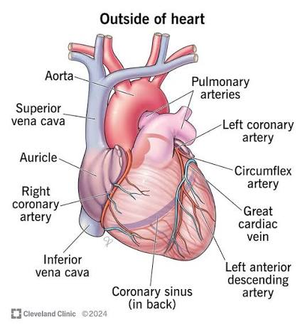
describe vena carva
Carries blood from body
—> right artium
deox blood
describe pulmonary artery
carries blood from R ventricle —> lungs
deox blood
describe pulmonary vein
carries blood from lungs —> left artium
ox blood
describe aorta
carries blood from L ventricle —> body
ox blood
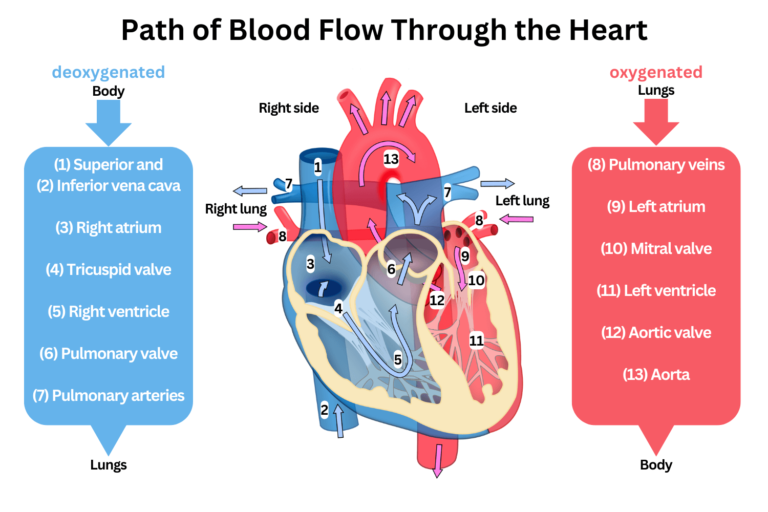
blood flow thru heart
Body → vena cava → right atrium → tricuspid valve → right ventricle → pulmonary semilunar valve → pulmonary artery → lungs → pulmonary vein → left atrium → bicuspid/mitral valve → left ventricle → aortic semilunar valve → aorta → body
list valves in heart
AV valves: atrioventricular valves
between atria + ventricles
stops blood flowing back into atria
Semilunar valves :
between ventricles and arteries
stops blood flowing back to ventricles
AV valves
Valve | Location |
|---|---|
Tricuspid valve RIGHT | Between right atrium and right ventricle |
Bicuspid / mitral valve LEFT | Between left atrium and left ventricle |
semilunar valves
Valve | Location |
|---|---|
Pulmonary semilunar valve RIGHT | Between right ventricle and pulmonary artery |
Aortic semilunar valve LEFT | Between left ventricle and aorta |
valve function
ensure one way flow
prevents backflow
Describe arteries
carry blood away from heart
Feature | Why |
|---|---|
Thick muscular wall | Withstands high pressure |
Thick elastic tissue | Stretches and recoils to maintain pressure |
Small/narrow lumen | Helps maintain high pressure |
Smooth endothelium | Reduces friction |
No valves along most arteries | Blood is already under high pressure |
Lumen = the hollow space inside a blood vessel where blood flows.
Endothelium = the thin inner lining of a blood vessel.
describe veins
carry blood towards the heart.
Feature | Why |
|---|---|
Thin muscular wall | Blood is at low pressure |
Less elastic tissue | Less recoil needed |
Large/wide lumen | Reduces resistance to blood flow |
Valves | Prevent backflow of blood |
Smooth endothelium | Reduces friction |
describe capillaries
tiny blood vessels that connect arteries/arterioles to veins/venules.
to exchange - move substances between blood and body cells
Examples of substances exchanged:
oxygen
carbon dioxide
glucose
amino acids
urea
water
capillary structure
Feature | Why |
|---|---|
Wall one cell thick | Short diffusion distance |
Made of endothelium only | Allows substances to pass through easily |
Very narrow lumen | Red blood cells pass close to cells |
Many branches / networks | Large surface area for exchange |
Gaps/pores between cells | Allow tissue fluid to form |
Arteries vs veins vs capillaries
Feature | Artery | Vein | Capillary |
|---|---|---|---|
Direction of blood flow | Away from heart | Towards heart | Between arteries and veins |
Pressure | High | Low | Low-medium |
Wall thickness | Thick | Thin | One cell thick |
Muscle | Lots | Little | None |
Elastic tissue | Lots | Little | None |
Lumen size | Narrow | Wide | Very narrow |
Valves | No, except near heart | Yes | No |
Main function | Transport blood under pressure | Return blood to heart | Exchange substances |
what does “circulatory system” mean?
A circulatory system is a system that transports substances around an organism.
It usually includes:
Part | Meaning |
|---|---|
Heart | Muscular pump that generates pressure |
Blood | Transport fluid |
Blood vessels | Tubes that carry blood around the body |
The point of a circulatory system is mass transport.
mass transport meaning
bulk movement of substances around an organism - usually fluid
Single circulatory system
Blood passes through the heart once during one complete circuit of the body.
fish:
Heart → gills → body → heart
single circulatory fish
The fish heart mainly receives and pumps deoxygenated blood.
The blood goes to the gills first to pick up oxygen. Then it goes straight from the gills to the rest of the body.
Why does pressure fall in fish?
When blood passes through the gill capillaries, pressure drops.
This means the blood reaches the body more slowly and at lower pressure than in mammals.
That is okay for fish because many fish have a lower metabolic demand than mammals, but it is less suitable for mammals.
Blood loses pressure in capillaries because:
capillaries are very narrow
there is resistance to blood flow
blood slows down to allow gas exchange
pressure is lost as blood passes through the capillary network
double circulatory system
Blood passes through the heart twice during one complete circuit of the body.
That is why it is called double circulation: the blood goes through the heart twice.
advantages of double circulatory system
what is cardiac cycle
The sequence of events that happens during one heartbeat.
events in a heart beat
the heart filling with blood
the atria contracting
the ventricles contracting
the heart relaxing again
stystole and diastole
Systole = squeeze; contract
Diastole = relax heart muscle
order of caridac cycle
1. Cardiac diastole
2. Atrial systole
3. Ventricular systole
4. Back to cardiac diastole
myogenic meaning
heart can generate its own electrical impulse without needed nerve impulse from brain
=independent
produced by muscle itself
Nerves can change the rate of the heartbeat, but the heartbeat itself is started by the heart’s own pacemaker tissue.
myogenic hstructures
Structure | Full name | Role |
|---|---|---|
SAN | Sinoatrial node | Starts the heartbeat |
AVN | Atrioventricular node | Delays and passes on the impulse |
Bundle of His | Conducting tissue in septum | Carries impulse down the septum |
Purkyne fibres | Conducting fibres in ventricle walls | Spread impulse through ventricles |
myogenic process
step | Key event | detail | keyworkds |
1 | SAN starts electrical impulse Causes atrial systole | SAN located in right atrium
SAN initiates wave of excitation actress atrial walls = causes atrial systole NOTE: impulse cant go straight thru ventricals as tissues between atria and ventricles are non conducting
| Pacemaker
Wave of excitation = electrical activity spreads thru cardiac muscle = contract |
2 | AVN receives + delays impulse (Atrioventricular node) | Impulse reaches AVN
Delay:
| |
3 | Bundle of hiss carries impulse down septum | BOH = conducting tissue Septum separates L and R Impulse:
So that ventricles contract from bottom upwards | BOH Apex |
4 | Purkyne fibres spread impulse thru ventricles | Purkyne fibres = branches of BOH PF spread impulse thru ventricle walls = ventricle systole
Forced blood into pulmonary artery (RV)+ aorta (LV) |
Myogenic stimulation vs nervous stimulation
myogenic control = heartbeat starts in heartFor example:
Situation | Effect |
|---|---|
Exercise | Heart rate increases |
Rest | Heart rate decreases |
But the brain does not normally start every heartbeat.
san generates electical impulse
nervous control = ns changes heartbeat
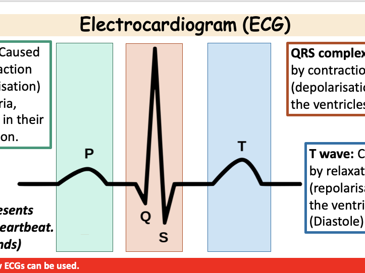
What is an ECG?
ECG stands for electrocardiogram.
An ECG trace is a graph showing the electrical activity of the heart.
The electrical activity causes heart muscle to contract, so ECG changes happen just before mechanical events like atrial systole or ventricular systole.
name parts of ecg
ECG part | What it shows | What happens after it |
|---|---|---|
P wave | Electrical activity spreading across atria | Atria contract |
QRS complex | Electrical activity spreading through ventricles | Ventricles contract |
T wave | Ventricles recovering electrically | Ventricles relax |
P wave = atria stimulated
QRS complex = ventricles stimulated
T wave = ventricles relax/recover
principles of ecg
blood moves from high to low pressure
Valve rule
Situation | Valve response |
|---|---|
Pressure behind valve is higher | Valve opens |
Pressure in front of valve is higher | Valve closes |
4.7
Xylem tissue
Main job of xylem
Xylem transports:
water
mineral ions
from the roots → stem → leaves.
This movement is mostly one-way, upwards through the plant.
xylem struc
Xylem vessels are long, hollow tubes made from dead cells that transport water and mineral ions through plants.
dead = less resistance (slow down movement)
no cytoplasm too
mature = dead
End walls break down = continuous tube
Thickened walls of lignin
waterproof
strenght
no colapse under tension
pits - small lignified gaps
sideays movement to near tissue
2. Phloem tissue
Phloem transports organic substances, mainly sucrose, around the plant.
This process is called translocation.
Translocation = movement of organic solutes, such as sucrose, through the phloem from sources to sinks.
source and sink
Source
A source is where sucrose is produced or released.- leabes
Sink
A sink is where sucrose is used or stored.
Examples:
roots
fruits
growing shoots
seeds
storage organs
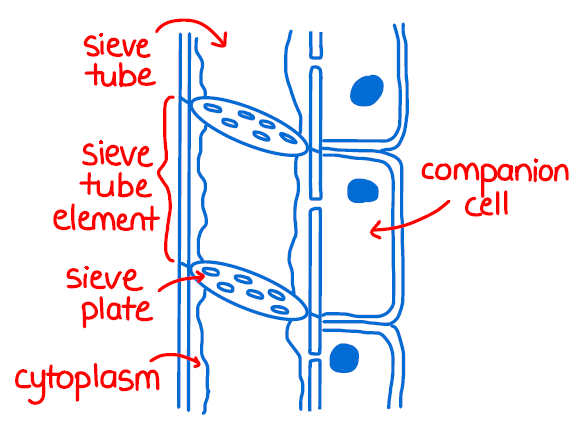
phloem struc
Phloem tissue is made mainly of:
sieve tube elements
companion cells
1. Sieve tube elements
Sieve tube elements are long living cells joined end to end to form tubes.
They transport sucrose solution through the plant.
They are alive, but they have very few organelles.
They usually have:
no nucleus
little cytoplasm
few organelles
Why this helps
There is more space inside the cell for sap to flow.
Sap = liquid in the phloem containing sucrose and other dissolved substances.
2. Sieve plates
Between sieve tube elements are sieve plates.
A sieve plate is an end wall with lots of pores.
Pores are tiny holes.
Why this helps
The pores allow phloem sap to move from one sieve tube element into the next.
So phloem forms a continuous transport pathway.
3. Companion cells
Companion cells are living cells next to sieve tube elements.
They have:
a nucleus
dense cytoplasm
many mitochondria
Why this helps
Companion cells control and support the sieve tube elements.
They provide energy for active transport of sucrose into and out of the phloem.
Key definition
Active transport = movement of substances against their concentration gradient using ATP.
ATP = energy-carrying molecule used by cells.
4. Many mitochondria in companion cells
Mitochondria are organelles where aerobic respiration happens.
Respiration releases ATP.
Why this helps
Phloem transport needs ATP because sucrose is loaded into phloem by active transport.
So companion cells need many mitochondria to supply ATP.
5. Plasmodesmata connect cells
Plasmodesmata are tiny channels between plant cells.
They connect companion cells and sieve tube elements.
Why this helps
They allow substances to move between the companion cell and sieve tube element.
This is important for loading sucrose into the sieve tube.
soil = higher wp thana cytop;as, of root hair cell
water = soil —? rhc
apoplastic
The apoplastic pathway is the movement of water through the cell walls and spaces between cells.
Important: water does not enter the cytoplasm in this pathway.
FAST:
doesnt need to cross plasma membranes
selective permeability
BUT CAS[ARIAN STRIP
The Casparian strip is a waterproof band of suberin in the cell walls of endodermal cells.
Suberin = a waterproof substance.
The Casparian strip blocks the apoplastic pathway because water cannot pass through it.
The Casparian strip forces water to leave the cell wall pathway and enter the symplastic pathway.
That means water must cross a plasma membrane.
This is useful because the plant can control which ions enter the xylem.
So the plant can selectively absorb mineral ions rather than letting everything from the soil enter the xylem.
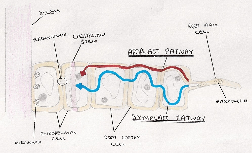
symplastic
The symplastic pathway is the movement of water through the cytoplasm of cells, via plasmodesmata.
Plasmodesmata are tiny cytoplasmic channels between neighbouring plant cells.
They allow substances to move directly from one cell’s cytoplasm into another.
cohesion
Cohesion is the attraction between water molecules.
Water molecules stick to each other because they form hydrogen bonds.
adhesion
Adhesion is the attraction between water molecules and the walls of the xylem.
Water sticks slightly to the xylem walls.
tension
Tension means a pulling force.
In xylem, water is under tension because it is being pulled upwards from the leaves.
cohesion tension model
Step 1: Water evaporates from mesophyll cells
Inside the leaf, water evaporates from the surface of mesophyll cells.
Mesophyll cells are photosynthetic cells inside the leaf.
Water changes from liquid water to water vapour.
Step 2: Water vapour diffuses out through stomata
The air spaces inside the leaf become humid because they contain lots of water vapour.
Usually, the air outside the leaf has less water vapour.
So water vapour diffuses:
leaf air spaces → outside air
through the stomata.
This is transpiration.
Step 3: Water loss lowers water potential in leaf cells
As water evaporates from mesophyll cell walls, those cells lose water.
This lowers their water potential.
So water moves into them from nearby cells by osmosis.
Step 4: Water is pulled out of the xylem
Water moves from the xylem into leaf cells to replace the water lost by evaporation.
This creates tension in the xylem.
The leaf is basically pulling water up the plant.
Step 5: Cohesion keeps water molecules together
Water molecules are cohesive because they form hydrogen bonds with each other.
So when water molecules at the top are pulled upwards, they pull the next water molecules with them.
This creates a continuous column of water in the xylem.
Step 6: The whole water column moves upwards
Because the water column is continuous, the pull is transmitted down the xylem.
So water moves:
roots → stem → leaves
This movement does not directly require ATP.
It is a passive process driven by water loss from the leaves.
Why xylem structure is perfect for this
Xylem vessels are:
dead
hollow
lignified
continuous tubes
narrow
This allows water to move with little resistance, and lignin prevents the vessels collapsing under tension.
Higher light intensity increases transpiration
In bright light, photosynthesis increases.
The plant needs carbon dioxide for photosynthesis.
So stomata open to allow carbon dioxide to diffuse into the leaf.
But when stomata open, water vapour can also diffuse out.
mass flow hypothesis
Translocation
Translocation is the movement of organic solutes through the phloem from sources to sinks.
The main organic solute is sucrose.
sucrose loaded into phloem