Gen Surg: Need to Know (Breast Surgery)
1/38
There's no tags or description
Looks like no tags are added yet.
Name | Mastery | Learn | Test | Matching | Spaced | Call with Kai |
|---|
No analytics yet
Send a link to your students to track their progress
39 Terms
4 weeks
Before breast surgery, a patient should stop smoking at least ___ _______ before the procedure to reduce the risk of wound dehiscence and fat necrosis
Cefazolin
What antibiotic is given before all breast surgeries involving an implant?
Intercostobrachial
What nerve is most commonly injured after an axillary node dissection?
-Causes numbness/paresthesia of medial upper arm
-Typically improves over months
Long Thoracic Nerve
What nerve, if injured during breast surgery, causes a winged scapula?
-Due to a traction injury or iatrogenic damage during axillary dissection
-Manage with PT
Seroma
Most common complication of breast surgery, where the risk increases with large tissue removal or no drain placement
-Occurs days to weeks after surgery
-Presentation: soft, non-tender swelling
-Management: aspiration, compression garment
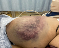
Hematoma
Rapid accumulation of blood in a surgical cavity, occurring 6-24 hours postop
-Presentation: sudden breast enlargement that is painful, skin may appear purple
-Tx: return to OR
Flap Necrosis
Ischemia of mastectomy skin flaps due to compromised blood supply, with smoking being the #1 risk factor
-Presentation: dusky purple skin with poor capillary refill
Breast Abscess
Common in breastfeeding women or a complication of mastitis
-Staph aureus is MC
-Presentation: fluctuant, red, indurated, tender nipple with discharge
-Dx: clinical, US to determine if abscess or mastitis
-Tx: I&D + dicloxacillin, bactrim if MRSA, continue breast feeding
Fibroadenoma
Benign tumor of glandular and fibrous tissue seen MC in women under 30
-Responsive to hormones, can see enlargement during pregnancy
-Presentation: single round, firm mass that is soft and rubbery. Mobile on exam
-Dx: clinical, US if under 30, mammogram if over 30. Biopsy if > 5 cm
-Tx: education and reassessment, decrease caffeine intake
Fibrocystic Disease
Multiple, painful breast masses that typically worsen during menses
-MC benign breast disorder
-Dx: US or mammogram, FNA shows straw colored fluid
-Tx: supportive bra, NSAIDs, caffeine reduction
No
Is there an increased risk of breast cancer with fibrocystic disease?
Intraductal Papilloma
Benign breast tumor arising from the epithelium of the lactiferous ducts, MC cause of pathologic unilateral nipple discharge
-Presentation: unilateral bloody or serous discharge, no palpable mass, no systemic sx
-Dx: US/mammography to start, core needle biopsy (contains epithelial and myoepithelial cells)
-Tx: microdochoectomy for solitary lesions, complete duct excision for multiple lesions
Both
Do intraductal papillomas contain epithelial, myoepithelial, or both types of cells?
Fat Necrosis
Fatty breast tissue is damaged, leading to the formation of firm, irregular masses. Often associated with trauma, surgery, or radiation therapy
-Presentation: firm, irregular, painless mass. Overlying skin may show redness, retraction, or bruising. Nipple retraction or dimpling may be present
-Dx: mammogram, US, core biopsy
-Tx: observation, work up for malignancy
Galactocele
Milk filled cysts within the breast due to obstruction on the lactiferous ducts
-Presentation: painless, smooth, mobile breast lump that typically occurs in lactating women
-Dx: US shows a cystic lesion with fat fluid level, aspiration to confirm
-Tx: conservative, resolves after breast feeding
Duct Ectasia
Inflammatory duct dilation with periductal fibrosis, which is common in perimeno and postmenopausal women
-Presentation: unilateral green, yellow, or bloody discharge. Nipple inversion and sometimes breast pain. May palpate a subareolar mass or fullness
-Dx: clinical + imaging
-Tx: conservative, pain control, smoking cessation
Smoking
What is the most important risk factor for mammary duct ectasia?
Gynecomastia
Development of palpable rubbery or firm disc of tissue under the nipple in males, due to an imbalance of estrogen and androgen
-Presentation: breasts
-Dx: clinical
-Tx: stop causative drug, treat underlying disease, SERMs for painful gynecomastia
Breast Cancer
Malignancy of the breast, with adenocarcinomas arising from epithelial cells of ducts or lobules being the most common
-Invasive ductal carcinoma is the most common
-Presentation: painless, firm, immobile breast mass (MC on upper outer quadrant), possible nipple retraction, skin dimpling, peau d’orange, or bloody nipple discharge. May present with axillary LAD.
-Dx: mammogram, sentinel node biopsy before axillary lymph node resection
-Tx: lumpectomy + radiation
Spine
Metastasis from breast cancer is often where?
Tamoxifen
In a patient with ER/PR positive breast cancer, what medication can they receive due to its response to hormonal therapy?
HER2 +
What type of receptor + breast cancer is treated with trastuzumab?
Chemotherapy
What is triple negative breast cancer treated with?
DCIS
Malignant cells confined to the ducts, no invasion through basement membrane
-Stage 0 is often found on mammogram with microcalcifications
-Tx: lumpectomy ± radiation, mastectomy if extensive
LCIS
Malignant cells confined to lobules
-Marker of increased risk for bilateral invasive cancer, usually found incidentally on biopsy
-Tx: observation + tamoxifen
IDC
Most common type of breast cancer, invasion of malignant ductal cells into surrounding tissue
-Firm, irregular, immobile mass. May cause skin/nipple retraction. Can metastasize via lymphatics
-Tx: surgery + radiation + chemo + hormone therapy
ILC
Malignant lobular cells invade the stroma
-Often bilateral or multicentric, may present as ill-defined thickening
-Tx: similar to IDC
Paget Disease
Ductal carcinoma cells invade nipple epidermis
-Eczematous nipple/areolar changes
-Tx: tx underlying carcinoma
Inflammatory Breast Cancer
Aggressive form of invasive ductal carcinoma that blocks dermal lymphatics
-Rapidly progressive, erythematous, warm, edematous (peau d’orange) breast. No discrete masses
-Tx: neoadjuvant chemo + surgery radiation
Radical Mastectomy
If other lymph nodes (beyond the sentinel node) are positive for cancer, what is the surgical approach necessary?
Kleinfelters
What genetic disease is related to male breast cancer?
Capsular Contracture
MC complication of breast implant surgery, which occurs when a fibrous scar capsule tightens around the implants
-RF: subglandular placement, hematoma, infection, radiation
-Sx: firm, painful augmented breast. No systemic symptoms
-Tx: capsulectomy
Implant Rupture
Rapid burst of a breast implant
-Presentation: rapid deflation and noticeable size decrease (saline), subtle contour changes (silicone)
-Dx: clinical if saline, MRI if silicone
-Tx: surgical removal and replacement
Textured
What type of implant is breast implant-associated anaplastic large cell lymphoma associated with?
-Sx: late onset, unilateral breast swelling, peri-implant seroma occurring greater than a year after implant
-Tx: capsulectomy and implant removal
Mastitis
Inflammation of the breast tissue that may or may not be associated with infection
-Presentation: periareolar pain, redness, swelling
-Dx: clinical, US if concerned about an abscess
-Tx: augmentin or dicloxacillin
IGM
Rare benign inflammatory breast disease of unknown etiology, often occurring within 5 years of pregnancy in an Asian woman
-Presentation: solitary peripheral tender inflammatory breast mass, can also present as multiple simultaneous peripheral masses with abscesses and/or overlying skin inflammation and ulceration
-Dx: US and core needle biopsy
-Tx: NSAIDs for pain, doxy with I&D if abscess is present
Phyllodes Tumor
Rare fibroepithelial breast tumor that is made of both epithelial and stromal elements, associated with Li-Fraumeni Syndrome
-Presentation: rapidly growing smooth, painless breast mass. Growth over weeks to months
-Dx: core needle biopsy
-Tx: wide excision
FNA
What technique should be used for a breast biopsy in these cases?
-Lesion is cystic
-Expecting benign pathology
-Mass is superficial, small, or easily palpable
-Need fast cytology
-Patient cannot tolerate larger biopsy
Core Needle
What technique should be used for a breast biopsy in these cases?
-Suspicious mass on imaging
-Solid mass
-Need to evaluate architecture
-Suspicion for invasive carcinoma
-Presence of microcalcifications
-Need for receptor testing