Histology Resp Lab Flashcards, Yosyraa
1/50
There's no tags or description
Looks like no tags are added yet.
Name | Mastery | Learn | Test | Matching | Spaced | Call with Kai |
|---|
No analytics yet
Send a link to your students to track their progress
51 Terms
Conducting portion
Respiratory portion
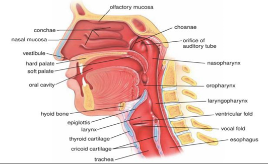
Nasal Cavity
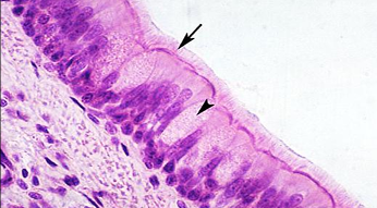
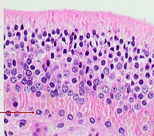
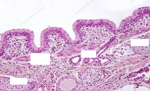
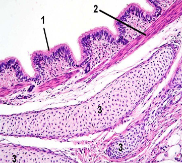
Pseudostratified columnar ciliated with goblet cells
(The epithelium lining of most of the conducting portion)

Pseudostratified columnar ciliated with goblet cells
(The epithelium lining of most of the conducting portion)
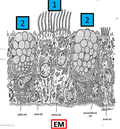
1- Ciliated Columnar cells
2- Mucous goblet cells
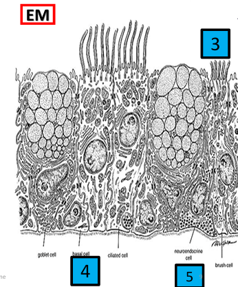
3- Brush cells (3%)
4- Basal cells (Stem cells)
5- Small granule cells (3%)
Olfactory epithelium
Modified pseudo-stratified columnar ciliated epithelium (NO GOBLET CELLS)
has 3 types of cells:
- Olfactory neurons
- Sustentacular cells (support)
- Basal cells (stem cells)
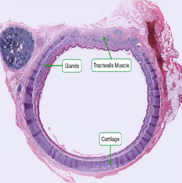
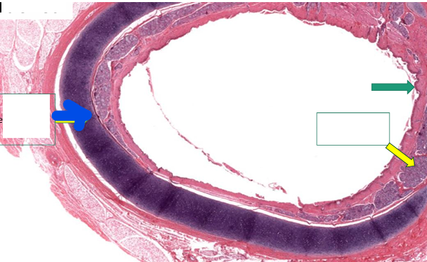
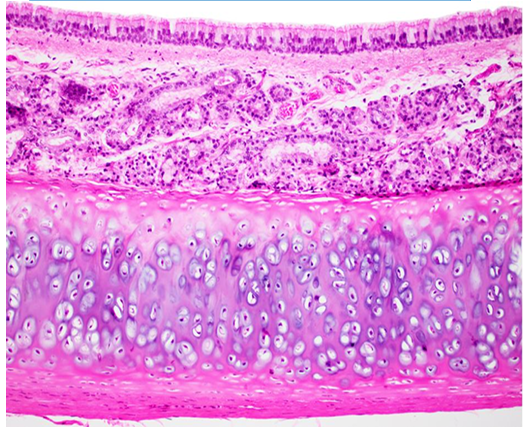
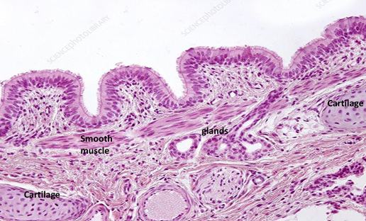
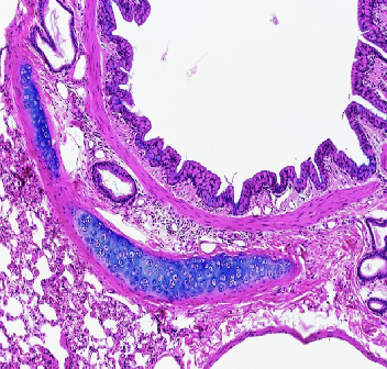
Trachea
(low power view)
Trachea
(low power view)
Blue- Hyaline cartilage
Green- epithelium
Yellow- Glands in submucosa
Trachea
Trachea
(medium power view)
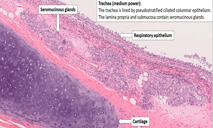
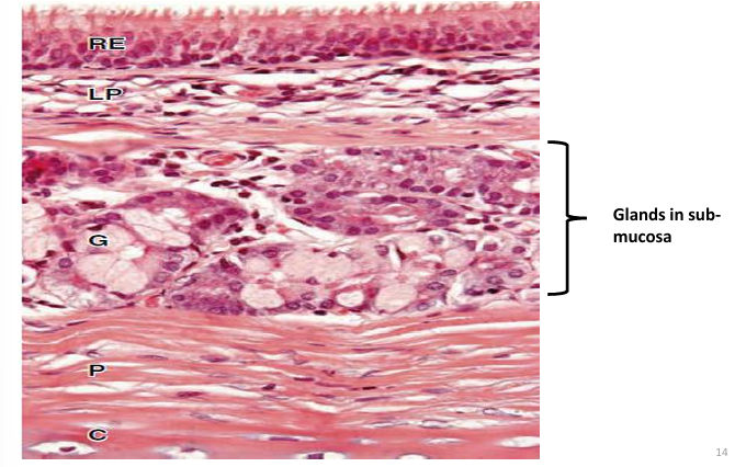
Trachea
upper part: submucosa
lower part: cartilage
Trachea
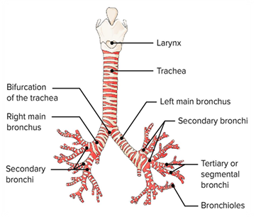
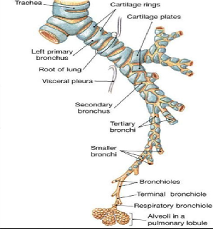
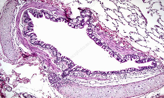
Bronchial Tree

Bronchial Tree
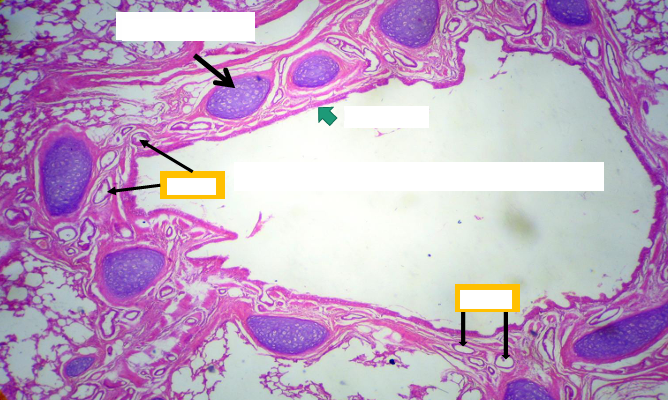
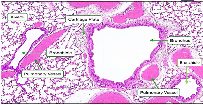
Section in the lung showing intrapulmonary bronchus
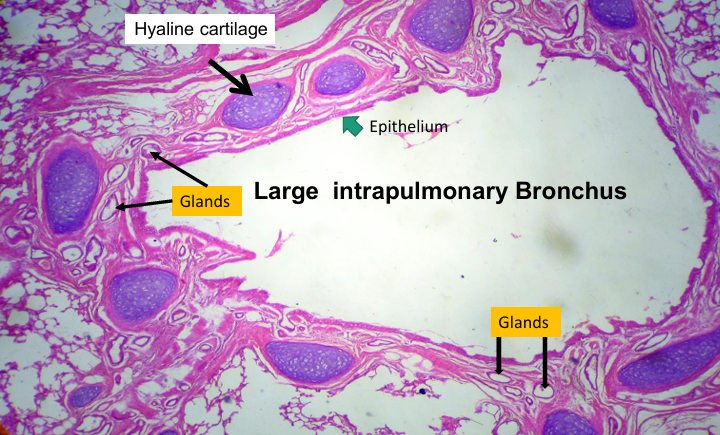
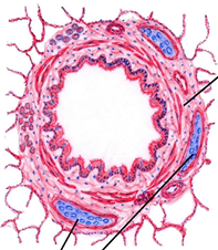
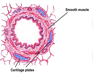
Bronchus
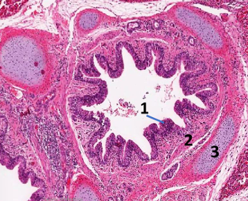
Intra pulmonary bronchus
1- epithelium, 2- smooth muscle, 3- cartilage
Intra pulmonary bronchus
Intra pulmonary bronchus
Intra pulmonary bronchus
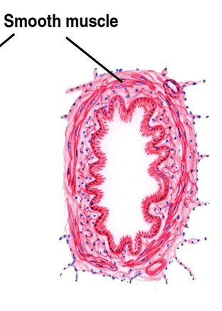
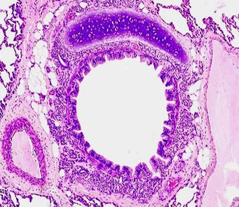
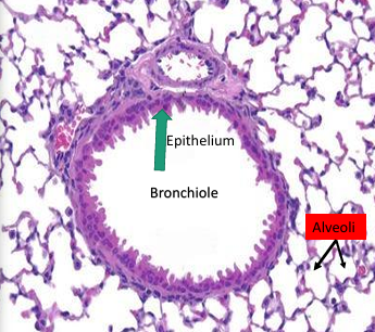
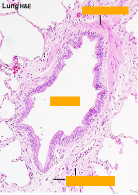
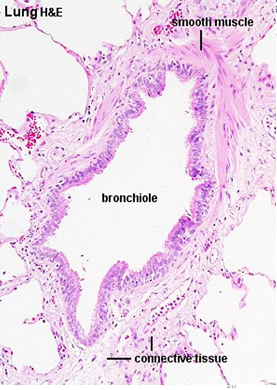
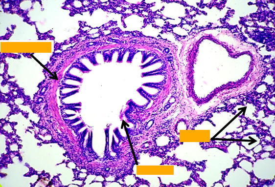
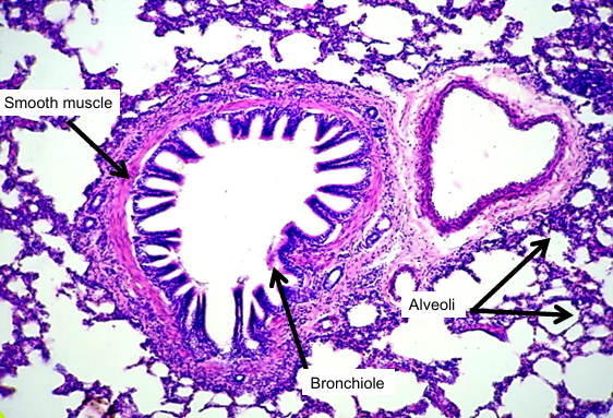
Bronchiole
Mucosa:
Epithelium: simple columnar to cuboidal partially ciliated with club cells
- Lamina propria: thin & contain elastic fibers.
Muscle layer: thin layer of circularly arranged smooth muscle.
- No goblet cells,
- No cartilage,
- No glands,
- No lymph follicles

Terminal Bronchiole and Clara Cells
Terminal Bronchiole and Clara Cells
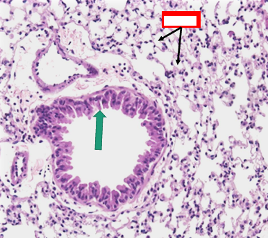
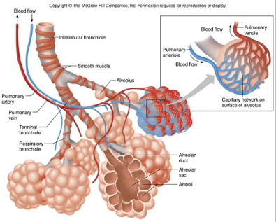
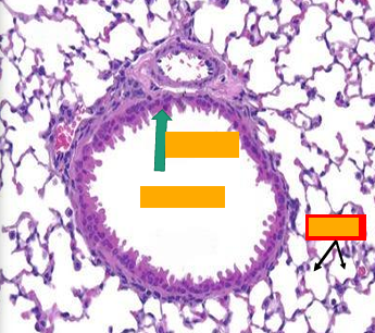
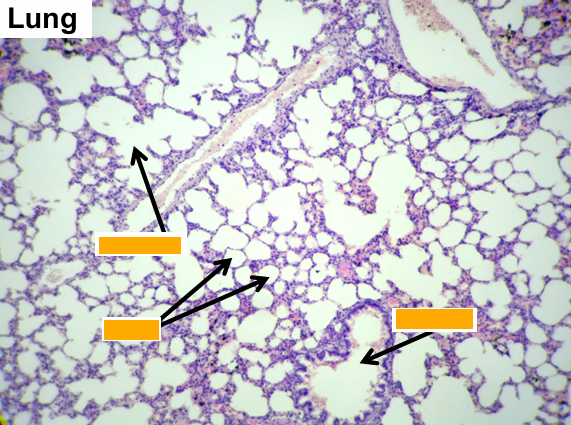
Section showing bronchiole and alveoli
Red = alveoli
Section showing bronchiole and alveoli
Red = alveoli
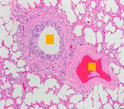
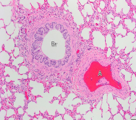
Section showing bronchiole and alveoli (white spaces all around)
Red A = Artery
Bronchiole
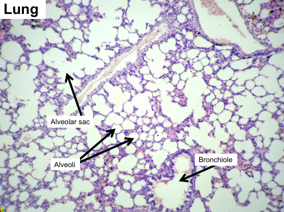
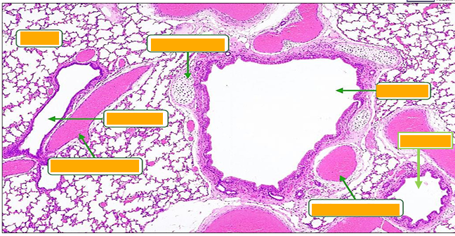
Bronchus and Bronchiole
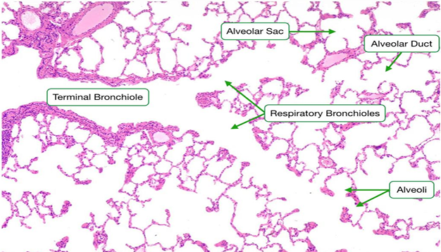
Respiratory portion consists of
1. Respiratory bronchiles
2. Alveolar ducts
3. Alveolar sacs
4. Alveoli
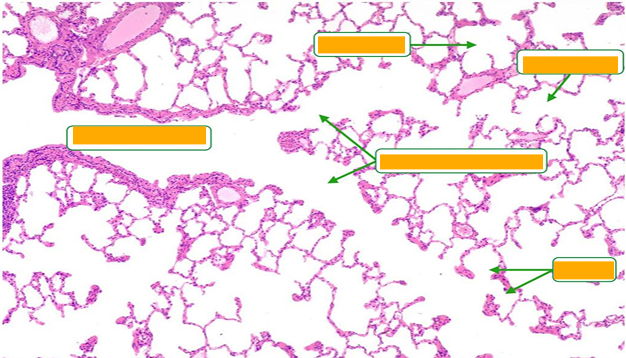
Lung
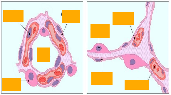
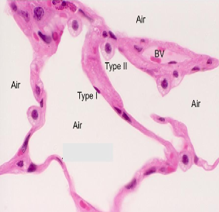
Alveolar epithelium
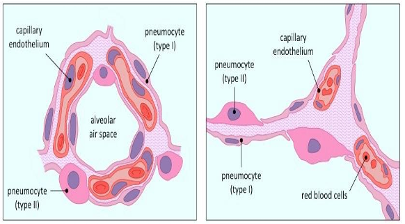
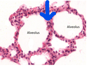
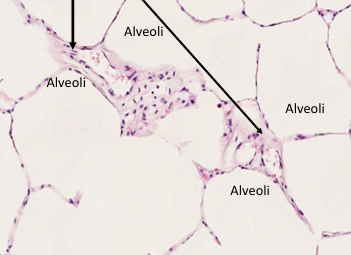
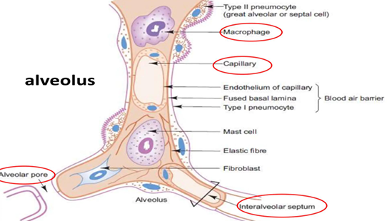
Alveoli
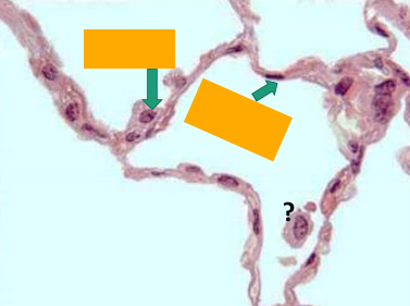
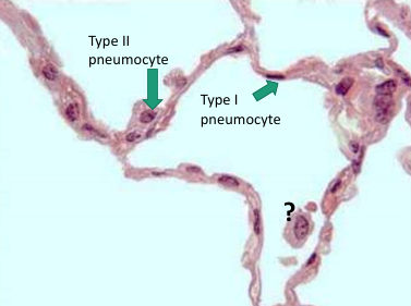
Arrow = Interalveolar septum
Arrow = Interalveolar septum
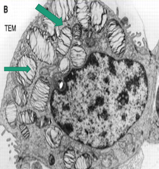
TEM photo of cells lining small bronchiole:
cubical ciliated cells and Clara cell
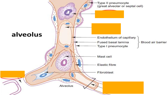
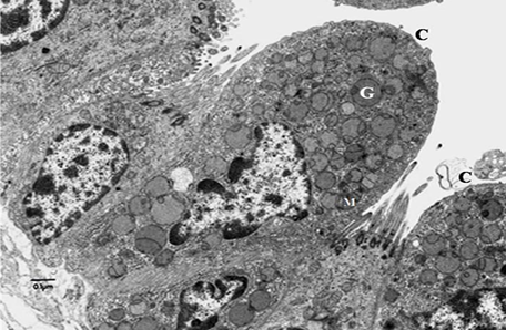
Type II pneumocyte
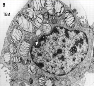
Type II pneumocyte
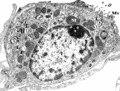
Type II pneumocyte
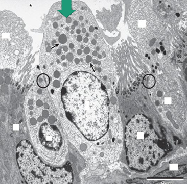
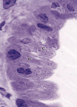
Identify labeled cell, sites and one specific feature?
Clara cell , mucosa of bronchiole, dome shaped apical surface and secretory granules
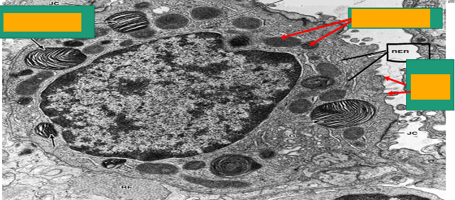
A -Identify the cell
B -Identify the labelled structure
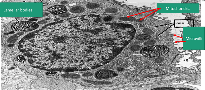
A- pneumocyte type II
B- Lamellar bodies
Identify labeled structure
Intrapulmonary bronchus
1. Epithelium
2. Smooth muscles spirally arranged
3. Plates of hyaline cartilage in adventitia

Terminal bronchiole
Intra pulmonary bronchus
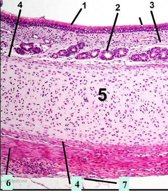
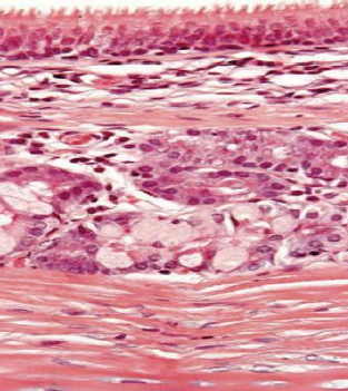
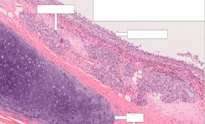
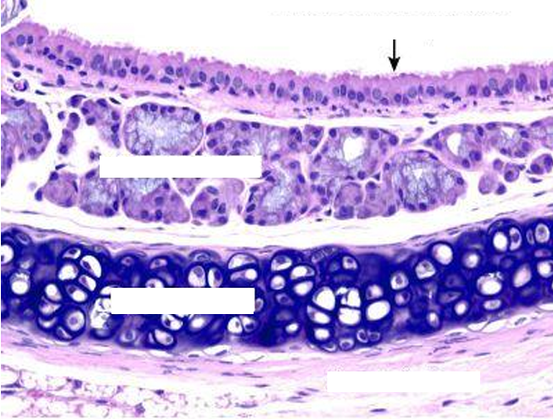
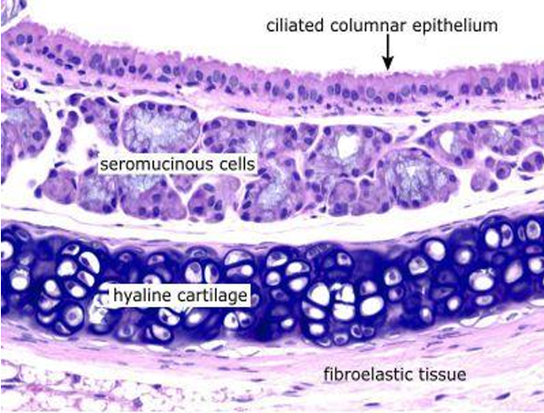
Trachea
1. Epithelium
2. Tracheal glands in submucosa
3. Elastic membrane
4. Perichondrium
5. Hyaline cartilage
6. Trachialis muscle
7. Adventitia

Lung alveoli and interalveolar septea