A&P Lab 1 (Chapter 5)
1/35
Earn XP
Description and Tags
The Integumentary System
Name | Mastery | Learn | Test | Matching | Spaced | Call with Kai |
|---|
No analytics yet
Send a link to your students to track their progress
36 Terms
What is the anatomical term for skin?
Cutaneous membrane
What is the 2 main layers of the skin?
Epidermis (on top)
Dermis (beneath, separated by a basement membrane)
What layers sits below both the Epidermis (top) and Dermis (beneath epidermis)?
Hypodermis (which secures skin to deeper muscle and bone via loose connective and adipose tissue)
What are the 6 accessory structures of the skin?
sweat glands
sebaceous glands
hair follicles
nails
sensory receptor
arrector pili muscles
The Epidermis
avascular (has no blood vessels)
cells receive oxygen and nutrients by diffusion from vessels in the deep dermis
The dominant cell type is the keratinocyte, which produces keratin (a tough, fibrous protein that gives the skin strength)
As keratinocytes migrate upward, they fill with keratin, harden, and eventually die, forming the protective outer surface.
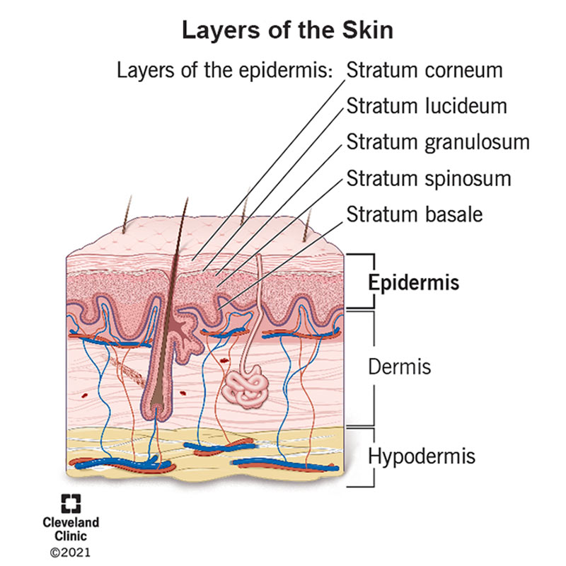
The epidermis is separated into 4-5 strata (layers) which allows the skin to keep recycling and building new protective barriers
What is avascular?
having no blood vessels
what is the dominant cell type of the epidermis?
keratinocyte
What do keratinocytes produce? What is Keratin?
keratin; a tough, fibrous protein giving the skin strength
What are the 5 epidermal strata (layers) (deep to superficial)?
Stratum Basale
2. Stratum Spinosum
Startum Granulosum
Stratum Lucidum
Stratum Corneum
(REMEMBER: Before Sun, Get Lotioned Completely)

What layer is the deepest layer?
Stratum Basale
Single row of stem cells on the basement membrane
Most mitotically active, continuously producing new keratinocytes to push upward to the surface to replace the dead surface cells.
Examples of these cells are melanocytes, tactile epithelial cells, and keratinocyte stem cells
What do melanocytes do?
produces melanin and the pigment (from the melanin) protects the underlying keratinocytes DNA from UV-radiation
What are the Tactile Epithelial cells? (in Basale)
sensory receptors that are paired with small neurons from the dermis
detect light touch concentrated in the finger tips, lips, base of hairs.
What are Keratinocyte cells? (in basale)
source of all cells populating the layers
where vitamin D synthesis begin
(whether this layer (basale) survives during an injury, determines whether skin can regenerate on its own)

Which layer is the thickest layer? (Hint: cell shape looks like spines)
Stratum Spinosum (10-14 layers)
Cells are still alive and mitotically active
Contains immune cells (phagocytes)
Contains Dendritic epidermal cells (immune phagocytes) that patrol for pathogens and protect skin and deeper tissues
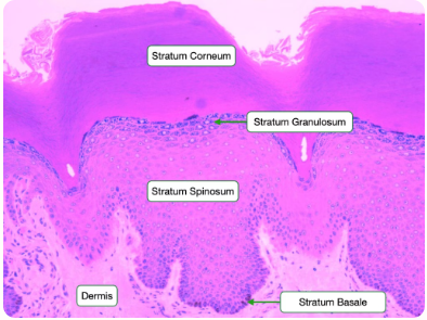
Which layer has 3-5 layers?
stratum granulosum (3-5 layers)
Contains prominent cytoplasmic granules with keratin bundles and lipid-based substances
Hydrophobic lipids are secreted and create a waterproof barrier
This lipid release cuts off nutrient supply, and the cell begins to die
Which layer is primarily thin but is only thick on the palms and soles (feet)?
stratum lucidum
thin, clear layer of dead keratinocytes found only on thick skin
ONLY thick in the palms and soles
provides protection
Which layer is the outermost layer?
stratum corneum
multiple layers of dead, flattened keratinocytes packed with keratin; primary barrier
This layer is the primary physical and chemical barrier between the body and the outside world
Whats the difference between thick and thin skin?
Thick skin
found ONLY on palms and soles
all FIVE epidermal layers
VERY THICK stratum corneum (the outermost layer)
MANY skin glands
Thin skin
covers MOST of the body
ONLY FOUR epidermal layers (No stratum lucidum - primarily thin skin but only thick on the palms and soles)
Includes hair follicles and sebaceous glands
A callus forms in either skin type when repetitive pressure stimulates the production of extra stratum corneum layers
What is the second major layer of the skin?
The dermis
The Dermis
Highly vascular (has blood vessels) connective tissue beneath the epidermis
Supplies blood to the avascular (has no blood vessels) epidermis
divided into 2 sub-layers:
Papillary Layer
Reticular layer
What are the 2 sub-layers of the dermis?
Papillary Layer
Thinner, more superficial
Loose connective tissue
Houses tactile corpuscles (Messiner’s) for light touch detection
Concentrated in fingertips, lips, and face
Reticular Layer
Deeper, thicker
Dense irregular connective tissue (collagen bundles provide strength)
Contains lamellar corpuscles (Pacinian) for deep pressure and vibration
+ Blood vessels, sweat glands, hair follicles, and sebaceous glands and adipose tissue
Injuries and Fingerprints on the Dermis layer
Injury requiring sutures
The reticular dermis provides the tensile strength (pulling and stretching) needed to hold wound edges together
Sutures only anchored in the epidermis layer CANNOT resist tearing forces from multiple directions
Proper anchoring into the reticular dermis minimizes scarring and ensure effective wound closure
Fingerprints
The ridges visible on fingertips are created by the interface between dermal papillae (small upward projections) and the overlying epidermis.
Sweat pores open along these epidermal ridges, depositing a thin film that creates residual fingerprints when you touch a surface.
How is skin color determined?
melanin
What does melanin produce?
melanocytes (in the stratum basale - the deepest layer)
What does melanin do? What are the 2 UV exposure trigger responses?
it absorbs UV radiation and protects the DNA of dividing keratinocytes from UV radiation
UV exposure triggers 2 responses:
immediate response: existing melanin in keratinocytes is oxidized, causing rapid darkening
delayed response: UV damage to melanocyte DNA stimulates new melanin production; develops slowly but lasts longer
Ex(s):
Freckles: localized increase in melanin production (more melanin, same # of melanocytes)
Moles (nevus): localized proliferation of melanocytes (more melanocytes, not just more melanin)
albinism (melanocytes present but fail to produce melanin; high UV-related DNA damage risk)
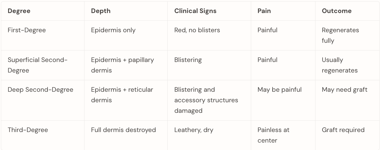
How are burns classified?
by depth and tissue damage
Critical variable is whether the stratum basale survives as this determines whether the skin can regenerate on its own or requires a skin graft
First degree burn
damage to the epidermis ONLY
skin appears red and painful; no blistering because the dermis is intact
stratum basale is unaffected, meaning the skin will regenerate completely without scarring
ex. sunburn
Superficial second-degree burn
epidermis and papillary dermis are damaged
blistering occurs as fluid leaks from damaged papillary capillaries
painful due to nerve endings remaining intact in the dermis
stratum basale may be partially spared
if enough viable cells survive along the basement membrane, regeneration without a skin graft is possible
Deep second degree burn
damage extends into the reticular dermis (beginning to destroy accessory structures - hair follicles, sweat glands)
still painful if some nerve endings survive.
Regeneration is less reliable, and grafting may be needed, depending on the extent
Third degree burns
the entire dermis is destroyed (including nerve endings, blood vessels, and accessory structures - hair follicles and sweat glands)
painless at the center because the nerve endings that transmit pain no longer exist
The stratum basale is completely gone and skin cannot regenerate on its own. grafting is required.
What are the 4 accessory structures?
hair, sweat glands, sebaceous (oil) glands, and nails
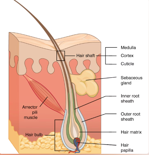
Integumentary Accessory Structure: Hair
covers the entire body except thick skin regions, the lips, and parts of the external genitalia
both parts are composed of stratified squamous keratinized epithelial cells
shaft: the visible portion projecting above the skin surface; made of columns of dead keratinized cells
root & hair bulb: embedded in the dermis; surrounded by a sensory neuron detecting hair movement
the root is enclosed in a hair follicle with an epithelial and dermal root sheath.
The hair bulb contains the hair matrix, which is the site of actively dividing keratinocytes producing new hair.
Arrector Pili: smooth muscle attached to the dermal root sheath; contraction pulls hair upright, causing goosebumps
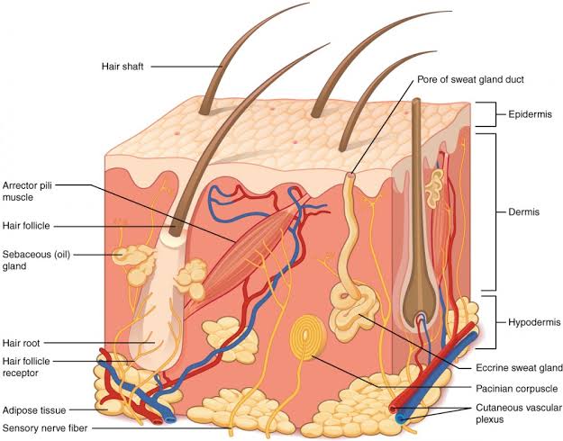
what 2 glands are comprised of the accessory structure, sweat glands?
eccrine gland
opens directly onto the skin surface
location: found all over the body surface (most numerous in palms and soles)
produces water and electrolyte-based substance
sebaceous glands: produce sebum, an oily secretion that lubricates hair and skin - found everywhere besides the palms and soles
apocrine gland
opens into hair follicles
location: concentrated in areas like the underarms and groin
produces a lipid-rich substance
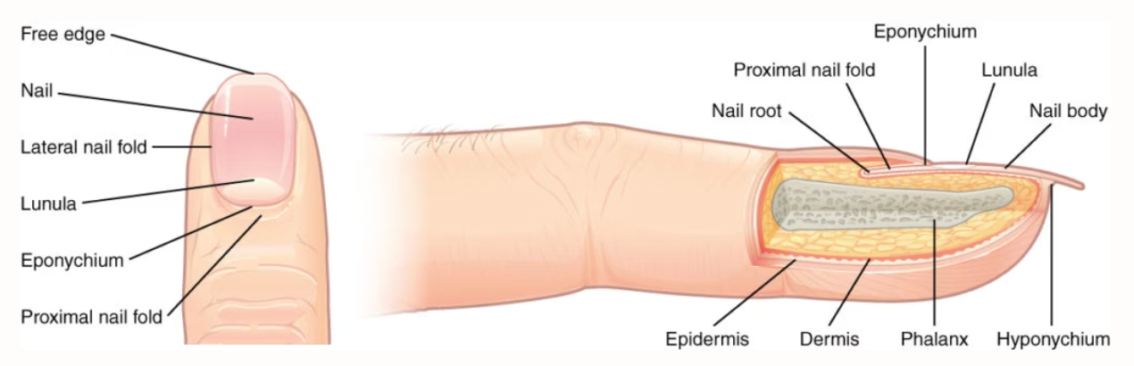
Nails
hard keritanized structures at digit tips
The nail plate includes visible nail body and nail root under the skin
growth occurs at the nail matrix beneath the nail root
The proximal nail fold produces the eponychium (cuticle); the distal free edge is secured by the hyponychium. Nails contain no melanocytes and are mostly translucent; the lunula (the proximal white half-moon) represents visible keratin accumulation from the matrix below.