Fundamentals Of Cell Biology Yr 1
1/65
Earn XP
Description and Tags
Sports and exercise science
Name | Mastery | Learn | Test | Matching | Spaced | Call with Kai |
|---|
No analytics yet
Send a link to your students to track their progress
66 Terms
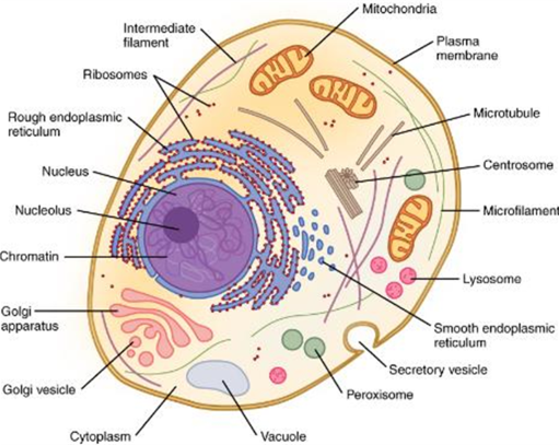
General cell structure
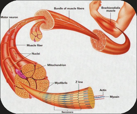
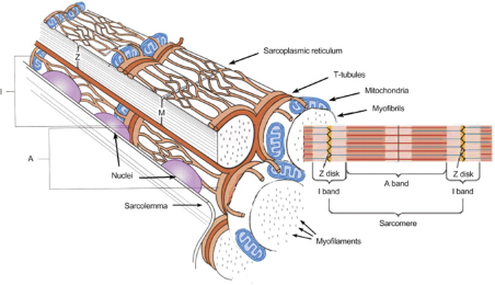
Muscle cell structure
Multinucleated - located close to the plasma membrane
Contain mitochondria
Microfibrils ( Actin/Myosin)
Sarcoplasmic reticulum
Myogenesis
How we build muscle from a single cell
Embryonic precursor cells (Myoblasts)
Fusion/ alignment of these cells
Constant cycles of de/regeneration
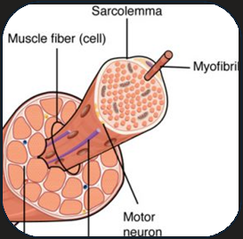
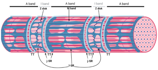
Cross section of muscle
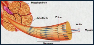
Longitudinal portion
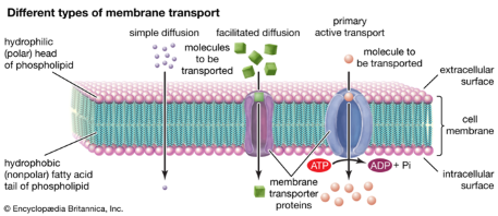
Plasma membrane
Fluid Mosaic Model:
Phospholipid bilayer - Hydrophilic head/Hydrophobic tails
Integral proteins act as carriers or channels
Selective permeability - H2O and O2 easy entry/ CO2 easy removal
Sarcolemma
Properties of excitability - allows for rapid distribution of signal throughout the muscle
Conduct electrical impulses during depolarisation
T tubules begin here and enter the muscle fibre
ER
Endoplasmic Reticulum (SER, RER & SR (muscle only))
RER = Protein
SER = steroids/lipids/phospholipids - important for metabolism
SR ( Sarcoplasmic reticulum)
Critical for excitation-contraction coupling
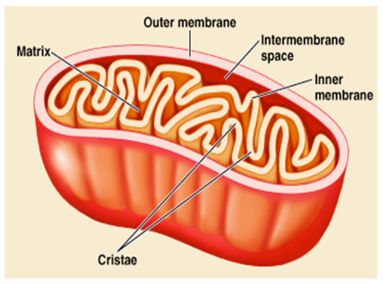
Mitochondria
The majority of ATP pathways
Regulates fats/CHO to produce energy
Double membrane (Cristae) - Increase SA for Electron Transport Chain
Cytoskeleton
80% of muscle cell space - 1% in other cells
Maintain cell shape/ structure
Microtubules - Substrate/organelle transport
Microfilaments - Allow contraction ( bind to myosin)
Intermediate filaments
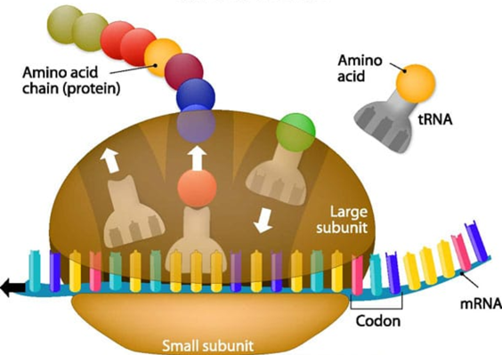
Ribosomes
Bind to the start of mRNA - tRNA delivers 1st amino acid ( Initiation)
The ribosome moves along mRNA, creating peptide bonds between Amino acids delivered by tRNA (elongation)
Ribosome reaches stop codon - Polypeptide chain released ( Termination)
Nucleus
Holds genetic info
Regulates gene expression
Allows the cell to respond to stimuli or damage
Myo-nuclei
Cannot divide
One cell has many nuclei - Supports adaptation of skeletal muscle
Muscle force generation / Contraction
Action potential at NMJ - Ach released
Ach → Sarcolemma → action potential, Propagated T tubules
Ca2+ released from SR
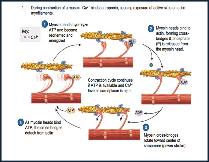
Ca2+ binds to troponin → exposing actin
Sliding filament theory
Myosin + Actin = Crossbridge
“Power stroke” - Pulls Actin to centre (Shortening Sarcomere)
Cross-bridge broken by ATP→ Myosin
Cocking myosin ( ATP hydrolysis → ADP =Pi
Ca2+ returns to SR
Tropomyosin block restored
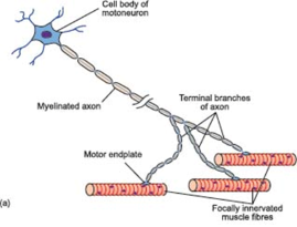
Motor unit
1 motor neuron can innervate MANY muscle fibres
BUT, 1 muscle fibre can only be innervated by 1 motor neurone
Localisation
The location of a protein or organelle within a cell is important
Central nuclei = muscle fibre repair
GLUT 4 = Important glucose transporter protein - location dictates function
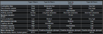
Muscle fibre types
Satellite cells
Located at the periphery of the fibre - fuse when necessary
Satellite cells can divide
Contribute to growth and repair - myogenesis
Muscular repair
Less severe than regeneration
Enable healthy maintenance of muscle
Damage through eccentric contractions
inflammatory response/satellite cell activation = strength recovery
Force can still be reduced for up to 7 days - Increased levels of satelite cells
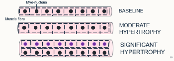
Contribution of Satellite cells to Hypertrophy
Nuclei can only govern a defined volume of cytoplasm
Growth requires more nuclei
Bigger muscles = more nuclei + available satellite cells

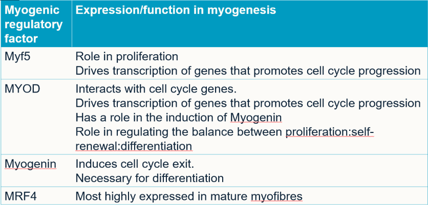
Myogenic Regulatory Factors
Stem cells
Cells that are capable of self-renewal, regulation & differentiation into specialised cell types
Embryonic stem cell
Pluripotent - Differentiate into many different cell types
Adult stem cell
Multipotent- Limited differentiation potential
Tissues that require stem cells
Epithelial lining ( SI absorption)
Endothelial (Lining of blood vessels - repair)
Connective tissue ( Bone, tendons, cartilage)
Cell differentiation
Starts early & progressively narrows options of what a cell can become
Terminal differentiation
Cell that becomes highly specialised & can no longer adapt
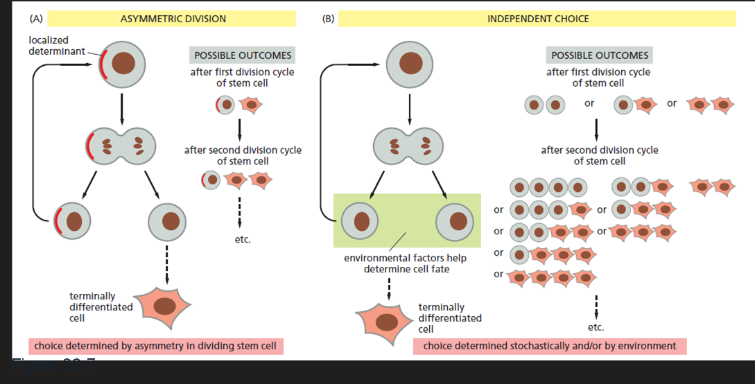
Types of cell division
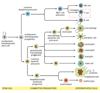
Hematopoietic stem cells (HSCs)
Multipotent
Located in bone marrow & responsible for Blood cell production
Hierarchy that dictates what cells they can become
Progenitor cells
Common myeloid progenitor ( CMP)
Rise to RBCs, platelets, & Myeloid cells
Common lymphoid progenitor ( CLP)
Rise to lymphocytes (B/T/ NK cells)
Erythropoiesis
Production of RBCs
Controlled by EPO - Liver
RBCs ( Erythrocytes) cannot grow, divide & have a limited lifespan
reduced O2/RBCs = Increased Erythropoiesis
Colony Stimulating Factor (CSF)
Stimulate the function of differentiated blood cell colonies formed from progenitor cells
EG. EPO acts on the erythropoietin progenitor
Function
Cell survival
Late in the hierarchy to facilitate differentiation
Early in the hierarchy to influence commitment
Colony Stimulating Factor (CSF) - Role in exercise
Maintain Blood cell function
Exercise = physiological stressor ( Increased O2 demand)
HSCs move into the bloodstream during exercise
Process is enhanced by endurance exercise (increased immune response) - Overtraining may reduce HSC activity
Volume & intensity = big factor to HSCs in blood - 70% = good intensity for mobilisation
Cell signalling
Process where cells respond to internal/External cues
Important for growth, metabolism, homeostasis, & Skill adaptation
Basic processing of Cell signalling
Signal (ligand) - Molecule that starts communication
Receptor - Protein in/on a cell that detects a signal
Signal pathway - Cascade of molecular events leading to a response
Changes in gene expression, protein activity & metabolism
Types of Cell signalling
Autocrine = Cell signals itself
Paracrine = Signal near cells
Endocrine = Signal travels a long distance through the bloodstream
Direct = Signal through junctions / cell-cell interactions
Receptors
Single molecule - protein, AA or steroid
Allows target cells to respond
Protein on cell surface / Receptor inside cell ( Small/ hydrophobic diffusion across membrane)
Types of cell surface receptor
Ion channel = changing ion/ excitability
G protein = binds to GPCR to change function - triggers interaction
Enzyme
2nd Messengers
Generated in large volumes following the 1st signal
Spread by binding/ altering protein behaviour
Acts as an on/off switch
Affected by phosphorylation - phosphate group removed
Specificity ( Cell signalling)
Cells receive 100s of signals
A cell may:
Respond to many signals
Require extracellular signals to promote processes
Express receptors/intracellular signalling pathways that respond to signals for cell regulation
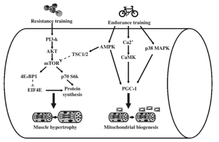
Cell signalling & its role in Exercise
Exercise = increases metabolic demand
Different types of exercise use different signalling pathways
AMPK
CaMK
PGC-1a
AMPK
Trigger = Cellular energy stress ( Low ATP/high AMP)
Function = Enhance glucose uptake & fatty acid oxidation - Restores energy balance
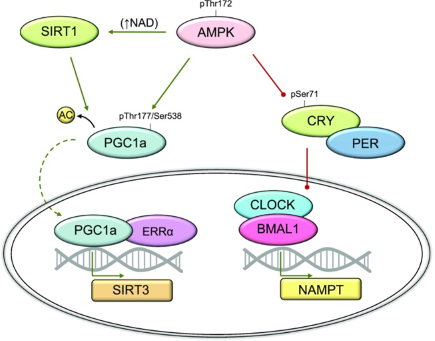
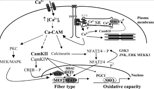
CaMK
Trigger = Increase Ca2+ during exercise
Function = Activate genes involved in mitochondrial biogenesis
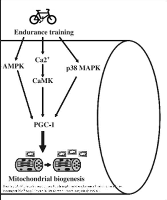
PGC-1a
Central regulator for mitochondrial biogenesis
Role = Enhance aerobic adaptation by increasing mitochondrial density or efficiency
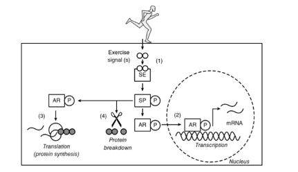
How change occurs from signalling pathways induced by exercise
mRNA & proteins
Related mRNA & synthesis involving mitochondria
Sensor proteins - detection
Signal pathways
Effector - Adaptation
Anaerobic signalling in skeletal muscle
Results in protein synthesis/ Less breakdown of proteins
Cell growth requires a positive protein balance
Stimulated by exercise/ Amino Acids
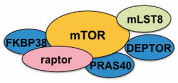
MTOR1
Enhances protein synthesis/ Less breakdown of proteins
Via:
Cap-dependent translation
Translation elongation
mRNA biogenesis
Ribosome biogenesis
Upstream regulators of anaerobic signalling in skeletal muscle
Insulin signalling
Amino Acids
Mechanical stimuli
AAs - activate Rag GTPases & recruit MTOR1
MS - PLD dependent increases in PA ( Binds to MTOR + signals)
MTOR1 inactivation
Nutrient deficient - Lack of AAs
Energy stress - AMPK activation
Negative feedback of S6K1
Pharmacological inhibition, redox stress
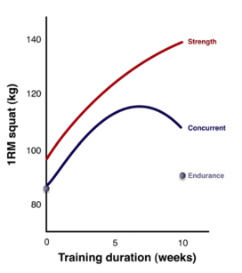
Competition of pathways
Fliiping does matter - Endurance better after strength
Innate immunity
Quick response to non- self antigens ( Barriers, inflammation , Phagocytosis)
Barriers preventing infection
Anatomic - Skin/ Membranes
Physiological - PH, Temp, chemical mediators
Inflammation
Endocytic/ phagocytic
Adaptive immunity
Mediated by T cells & antibodies
Recognises non-self antigens - activate relevant pathways
APCs are key to T cell antigens
MHCs:
Surfaces of APCs - express proteins
T cells express T receptors
Relevant immune cells
Dendritic cells - APCs - Induce immune response
Macrophage - Phagocytosis/inflammation
Neutrophil - guided by cytokine factors - 1st cell to arrive - Phagocytosis
NK cells - Kill virus cells
Exercise effect on immune system
Moderate/vigorous exercise for 60 mins = Increase mobilisation
Open window theory:
Temporary decrease in immunity post-exercise
Decreased respiratory infections in active individuals/ Increase in elite
Insulin action
Stimulate glucose uptake into skeletal muscle & adipose
Inhibits glucose production (Gluconeogenesis/ Glycogenesis) - Promotes liver storage
Promotes fat storage - Enhanced Lipogenesis/Lipolysis
Inhibits protein breakdown
Increase blood flow and perfusion
Appetite suppressant
Insuline synthesis pathway
Insulin/ exercise similarity
Insulin/ muscle contraction has similar pathways - GLUT 4 translocation
Synergistic effect on tissue glucose uptake
Insulin signalling pathway
Insulin binds to receptors (IR)
Insulin activates the Tyrosine Kinase domain of the beta subunit of IR
Insulin receptor substrate proteins (IRS) become tyrosine phosphorylated
PI3K becomes activated and converts PIP2 to PIP3
PIP3 recruits Akt
Akt becomes phosphorylated on 2 different sites
Cross-talk between Akt and Rac1
Reorganisation of the actin skeleton
GLUT4 translocation to the cell membrane
Key nuclear receptor in Skeletal muscle
PGC 1a - Regulates mitochondrial biogenesis
Oestrogen receptors - Energy metabolism & Muscle endurance
Androgen receptors - Muscle growth
Oestrogen recpetors
Mediate effects of oestrogen:
Mitochondrial function
Muscle repair
Muscle strength/contraction
Metabolic homeostasis
Androgen receptors
Binds androgen (Test)
Ligand induces AR translocation to the nucleus
Binds androgen response elements to DNA
Regulates genes involved in muscle protein synthesis
Dependent on muscle growth - Men have more than women
Nuclear receptor 1
Ligand = Steroid hormone
In the cytoplasm, when inactive
Bound to chaperone proteins
Hormone binding to receptor = movement to the nucleus
In nucleus = Bind to HREs on DNA - regulates transcription
Eg - Androgen receptor
Nuclear receptor 2
Ligand = nonsteroid hormone - Thyroid
Nucleus, even when inactive
No ligand = Bound to DNA with compressor proteins that suppress genes
Ligand binds = Compressor released = Gene activation
Eg. Thyroid hormone receptor
Nuclear receptor 3
Ligand = Unknown/ respond to metabolism/Nutrients
Similar to type 1 - do not require ligand
Eg - Rev-Erb
Nuclear receptor 4
Ligand = Unknown
Do not bind to DNA
Interacts with binding proteins/ Transcription factors
Eg - SF1