anatomy exam 4: the heart
1/73
There's no tags or description
Looks like no tags are added yet.
Name | Mastery | Learn | Test | Matching | Spaced | Call with Kai |
|---|
No analytics yet
Send a link to your students to track their progress
74 Terms
the heart is a ____ with ____ functions
muscular pump; 2
what are the 2 functions of the heart?
pulmonary circuit and systemic circuit
function of the heart that involves the pulmonary circuit
right side of the heart picks up oxygen-poor blood from body tissues and pumps it to the lungs. Goal: for oxygen-poor blood to pick up oxygen and get rid of carbon dioxide.
function of the heart that involves the systemic circuit
left side of the heart receives oxygen-rich blood that is returning from the lungs and pumps this blood throughout the body. Goal: to nourish and supply oxygen to the body tissues.
what makes up the pulmonary circuit?
blood vessels that carry blood to and from lungs.
what makes up the systemic circuit?
blood vessels that transport blood to and from all body tissues and back to the heart.
how many receiving chambers (atria) does the heart have? what is their names?
2; right atrium and left atrium.
what purpose does the atria serve?
they receive the blood that returns from the systemic and pulmonary circuits.
what are the two main pumping chambers of the heart?
the right ventricle and left ventricle.
what are the functions of the ventricles?
pump blood around the two circuits.
pericardium
a triple-layered sac around the heart
name of the outer layer of this sac (pericardium)
fibrous pericardium
fibrous pericardium
outer most layer of the pericardium. attaches to the diaphragm inferiorly and superiorly to the roots of the large vessels that leave and enter the heart. holds the heart in place and keeps it from overfilling with blood.
what is deep to the fibrous pericardium?
double-layered serous pericardium.
what makes up the serous pericardium?
parietal pericardium and visceral pericardium (aka epicardium)
parietal pericardium
adheres to the inner surface of the fibrous pericardium
visceral pericardium is also known as
epicardium
what are the 3 layers of the heart wall
epicardium, myocardium, and endocardium
epicardium
visceral layer (lines the surface of the organ) of pericardium
myocardium
cardiac muscle tissue
endocardium
lines the chambers and covers the valves. composed of simple squamous epithelium
do ventricles have larger/thicker walls than atriums? what are the ventricles separated by?
ventricles have larger/thicker walls. separated by the interventricular septum (myocardium)
external grooves on the heart
coronary sulcus (groove), anterior interventricular sulcus, posterior interventricular sulcus.
arteries
carry blood away from heart
veins
carry blood towards the heart
how many pulmonary arteries and pulmonary veins does the heart have?
2 pulmonary arteries, 4 pulmonary veins.
which ventricle requires a lot of force?
the left ventricle requires a lot of force because it is pumping oxygenated blood to the whole body. right ventricle does not need a lot of force because the lungs are right there.
what is right atrioventricular valve also known as?
tricuspid
what is left atrioventricular valve also known as?
mitral
what conducts electrical signals through the heart?
specialized cardiac muscle cells known as pacemaker cells.
left anterior descending artery (LAD)
supplies blood and nutrients to interventricular septum and anterior walls of both ventricles
what serves as the primary anatomical pathway for the left anterior descending artery (LAD)?
anterior interventricular sulcus
the coronary sulcus houses which arteries?
right coronary artery (RCA) and circumflex artery.
the right coronary artery branches into what
right marginal artery and posterior interventricular artery (PDA).
what does right coronary artery supply blood and nutrients to?
supplies the right atrium and most of the right ventricle
what is anterior interventricular artery also known as?
left anterior descending artery (LAD)
where is the great cardiac vein located on the heart?
anterior interventric sulcus
where is the middle cardiac vein located on the heart?
posterior interventric sulcus
where is the small cardiac vein located on the heart?
inferior right margin
where does the great, middle, and small cardiac veins dump into?
coronary sinus (posterior of coronary sulcus)
what are two coronary artery diseases?
atherosclerosis and angina pectoris
atherosclerosis
fatty deposits in the inner lining of the arteries that can occlude (block) blood flow
angina pectoris
chest pain from tissue hypoxia in the myocardium or from a spastic coronary artery
what causes myocardial infarction (also known as heart attack)?
blocked lumen in branch of left coronary artery. an irreversible death of heart muscle. blockage causes tissue death
does one side of the heart contract first?
no, both sides of the heart contract simultaneously.
what is the path in which blood flows from the heart to the body?
blood flows out of the heart through arteries to smaller arteries until the blood reaches the capillaries of the organs.
what is the path in which blood flows from the body to the heart?
blood flows out of the capillaries, in to small veins, and moves into larger veins as it returns to the heart.
what is the term that represents the hollow space inside blood vessels which blood flows?
lumen
is the lumen of a vein or artery larger?
the lumen of a vein is larger
what are the 3 tissue layers that make up blood vessels (arteries and veins)? [listed from outer to inner layer]
tunica externa (outer connective tissue layer), tunica media (middle smooth muscle layer), and tunica intima (inner endothelial layer)
what are two visible differences between arteries and veins?
the lumen of the vein is larger, and the tunica media is thicker in arteries.
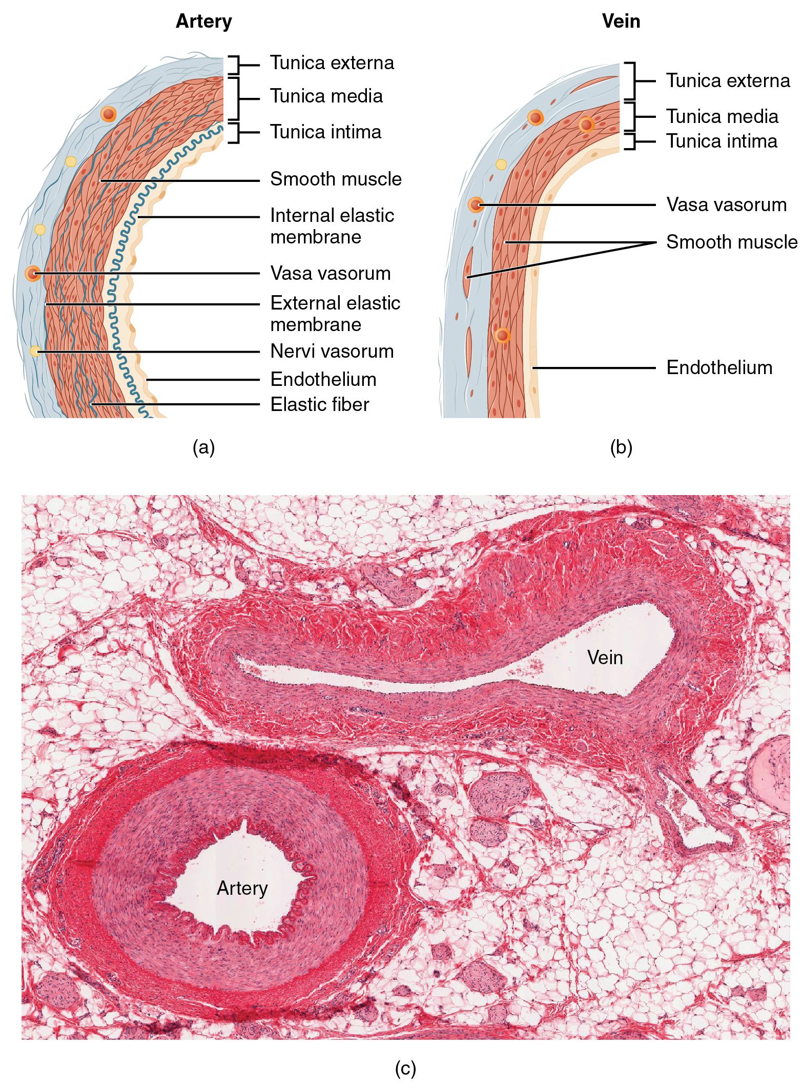
what is a special characteristic of veins?
they have venous valves! these are one-way bicuspid (two-flap) leaflets that prevent blood from flowing backward against gravity. these valves ensure efficient blood return to the heart!
endothelial cells in capillaries allow?
gas and nutrient exchange. endothelial cells are simple squamous cells.
what are the different types of arteries listed largest to smallest?
elastic arteries, muscular arteries, and arterioles.
what type of muscle is found in elastic arteries, muscular arteries, and arterioles?
smooth muscle! this enables involuntary movement
elastic arteries
large lumens, lots of elastin. e.g.: aorta. elastin allows it to stretch and recoil during heart contractions to maintain blood pressure.
muscular arteries
thick tunica media
arterioles
lead into capillaries.
what is always present in the arteriolar wall?
smooth muscle tone which is the continuous, low-level, involuntary contraction of smooth muscle tissue. helps regulate blood pressure.
how large are capillaries?
just large enough to allow single-file passages of RBCs.
what are capillaries made of?
endothelium and basement membrane
precapillary sphincters are isolated to the
mesenteric vasculature (associated with the intestines)
is the vascular shunt (which is the metarteriole and thoroughfare channel) considered true capillaries?
no, they are not considered true capillaries.
does blood flow through true capillaries when the sphincters are open or closed?
when the sphincters are open
what are the 4 routes molecules can pass into and out of capillaries?
diffusion through endothelial cell membranes (O2, CO2), intercellular clefts (spaces b/w endothelial cells), pinocytotic vesicles (windows/holes through cells), fenestrations
what is the difference between endothelial cells and epithelial cells?
endothelial cells cover the blood vessel inner surface, while epithelial cells cover outer surface of the internal organs and the body
what is the exception to capillary permeability?
blood brain barrier. there is no fenestrations or clefts. there are complete tight junctions. glucose is “ushered” across the walls.
what are the 3 types of capillaries?
continuous capillary, fenestrated capillary, and sinusoid capillary.
characteristics of continuous capillaries?
least permeable, and most common (e.g., skin, muscle).
characteristics of fenestrated capillaries?
large fenestrations (pores) increase permeability. occurs in areas of active absorption or filtration (e.g., kidney, small intestine).
characteristics of sinusoid capillaries?
most permeable. occurs in special locations (e.g., liver, bone marrow, spleen). they have an incomplete basement membrane.
veins acts as a —— for blood
storehouse.
65% of blood is in the venous circulation at any given moment. low pressure system (there are mechanisms, like venous valves, that help push blood back up to the heart).
what are the three types of veins ordered largest to smallest?
veins, venules, and post-capillary venules.
how does venous blood fight gravity?
valves and skeletal muscle pump
valves are mainly located in limbs. similar structure as the semilunar valves in the heart.
skeletal muscle pump: skeletal muscles contract and push blood up a vein (like toothpaste); back flow is prevented by valves.
veins are a low-pressure conduit system