Lab Midterm
1/235
There's no tags or description
Looks like no tags are added yet.
Name | Mastery | Learn | Test | Matching | Spaced | Call with Kai |
|---|
No analytics yet
Send a link to your students to track their progress
236 Terms
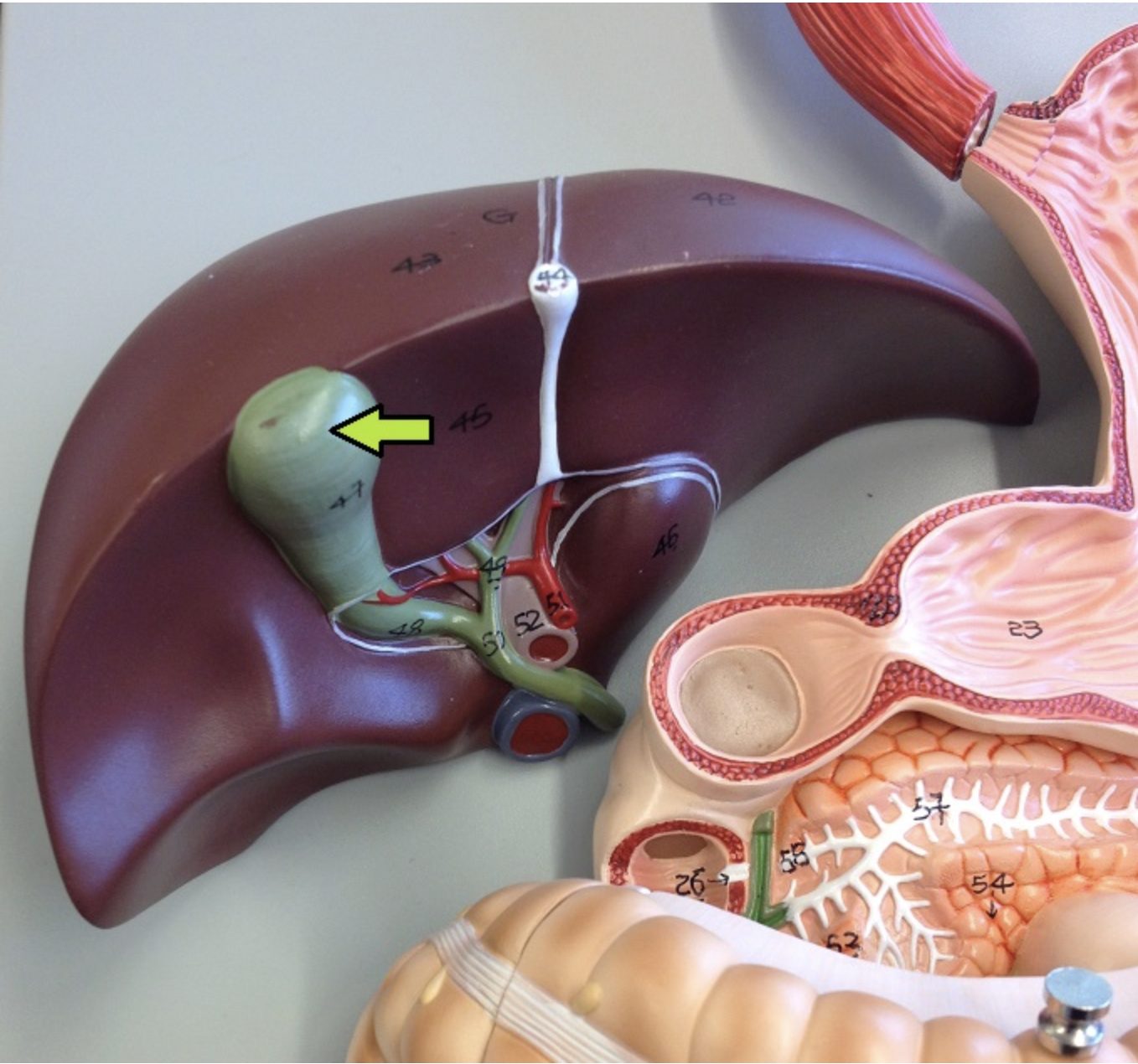
identify the major organ
gallbladder
a professional fighter hit in the “mental” region might have damage to the
a. nose
b. shoulder
c. knee
d. chin
e. ear
d. chin
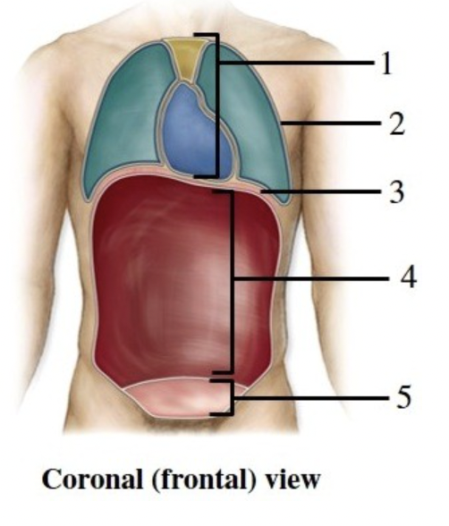
the figure shows a frontal view of a human. What does number 1 indicate?
a. pelvic cavity
b. mediastinum
c. pericardial cavity
d. peritoneal cavity
e. pleural cavity
b. mediastinum
you’re looking through a microscope with an ocular lens that magnifies the image 10X. The yellow-ringed (10X) objective lens is aimed at the specimen. What is the total magnification?
a. 10X
b. 100X
c. 40X
d. 50X
e. 400X
b. 100X
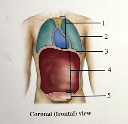
The figure shows a frontal view of a human. What does number 5 indicate?
a. abdominal cavity
b. mediastinum
c. pleural cavity
d. pelvic cavity
e. pericardial cavity
d. pelvic cavity
What is the anatomic term for the “foot”?
a. popliteal
b. acromial
c. patellar
d. pubic
e. pedal
e. pedal
Identify the serous membrane. Be sure to include whether it is visceral or parietal layer in your answer.
visceral pleura
identify the serous membrane. be sure to include whether it is visceral or parietal layer in your answer.
parietal peritoneum
the directional term that means “in back of” or “towards the back surface” is
a. cephalic
b. anterior
c. caudal
d. posterior
e. proximal
d. posterior
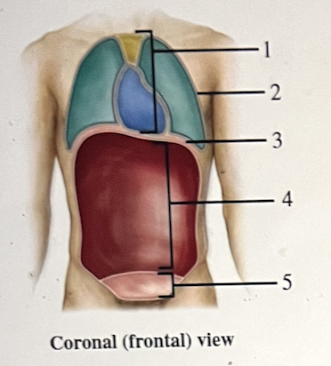
The figure shows the frontal view of a human. What does number 2 indicate?
a. abdominal cavity
b. pelvic cavity
c. thoracic cavity
d. mediastinum
e. cranial cavity
c. thoracic cavity
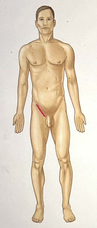
identify the body region
inguinal region
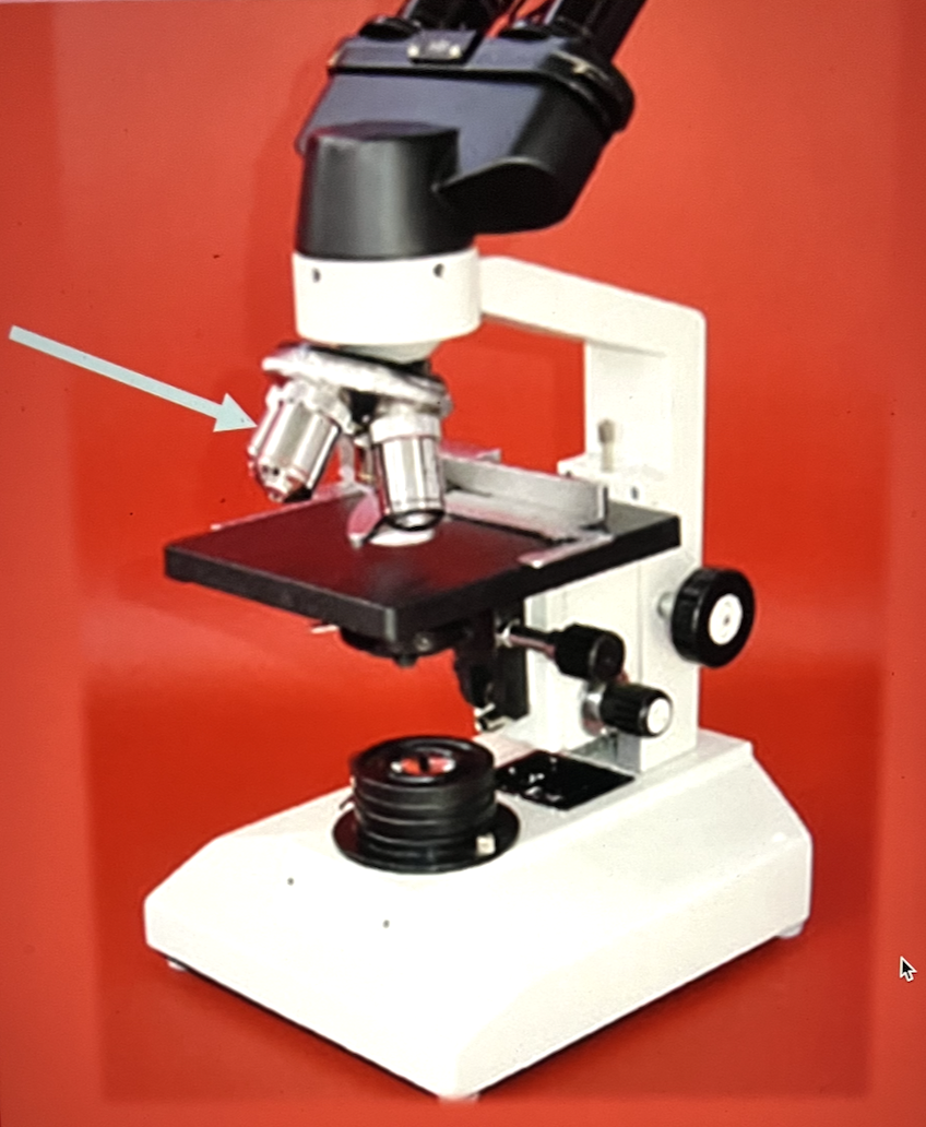
identify the name
objective lens
If the objective lens is set to the 40X power, what is the overall magnification of the image
400
the limbs of the body are attached to the axis and make up the
a. thoracic region
b. abdominal region
c. axial region
d. ante-brachial region
e. appendicular region
e. appendicular region
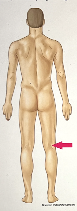
identify the body region
popliteal region
the anatomic term for the “cheek” is
a. cervical
b. buccal
c. sacral
d. crural
e. pelvic
b. buccal
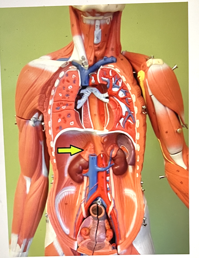
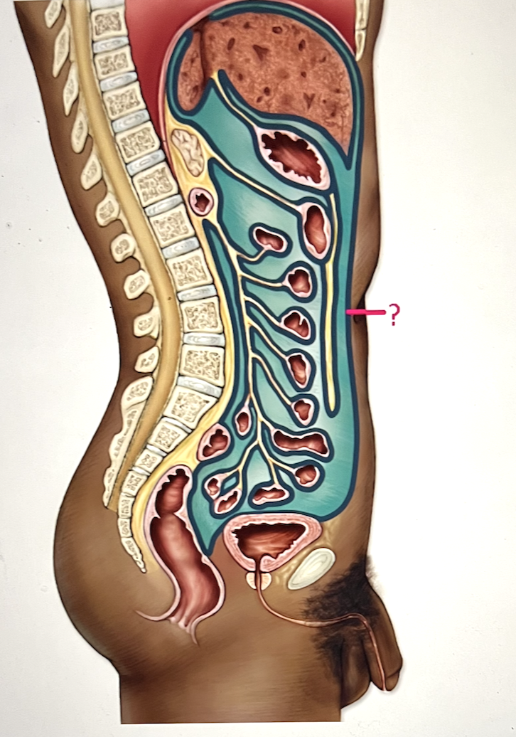
identify the major organ
Right adrenal gland
the appendix is in the right iliac region, and is therefore located in the ____ quadrant
right lower quadrant
identify the body region
antecubital region
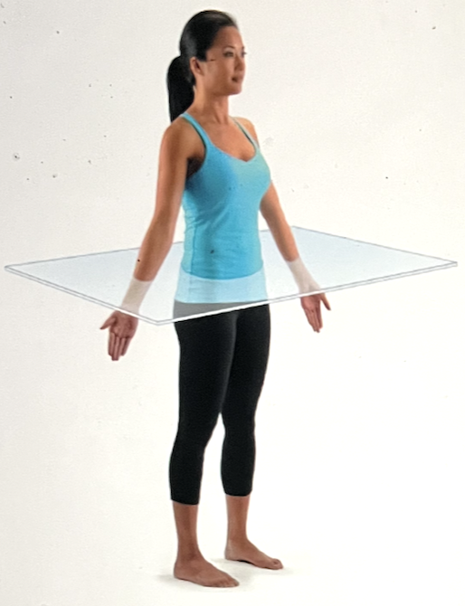
identify the body plane
transverse plane
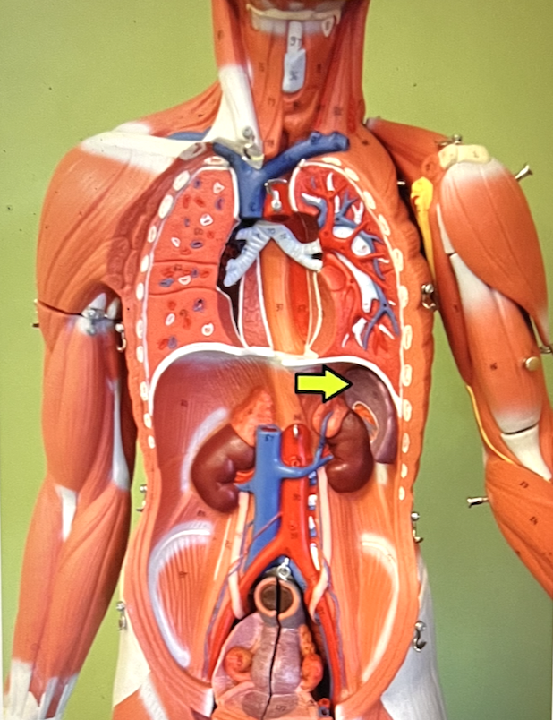
identify the major organ
spleen
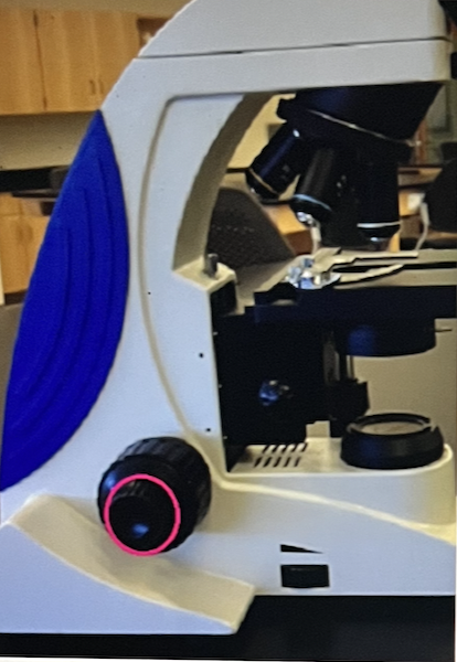
identify the microscope part
fine adjustment knob
The anatomic position allows all observers to have a common point of reference.
True/False
True
what is the anatomic term for the “hip” region?
a. sural
b. coxal
c. sternal
d. dorsal
e. crural
b. coxal
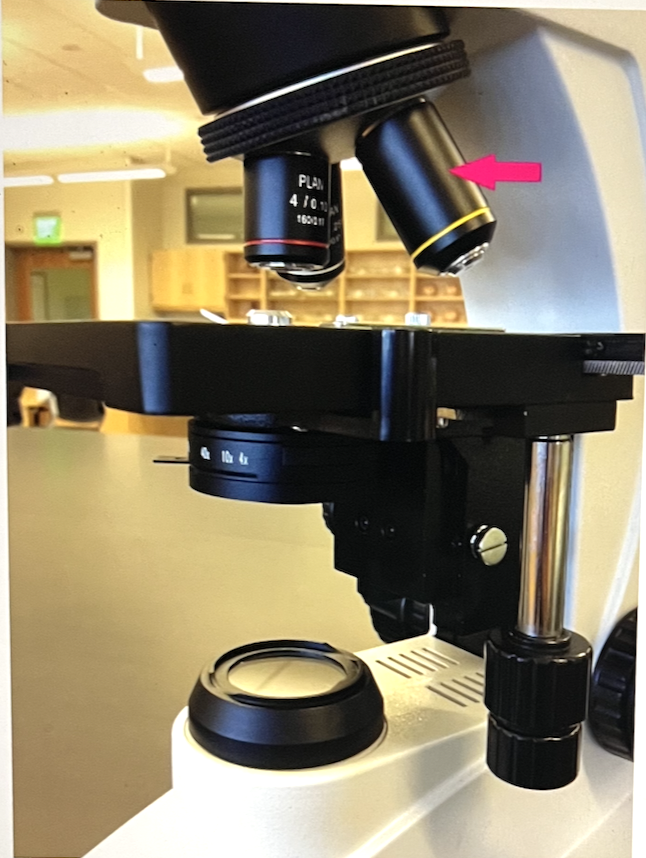
Identify the microscope part
objective lens
The serous fluid that helps in cardiac function is located
a. inside the heart’s chambers
b. between the visceral pericardium and the cardiac muscle
c. in the pericardial cavity, between the parietal and visceral pericardial layers
d. between the parietal pericardium and the sternum
c. in the pericardial cavity, between the parietal and visceral pericardial layers
The adbominopelvic quadrants are formed by passing one horixontal and one vertical line through the
a. gluteal region
b. crural region
c. umbilicus
d. patellar region
e. antebrachial region
c. umbilicus
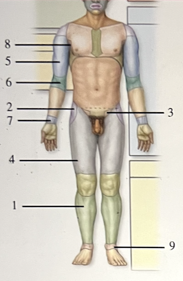
The figure shows an anterior view of a human in the anatomic position. Which number indicates the inguinal region?
a. 1
b. 2
c. 3
d. 4
e. 5
c. 3
the substage condensor is used to
a. concentrate light on the specimen
b. provide the light for illumination
c. reposition the slide on the stage
d. enlarge or reduce the field of view
a. concentrate light on the specimen
when do you use the coarse adjustment knob?
a. every time you switch between objective lenses
b. to see depth/3-D in the slide
c. to bring the object into sharp focus
d. only with the scanning (4X) objective lens to first bring the object into view
d. only with the scanning (4X) objective lens to first bring the object into view
the muscular partition that separates the thoracic and abdominopelvic cavities is the _____
diaphragm
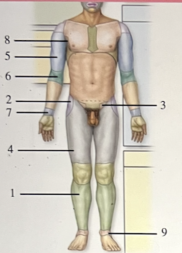
The figure shows an anterior view of a human in the anatomic position. What does number 2 indicate?
a. sacral
b. carpal
c. axillary
d. coxal
e. antecubital
d. coxal
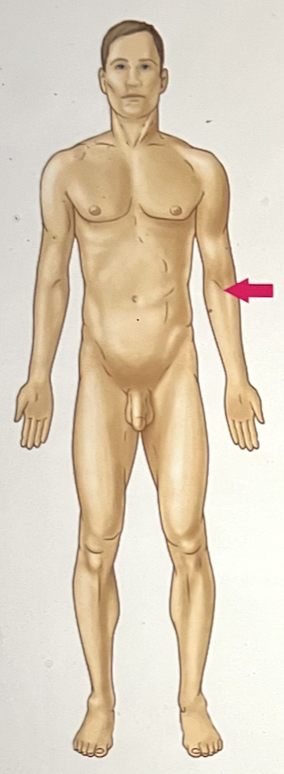
the antecubital region is _____ to the brachial region
distal
The level of organization one step more complex than the organ level is the _____ level
organ system
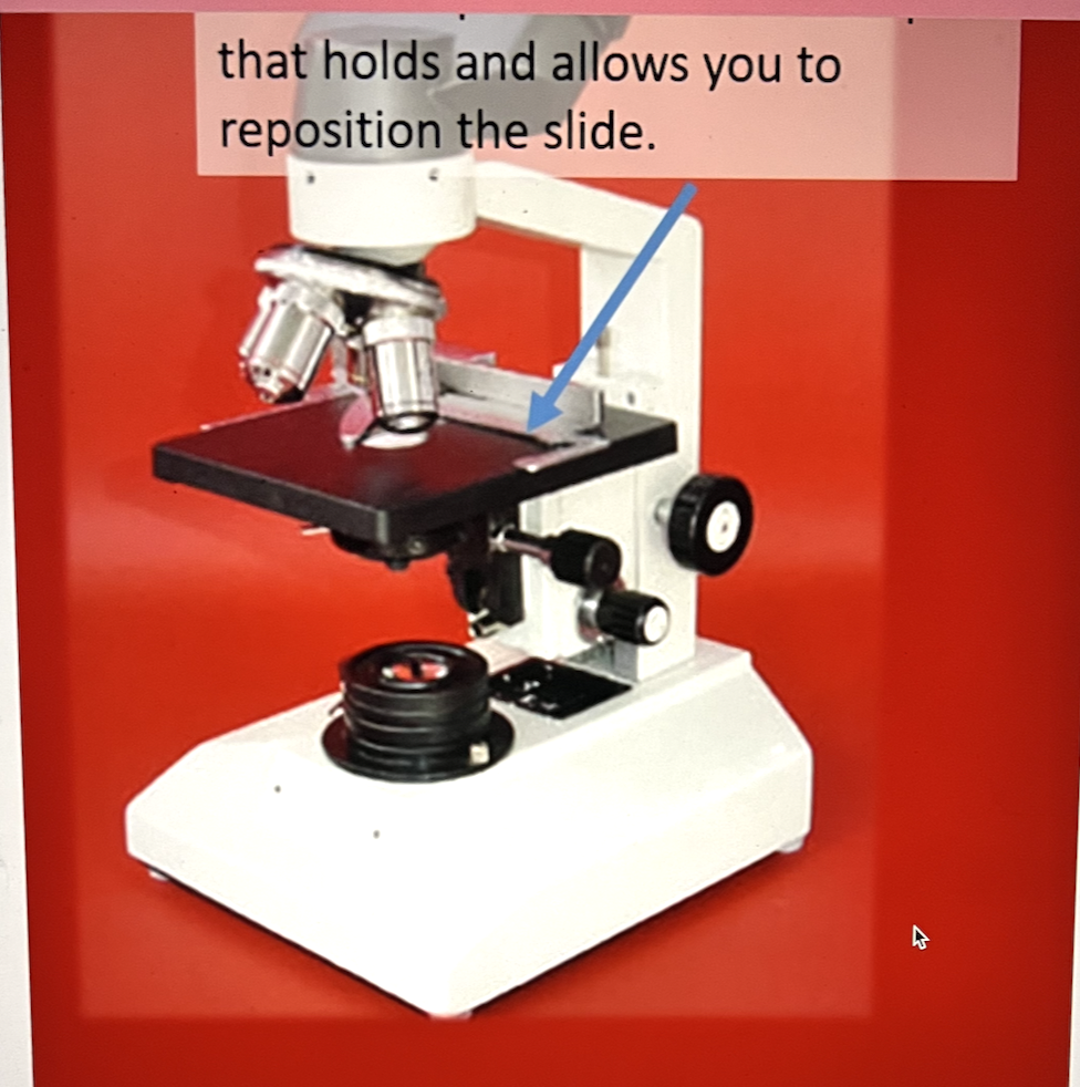
Name the part of the microscope indicated in the image below.
mechanical stage
The organ system that transports and filters interstitial fluid while also participating in immune responses is the ____ system.
lymphatic
The pituitary, thyroid, and adrenal glands are typically grouped within the _____ system.
endocrine
An “inguinal” hernia is in the region of the
a. thigh
b. umbilicual
c. groin
d. calf
e. shoulder
c. groin
the median space in the thoracic cavity between the lungs is called the
a. mediastinum
b. hypochondriac space
c. pericardial cavity
d. peritoneal cavity
e. pleural cavity
a. mediastinum
when a cell is dividing the genetic material is in the compact form of ____
chromosomes
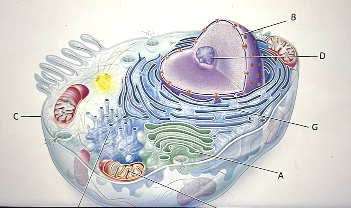
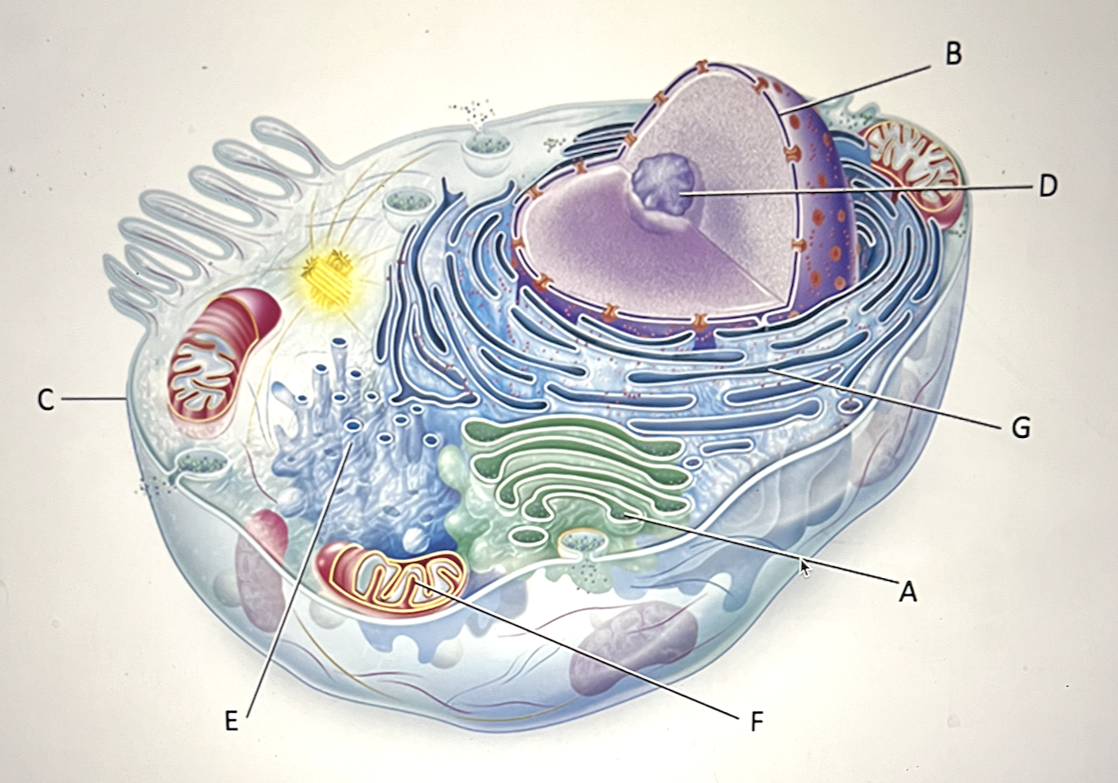
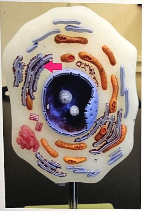
Which statement best describes letter C?
a. central area of the cell containing centrioles
b. group of membrane-enclosed sacs; modifies and packages proteins for export or use in the cell
c. series of membrane-enclosed spaces that are the site of lipid and cholesterol synthesis and metabolism, in addition to drug and toxin detoxification
d. structure surrounding the cell that creates a boundary between the cell and the external environment; regulates the movement of substances in and out of the cell; composed of a phospholipid bilayer and many other components
d. structure surrounding the cell that creates a boundary between the cell and the external environment; regulates the movement of substances in and out of the cell; composed of a phospholipid bilayer and many other components
In humans, the only cell that bears a flagellum is the ____ cell
a. sperm
b. oocyte
c. kidney
d. brain
e. red blood
a. sperm
DNA directs the synthesis of proteins, including enzymes that catalyze the synthesis of several important molecules.
True/False
True
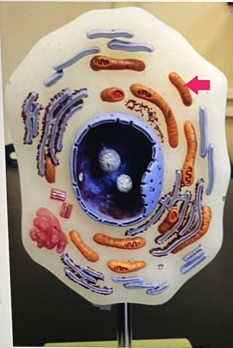
Identify the cell part
mitochondria
Everything packaged by the Golgi apparatus for secretion leaves the cell within a vesicle
True/False
True
The two identical cells that arise from mitosis are called ____ cells
daughter
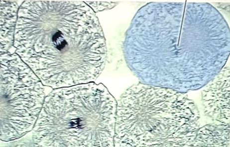
The phase of mitosis that begins with the arrival of a group of single-stranded chromosomes at each pole of
a. telophase
b. S phase
c. anaphase
d. metaphase
e. prophase
a. telophase
another name for the intracellular fluid is
a. cisternae
b. intercellular matrix
c. interstitial fluid
d. cytosol
e. cytoplasm
d. cytosol
the largest internal cellular structure is known as the _____ and is the cell’s control center
a. golgi apparatus
b. nucleus
c. lysosome
d. cytosol
e. smooth ER
b. nucleus
ribosomes that are attached to the Rough Endoplasmic Reiticulum are called “free ribosomes”
True/False
False
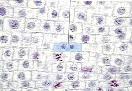
Identify the stage of mitosis
telophase
which of the following serves to increase the surface area of a cell?
a. microvilli
b. microfilaments
c. the nucleolus
d. centrioles
a. mivrovilli
two perpendicularly oriented ____ from the centrosomes, which is responsible for organizing microtubules during cell division
centrioles
Mucus is moved along the lining of the trachea by extensions from cell membranes known as
a. cilia
b. stereovili
c. microvili
d. flagella
a. cilia
the _____ is responsible for forming the outer, limiting barrier of a cell
a. mitochondrion
b. centrosome
c. peroxisome
d. ribosome
e. plasma membrane
e. plasma membrane
Apoptosis is best described as
a. the process of immune cells recognizing an infected cell as “foreign”
b. the process of an aging cell becoming cancerous
c. the destruction of a cell through mechanical damage
d. a process where cells destroy themselves
d. a process where cells destroy themselves
Identify the stage of mitosis at the pointer
metaphase
which of the following shows the correct sequence of mitosis?
a. metaphase - prophase - anaphase - telophase
b. metaphase - telophase - anaphase - prophase
c. prophase - anaphase - metaphase - telophase
d. prophase - metaphase - anaphase - telophase
e. telophase - metaphase - prophase - anaphase
d. prophase - metaphase - anaphase - telophase
____ is the division of the cytoplasm during cell division
cytokinesis
the ____ are responsible for synthesizing most of a human body cell’s ATP
a. microfilaments
b. ribosomes
c. lysosomes
d. mitochondria
e. nucleoli
d. mitochondria
The organelles responsible for organizing microtubules that are part of the mitotic spindle are called
a. centrioles
b. nucleoli
c. microvili
d. vesicles
e. cilia
a. centrioles
Identify the organelle that provides digestive enzymes for autolysis
a. lysosomes
b. peroxisomes
c. golgi apparatus
d. smooth ER
e. mitochondria
a. lysosomes
which of the following serve to increase the surface area of a cell for absorption and secretion?
a. cilia
b. microvili
c. cilia and microvili
d. cilia and flagella
e. flagella
b. microvili
the duplicated chromosomes that appears during prophase consists of two genetically identical structures called sister _____
chromatids
_____ is the general term for all cellular contents located between the plasma membrane and the nucleus
cytoplasm
The function of the nucleolus is to make
a. the secretions that will be packaged by the Golgi apparatus
b. histones
c. DNA molecules
d. the ddeoxyribose sugar
e. the subunits of ribosomes
e. the subunits of ribosomes
The folds of the internal membrane of a mitochondrion are called
a. cisternae
b. cristae
c. vacuoles
d. matrix
e. vesicles
b. cristae
the phase of mitosis that begins as spindle fibers pull sister chromatids apart at the centromere is
a. anaphase
b. metaphase
c. telophase
d. interphase
e. prophase
a. anaphase
the most abundant lipid of the membrane consists of a head and two tails. This type of lipid is
a. cholesterol
b. glycolipid
c. a steroid
d. glycoprotein
e. a phospholipid
e. a phospholipid
chromosomes are loose, and chromatin is tightly packed genetic material
True/False
false
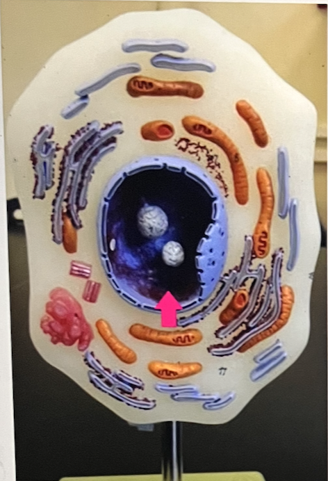
series of membrane-enclosed spaces that modify proteins made by the ribosomes; has ribosomes on the surface
a. E
b. C
c. F
d. A
e. D
f. G
g. B
f. G
because they produce ribosome subunits, one would expect to find large numbers of nucleoli in cells that synthesize
a. pigments
b. steroid hormones
c. solubility-enhancing substances
d. proteins
e. energy sources
d. proteins
identify the cell part…..this answer is NOT “nucleus” (hint: genetic material in nondividing cell”
chromatin
The term used to describe the fluid portion only within a cell is ____, or intracellular fluid
cytosol
identify the cell part
rough endoplasmic reticulum
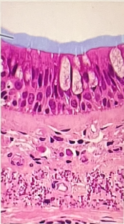
which structures are highlighted?
a. microvilli
b. goblet cells
c. basement membrane
d. cilia
e. nuclei
d. cilia
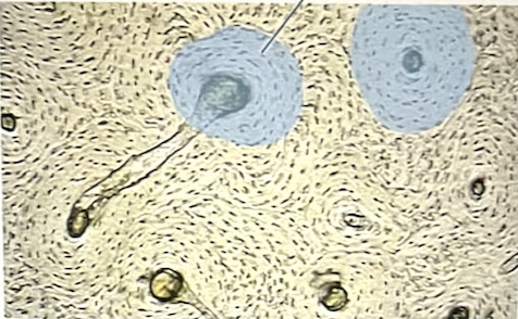
identify the highlighted SPACES that the cells are sitting in
lacunae
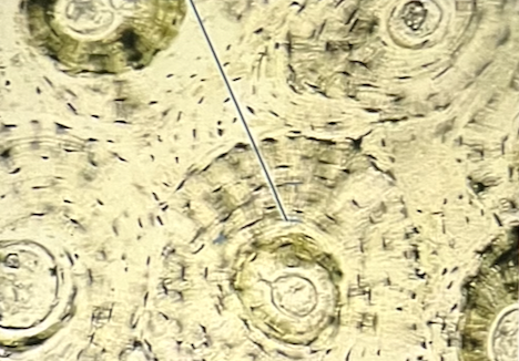
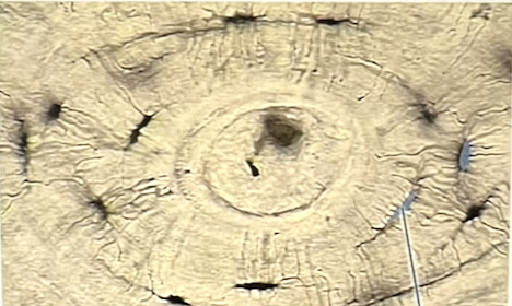
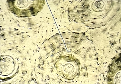
identify the specific tissue
a. dense regular CT
b. osseous tissue
c. elastic cartilage
d. blood
e. reticular CT
b. osseous tissue
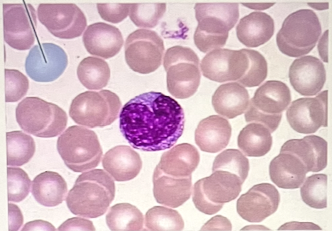
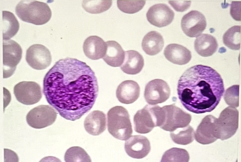
identify the highlighted cell type at the pointer (Give complete answer - NO abbreviations)
erythrocyte
which of the following is not a characteristic of nerve tissue?
a. it is found in the brain, spinal cord and nerves
b. some of its cells are support cells that are not excitable
c. its intercellular spaces are filled with collagen
d. it contains cells that respond by transmitting nerve impulses
e. its functional cells are excitable and respond to changes in their surroundings
c. its intercellular spaces are filled with collagen
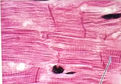
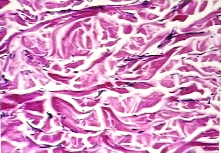
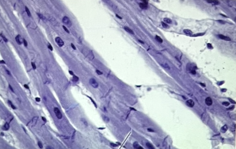
identify the specific muscle tissue
cardiac muscle tissue
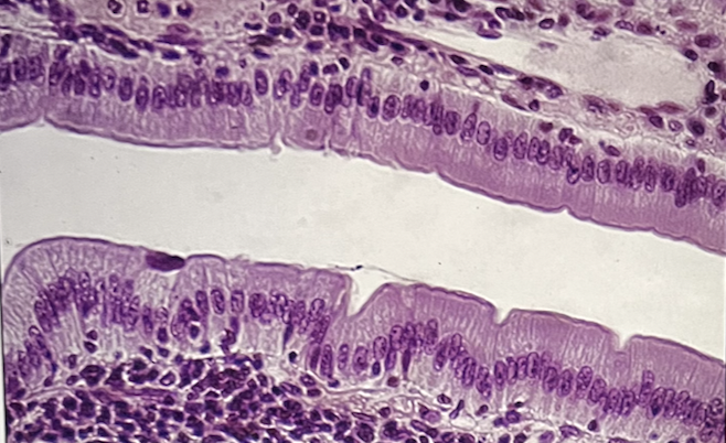
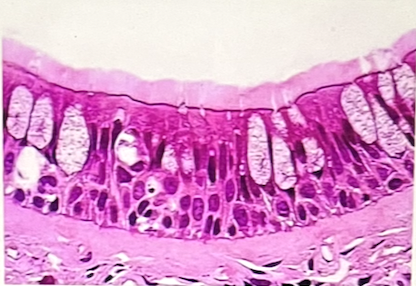
which epithelial type lines the small intestine
simple columnar epithelium
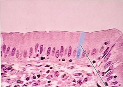
identify the highlighted epithelium at the pointer
simple columnar epithelium
identify the highlighted epithelium at the pointer
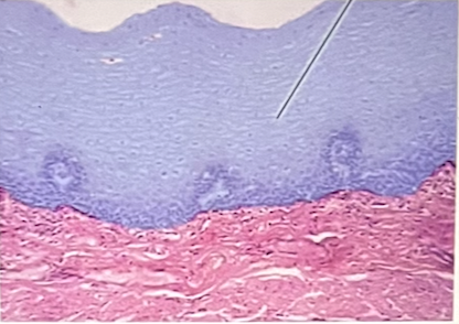
stratified squamous epithelium
identify the specific tissue
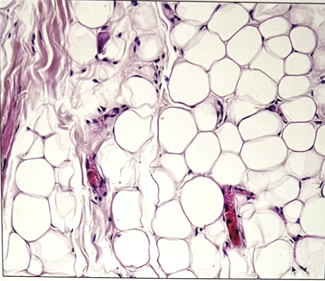
adipose tissue
identify the specific connective tissue in the picture
dense regular connective tissue
where is this epithelial type located?
a. urinary bladder
b. thick skin
c. esophagus
d. trachea
e. oral cavity
a. urinary bladder
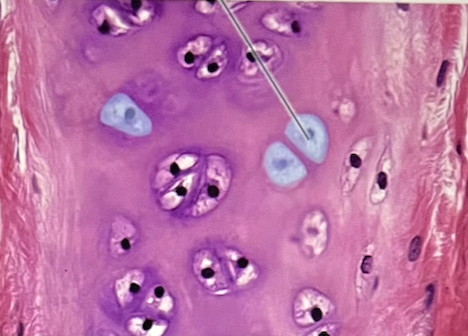
identify the CELL (at the pointer) contained within the highlighted spaces
osteocyte
Identify the specific tissue type
smooth muscle tissue
identify the highlighted tissue in picture
osseous tissue
Identify this tissue
a. stratified cuboidal epithelial tissue
b. simple cuboidal epithelial tissue
c. simple columnar epithelial tissue
d. pseudo-stratified ciliated columnar epithelial tissue
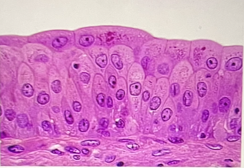
c. simple columnar epithelial tissue
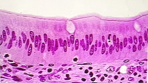
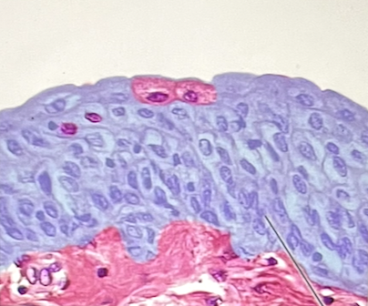
Identify the epithelium
a. simple columnar epithelium
b. simple squamous epithelium
c. stratified squamous epithelium
d. pseudostratified columnar epithelium
e. simple cuboidal epithelium
d. pseudostratified columnar epithelium
identify the highlighted structures at the pointer
platelets
Identify the highlighted epithelium
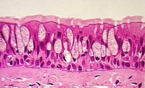
pseudostratified columnar epithelium
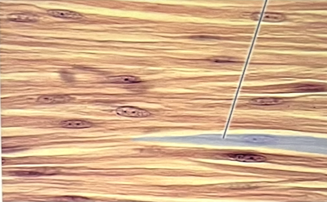
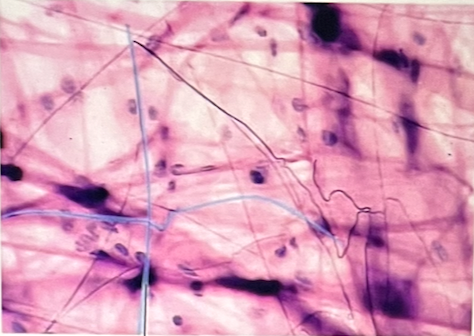
Identify the specific connective tissue in the picture
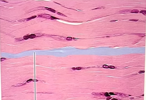
dense irregular connective tissue
Identify 1 characteristic that describes this muscle tissue
striated
Identify the highlighted spaces that the cells are sitting in
a. central canals
b. canaliculi
c. adipocytes
d. lacunae
e. lamellae
d. lacunae
identify the highlighted epithelium found lining the urinary bladder
transitional epithelium
identify the specific tissue in picture
areolar connective tissue
smooth muscle is found in the wall of the
a. stomach and intestine
b. kidney and liver
c. heart
d. kidney
e. stomach
a. stomach and intestine