Radiology - Pulmonary & Neurology
1/28
There's no tags or description
Looks like no tags are added yet.
Name | Mastery | Learn | Test | Matching | Spaced | Call with Kai |
|---|
No analytics yet
Send a link to your students to track their progress
29 Terms
age 15, gender M, pt is acutely breathless and might have a pneumothorax. CXR nl, no consolidation collapse, pneumothorax, or effusion. Nl mediastinal contour and pulmonary vasculature
Normal chest radiograph
Mail 25-year-old has a chief complaint of throat, pain, body ache, fever and left side of chest pain for two days no history of breathing difficulty or cough. Pain is not radiating to the left arm or neck. patient has poetic chest pain that is a stabbing feeling and is worse on inspiration
Pneumonia
55-year-old male is a smoker presents with chest pain. Fever and cough. Clinicians are worried about seeing cancer on a chest x-ray.
Opacity on xray, air bronchogram needed next
What does this air bronchogram indicate for the pt
Solid upper left lobe “hepatization”. Pt has lobar PNA
65-year-old female presented with one week history of difficulty passing urine, constipation, and back pain. Patient was found to be in a hyponatremic hyperosmolar and euvolemic state. Serum osmolality is low but urine osmolality is high, urine sodium is high. Pt suspected to have SIADH. no respiratory sx, no smoking hx or fhx of ca.
Non small cell lung carcinoma
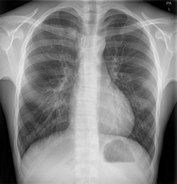
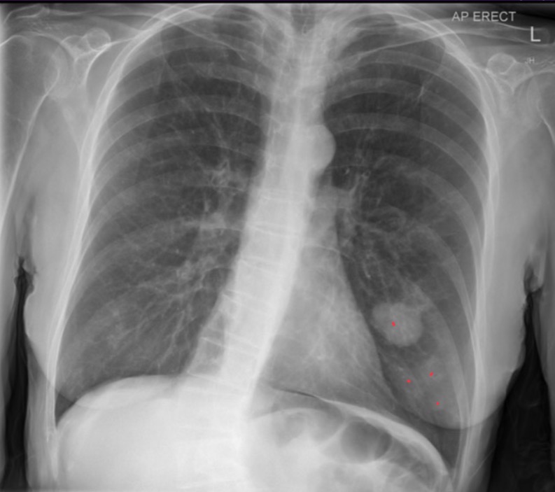
25-year-old female with no background available
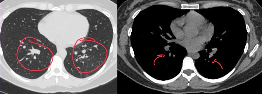
Radiolucent area in right lung field with compressive atelectasis of right lung and mediastinal shift to the left consistent with pneumothorax
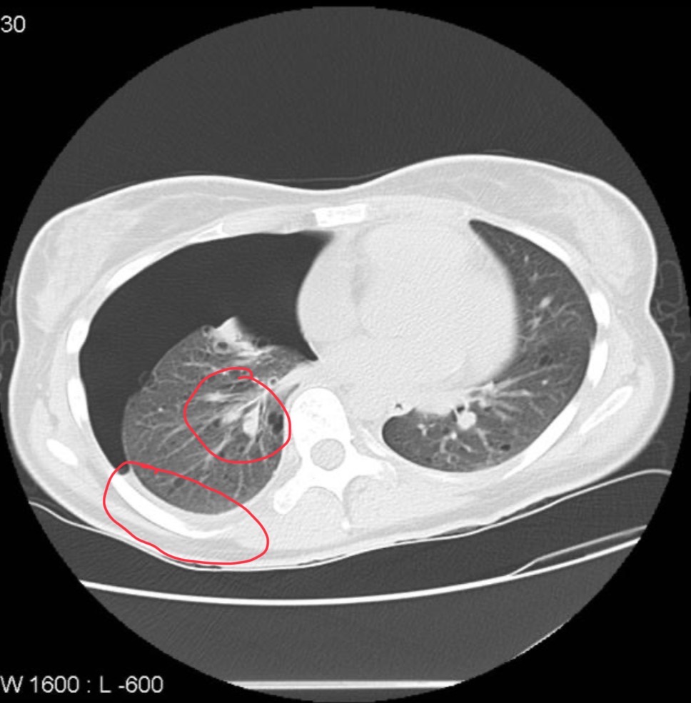
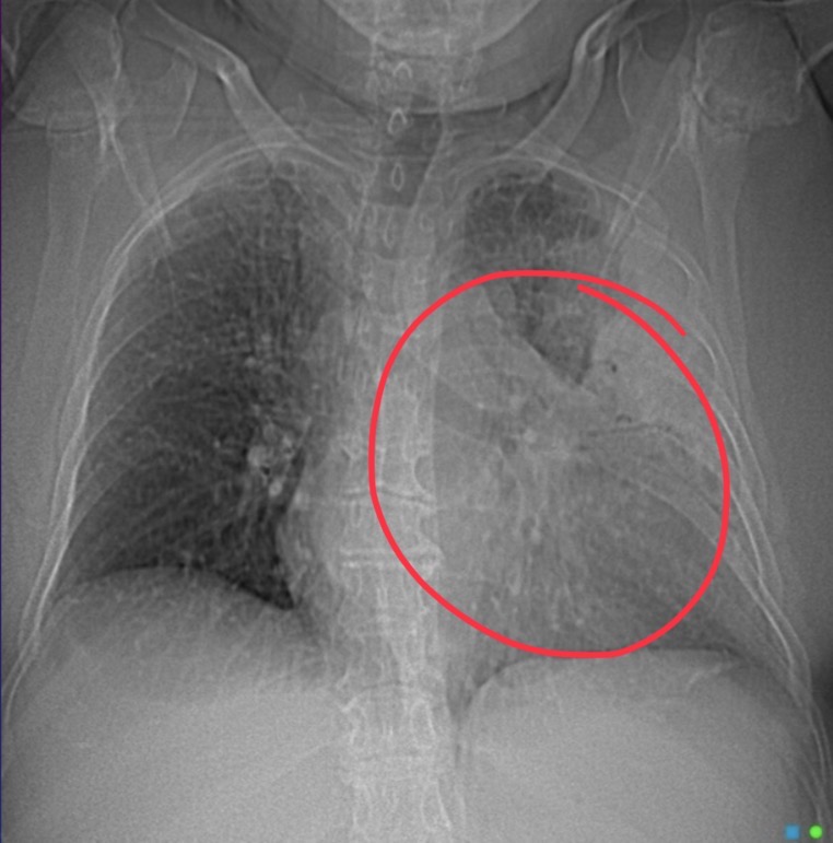
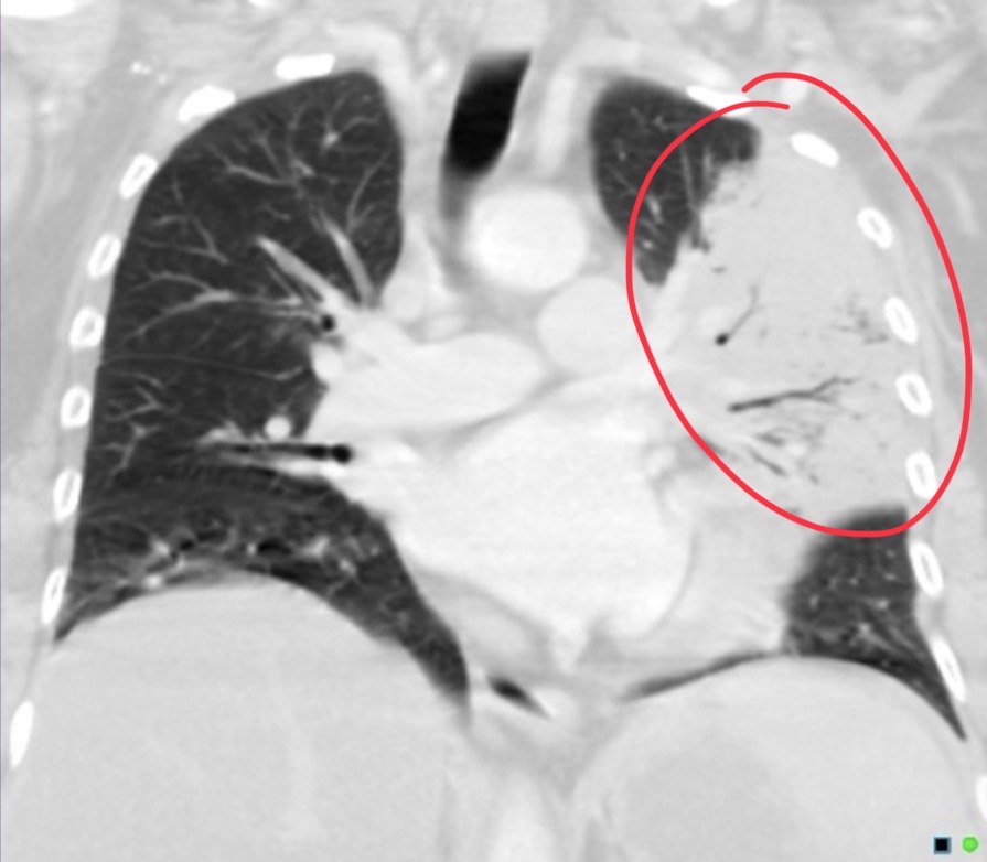
CT of pt
Shows pneumothorax in the right lung with mild plural fusion mediastinum push the left multiple mass lesions then the wild air density cyst surrounded by normal lung tissue.
Pt has tuberous sclerosis with lymphangioleiomyomatosis
40 yr old male is wheezing and coughing
Opacity seen in right upper lobe boarded by thin radiolucent streaks
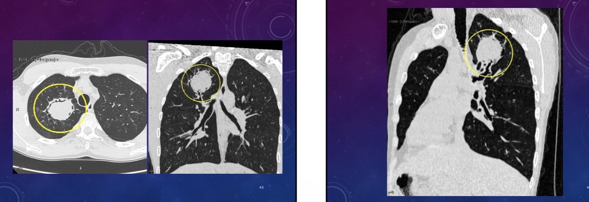
What does ct show
aspergilloma. Fungus ball that develops because of formation of hyphae inside lung cyst or an existing cavity (from tb or sarcoidosis) usually in the upper lobe.
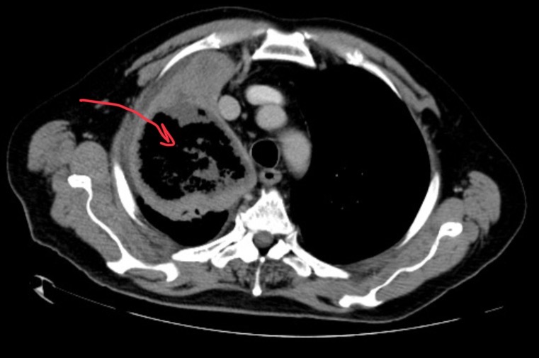
60-year-old male has been treated for AML. Patient has cough, fever and dyspnea.
Atoll sign = air inclusions without fluid levels
Pt has fungal PNA in immunocompromised pt. Pt had lobectomy and pathology was mucormycosis
25-year-old female has worsening of a recurrent cough. She has a history of overseas travel and exposure to unsanitary conditions and animal droppings.
Mass with internal calcifications in left lower lobe
CT of pt shows
pulm hydatid cyst. Broncho alveolar lavage showed pos echinococcus titers
20 year-old female presents with tremor and dystonia
Panda sign = Wilson disease aka hepatolenticular degeneration
6 year-old male has progressive, bilateral visual impairment, and poor motor progression for his age
Adrenoleukodystrophy
Damaged ponds dilated lateral ventricles, prior lesion, white matter, deviation hyperintensities, and left temporal and occipital areas.
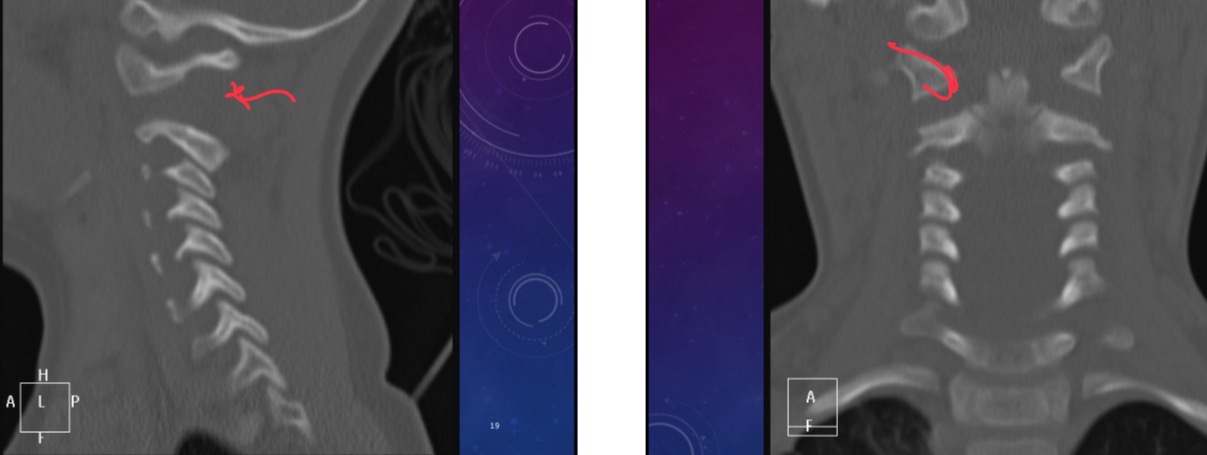
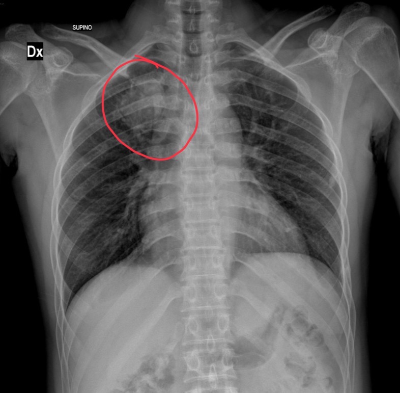
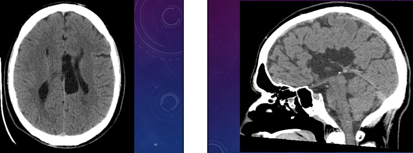
Three-year-old female was in a motor vehicle accident and is in GCS 3. (Coma). What does this cervical spine xr show
Widened interspinous distance between C1/C2
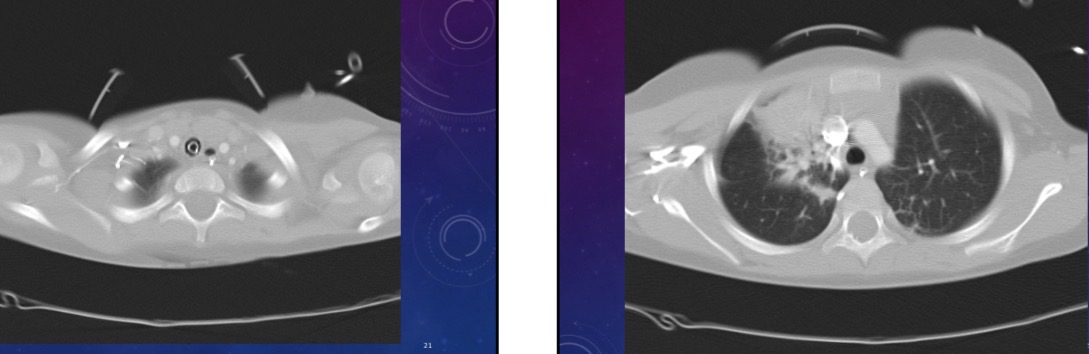
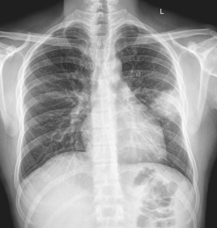
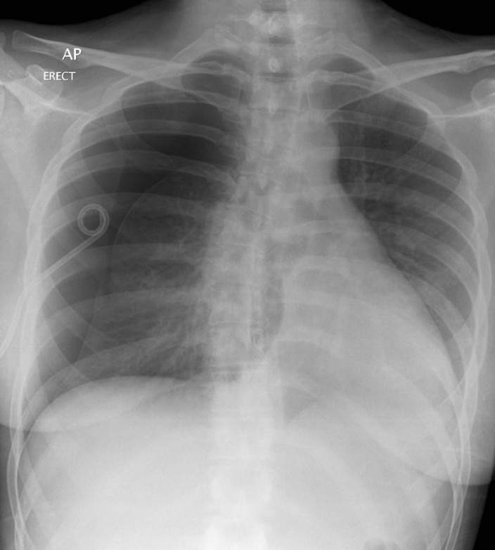
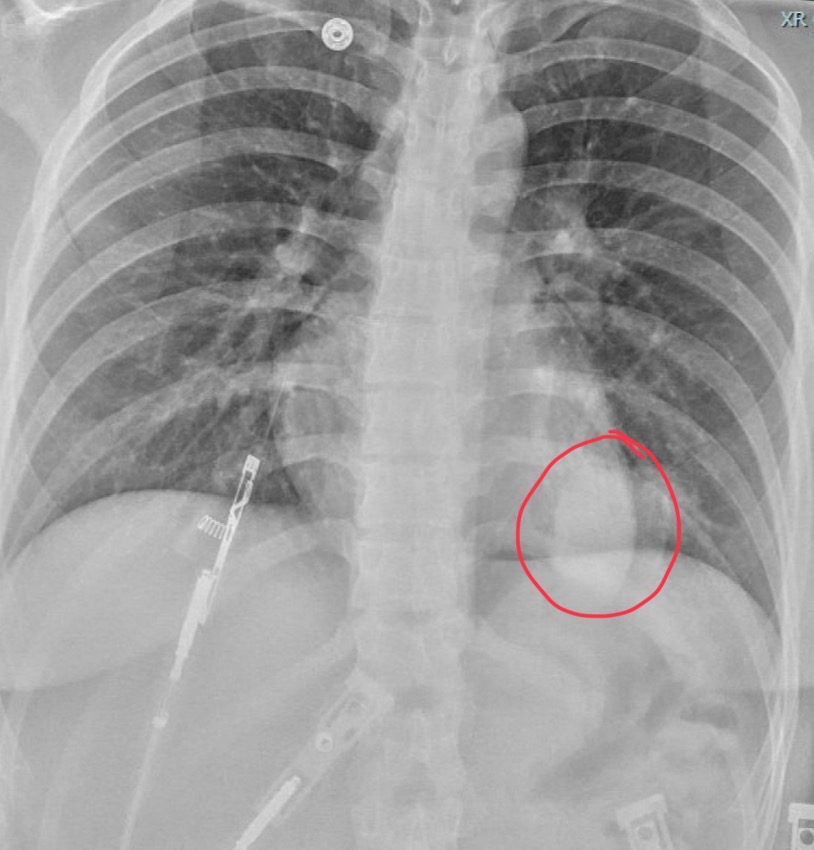
What does chest scan show
Left pneumothorax, atelectasis in right upper and left lower lobes
55-year-old woman presents with left facial droop
Epidermoid cyst. Pushing on pons and causing blood to pool there which compresses on facial nerve
20 year-old female presents with convulsions
Neurocysticercosis - vesicular stage
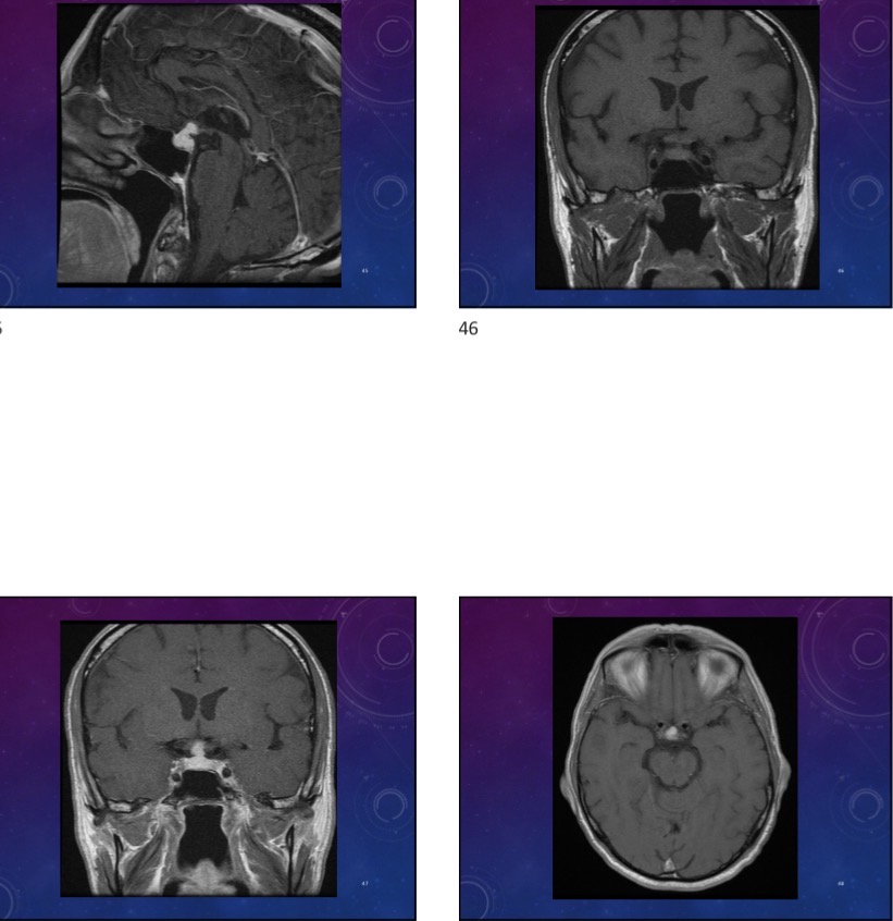
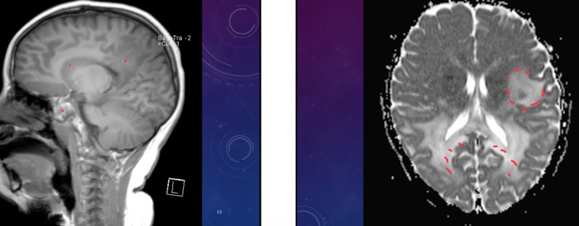
65-year-old male has a past history of CLL and is in remission is currently unwell and has a visual changes
Panpituitary lymphocytic hypophysitis bc of CLL.
Reactive chronic t lymphocytic infiltrate and fibrosis
Moderately thickened and lobulated stalk, superior extension to the hypothalamus. Pituitary gland and stalk are replaced by vividly homogenous enhancing tissue.
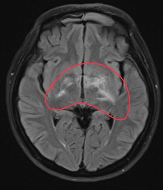
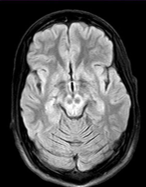
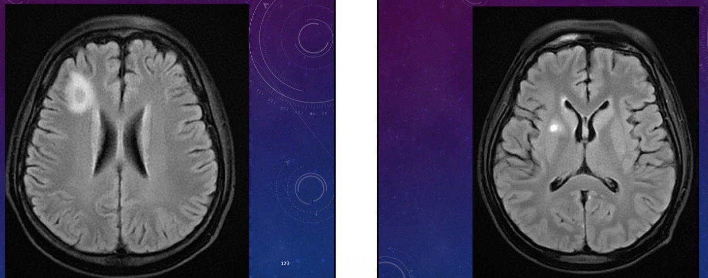
55 year-old female developed patchy vision with white specks seen bilaterally for a few weeks. Currently reports loss of vision in both eyes. There is hyper intensity around the bilateral hypothalamus, left globes pallidus, left post internal capsule, b/l optic tracts, left incision, right midbrain.
MS
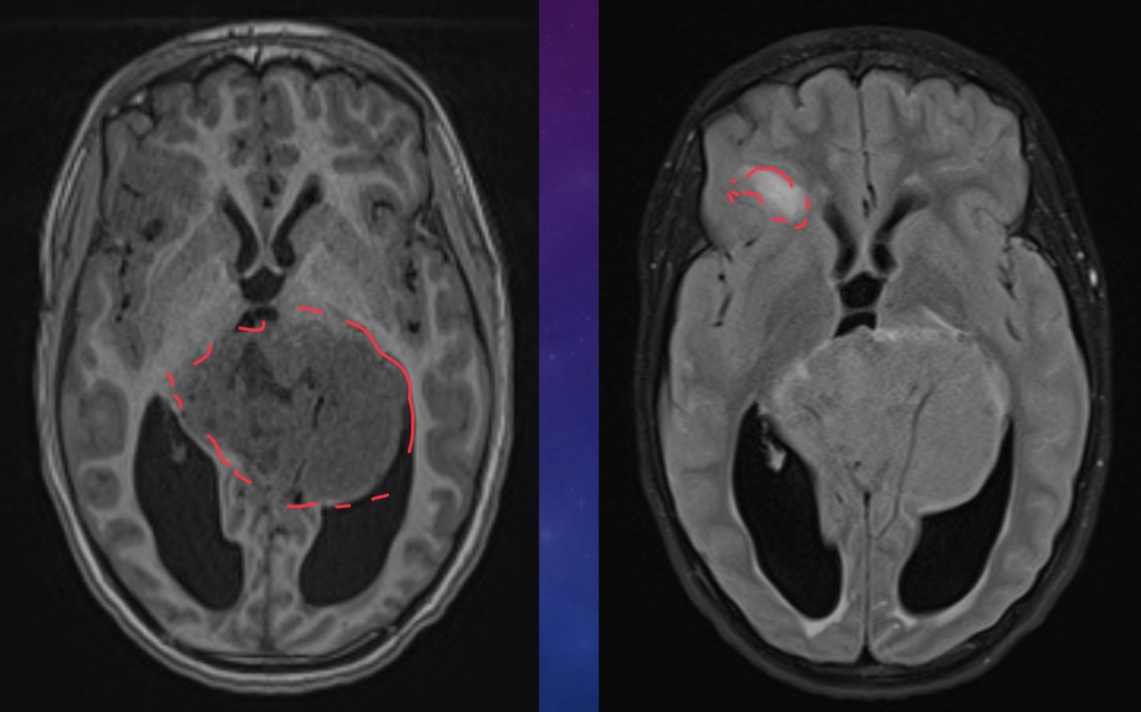
10-year-old male has a history of intermittent headaches for the past five months.
Pineoblastoma
Pineal gland tumor with mets to the frontal lobe
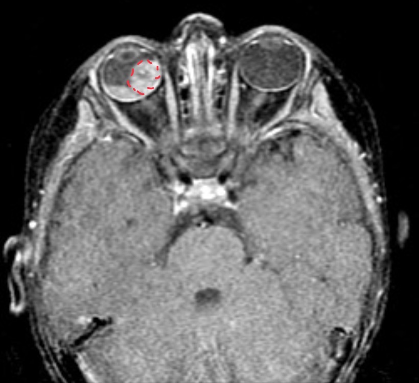
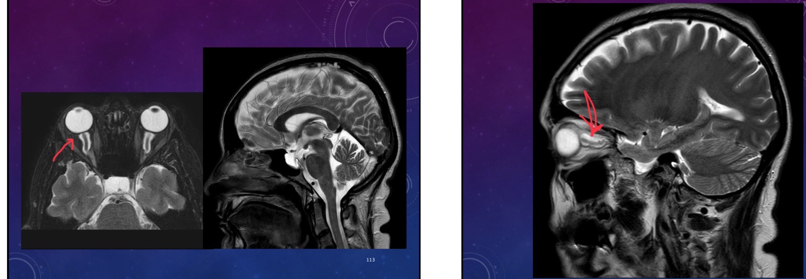
Child presents with no available information
Retinoblastoma w retinal detachment
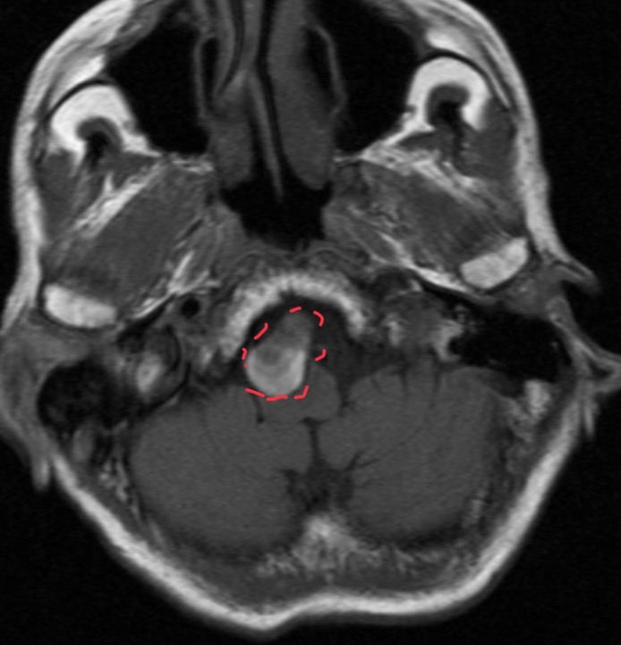
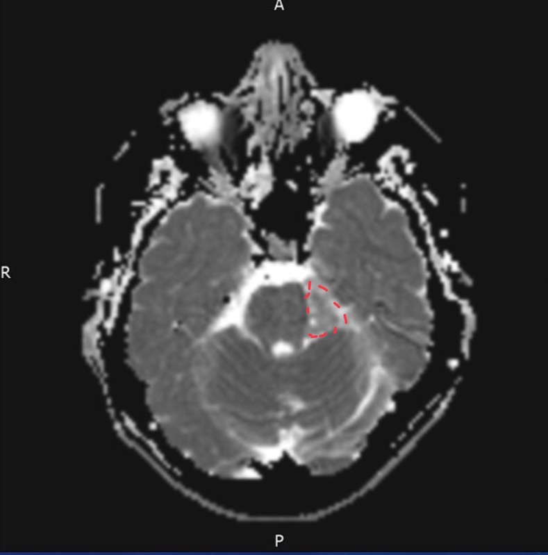
55-year-old female presents with occipital headaches and neck stiffness
Thrombosed Aneurysm of postero-inferior cerebellar artery
55 year-old male is following up with neurosurgery for a pre op scan
Spoke wheel sign = meningioma
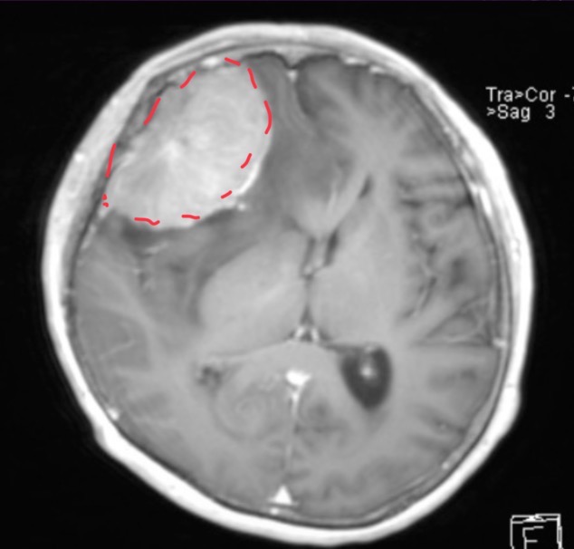
25-year-old male presents with behavioral abnormalities
corpus callosum agenesis with interhemispheric cyst (open area where corpus callosum wa supposed to be)
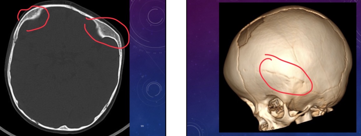
15 month old female has a cranial deformity.
Cranial bone invasion into brain tissue. Left coronal suture fused = harlequin eye deformity.
Frontal plagiocephaly, craniosynostosis
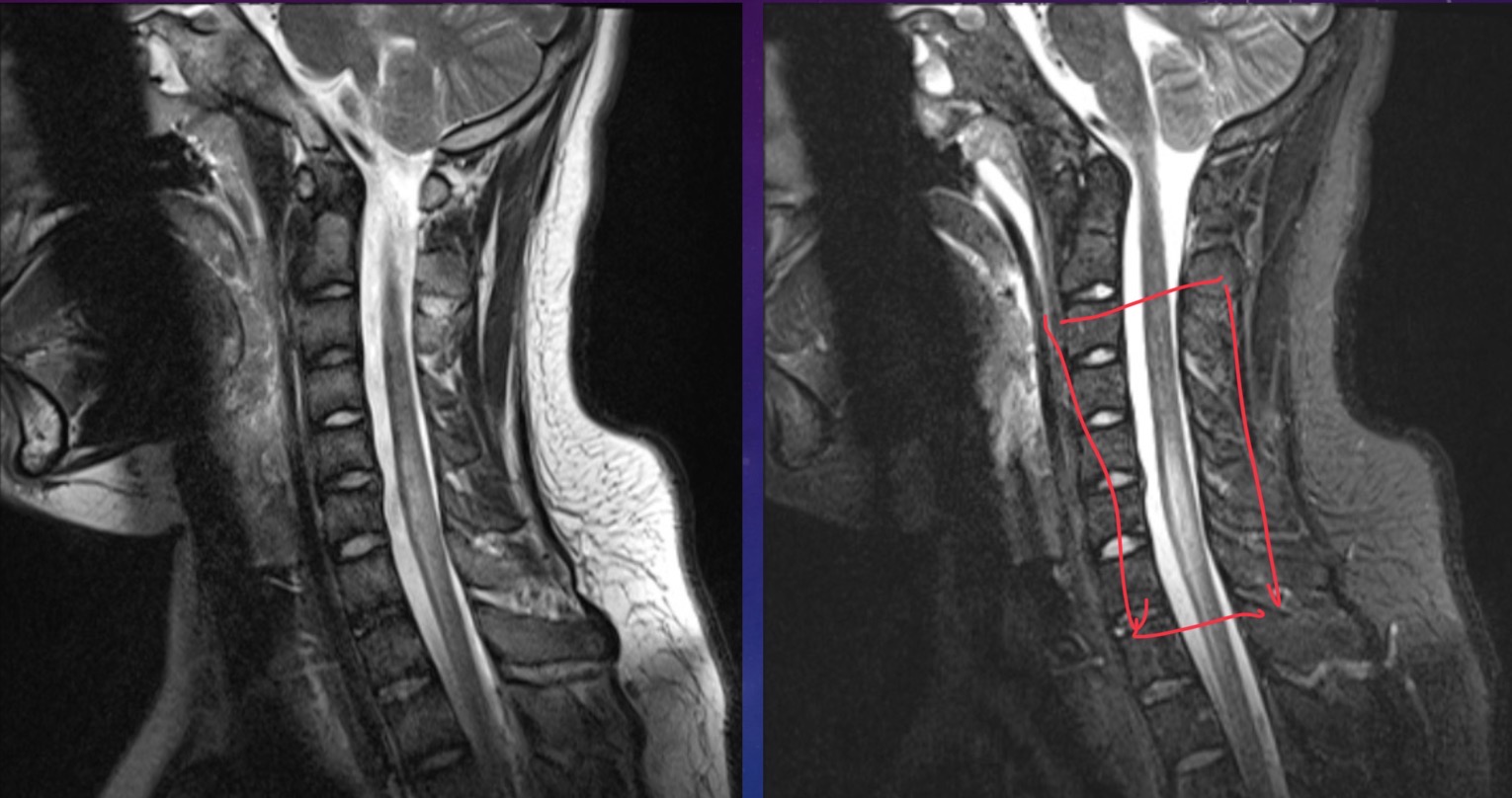
12-year-old male is a secondary school student with acute onset tetraparesis. He has a rapid ascending paralysis with neuromuscular weakness with brisk reflexes. Patient also has cervical myelitis
Cervical transverse myelitis. Canal looks narrower and bulges forward
35-year-old female presents with a chronic headache and upper and lower limb numbness
Idiopathic intracranial hypertension
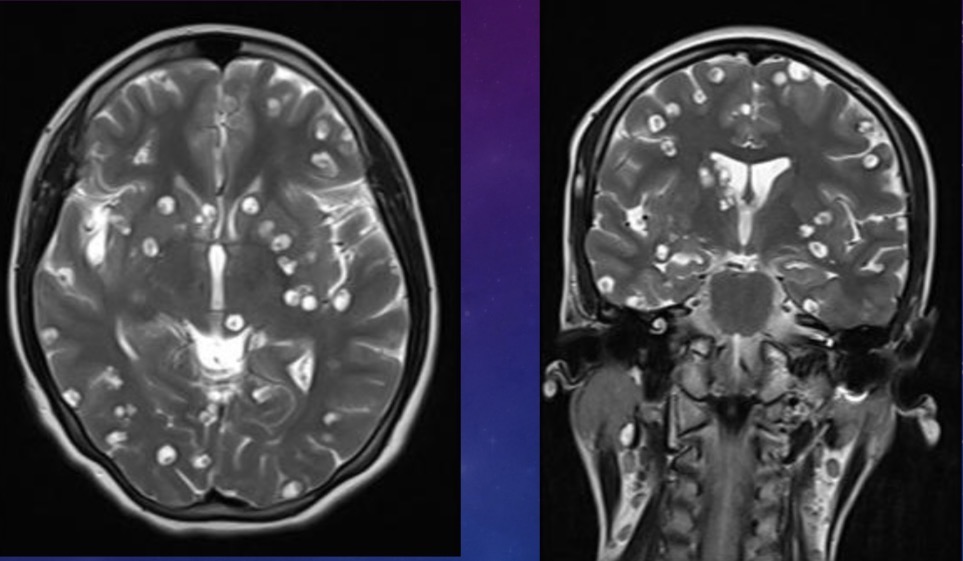
30-year-old female presents with fever and AMS for five days with underlying immunosuppressive illness and stem cell transplant. Scan shows multiple ring enhancing lesions
CNS Toxoplasmosis