Lec 7: Protein Structure
 Call Kai
 Learn
Learn Practice Test
 Spaced Repetition
 Match
Match Flashcards
 Knowt Play
1/31

There's no tags or description
Looks like no tags are added yet.
Name | Mastery | Learn | Test | Matching | Spaced | Call with Kai |
|---|
No analytics yet
Send a link to your students to track their progress
32 Terms
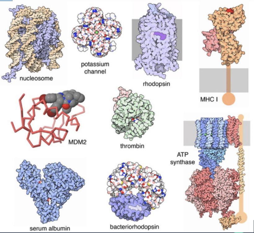
Why study Proteins?
what are proteins?
where do they occur?
how much of cellular dry weight do they constitute
what are they product of?
Proteins are the most abundant biological macromolecules
Occur in all cells and all parts of cells
Constitute >50% of cellular dry weight
Thousands of different kinds in a single cell
Products of genes → agents of biological function
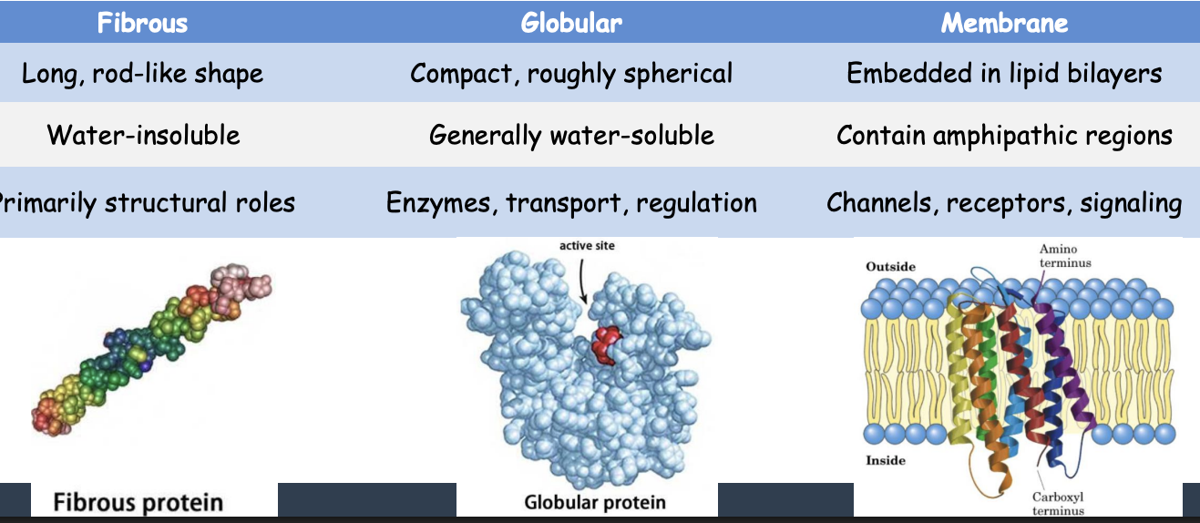
3 Structural Classes
describe their shape
water soluble or insoluble
roles
proteins can be generally categorized by shape and solubility
Fibrous
Globular
Membrane
4 Levels of Protein structure
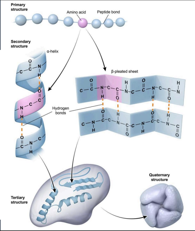
protein structure architecture includes four levels:
Primary (linear amino acid sequence): Determined by gene sequence
Secondary (α-helices and β-sheets): Stabilized by backbone H-bonds
Tertiary (3D folding of single peptide): Stabilized by multiple interactions
Quaternary (subunit organization): Assembly of multiple polypeptide subunits -Stabilized by same forces as tertiary
Protein Structure Image
Non-Covalent Forces and Folding
what are secondary structures stabilized by?
what about tertiary and quaternary?
what does each level depend on?
what is folding dictated by?
Secondary structures are stabilized by H-bonds. Tertiary and quaternary structures are stabilized by H-bonds, ionic interactions, van der Waals forces, hydrophobic interactions, and disulfide bonds.
Each level depends on the level below it.
Folding is dictated by the order of AAs in the primary structure.
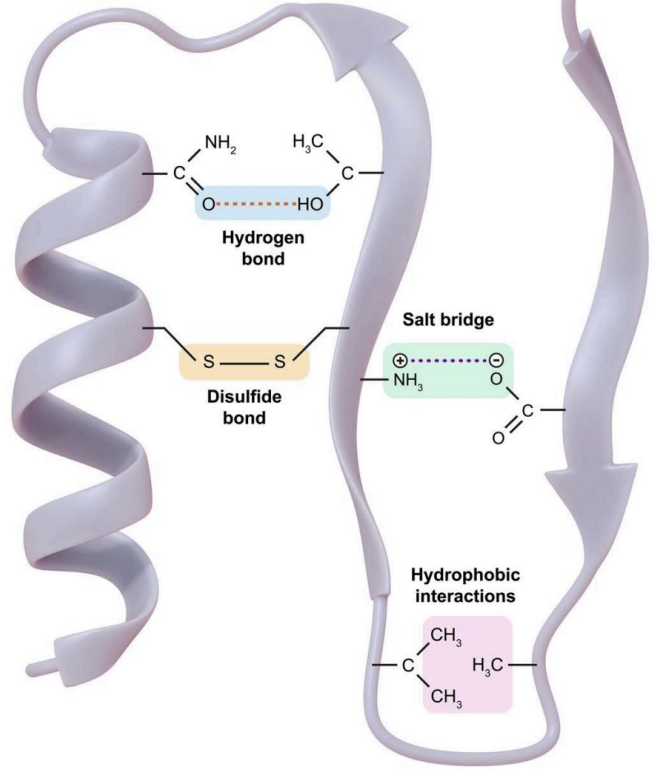
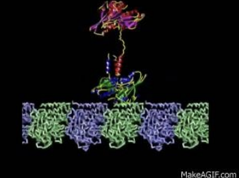
Seven Principles Linking Structure and Function (1-3)
1. FUNCTION DEPENDS ON STRUCTURE
A protein's shape determines what it can do
2. STRUCTURE DEPENDS ON SEQUENCE + WEAK FORCES
Primary sequence encodes the fold while non-covalent forces drive folding
3. THE NUMBER OF FOLDING PATTERNS IS LARGE BUT FINITE
Only ~1,400 distinct folds exist, and new proteins typically adopt one of these established patterns.
Seven Principles Linking Structure and Function (4-6)
4. GLOBULAR PROTEINS ARE MARGINALLY STABLE
Net ΔG of folding: only -20 to -40 kJ/mol
Compare: single covalent bond ≈ -350 kJ/mol
5. MARGINAL STABILITY FACILITATES MOTION
Proteins exhibit conformational dynamics, continuously transitioning between closely related structural states.
6. MOTION ENABLES FUNCTION
Conformational changes are essential for enzyme catalysis, signal transduction and molecular transport
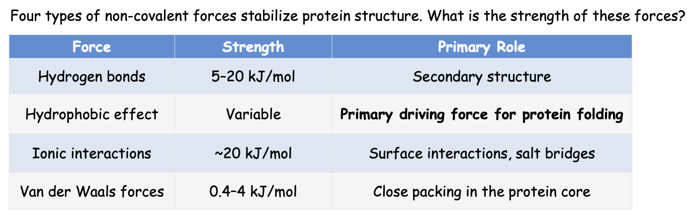
Non-Covalent Forces:
name the 4 forces
strength
primary role
what is secondary and tertiary structure made out of
Net ΔG of folding?
Secondary structure: primarily backbone H-bonds
Tertiary structure: collective action of ALL forces
Net ΔG of folding: -20 to -40 kJ/mol (marginally stable)
Hydrogen Bonds: The Most Versatile Force
who do they form with
function? how do they stabilize secondary structure and support tertiary structure?
strength kj/mol
surface:
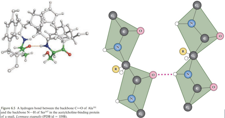
Hydrogen bonds form between donor (N–H) and acceptor (C=O) groups
Backbone H-bonds stabilize secondary structure; side chains support tertiary structure
Weak individually (~5–20 kJ/mol) but are collectively strong - Directional (strongest when linear)
Surface: H-bonds with water
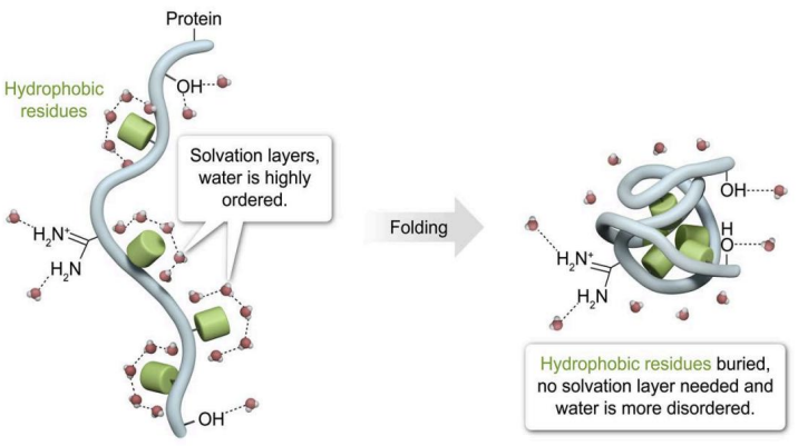
The Hydrophobic Effect: The Dominant Driver of Protein Folding
what do non polar side chains cause?
what does burying nonpolar residues in the protein core do?
what is the primary driving force for protein folding?
Nonpolar side chains cause ordered water “cages” that are entropically unfavorable.
Burying nonpolar residues in the protein core releases ordered water, gives an entropy gain and forms the hydrophobic core
Burial of hydrophobic residues is the primary driving force for protein folding
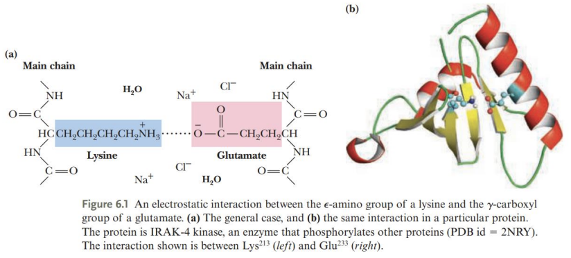
Ionic Interactions: Charged Partners
where do they form?
where are they found?
what is unique about them?
Ionic interactions form between oppositely charged side chains (salt bridges / ion pairs)
Found on protein surfaces, since burying charges in the core is unfavorable
pH-dependent: changes in pH can disrupt ionic interactions, causing conformational changes or denaturation
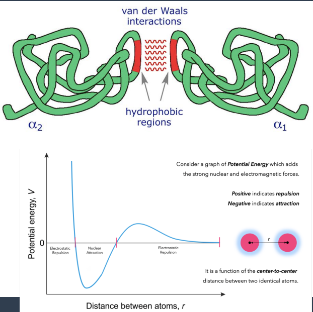
Van der Waals Forces: The Packing Force
are the forces weak or strong? who are they between?
arise from what?
strength in kj/mol
what is unique about them?
function?
who do they work together with?
Weak attractive forces between ALL atoms
Arise from transient dipoles (electron fluctuations)
Strength: 0.4-4 kJ/mol per interaction
Distance-dependent: optimal at ~3-4 Å
Individually very weak but are collectively significant
Contribute to close packing of hydrophobic core
Work together with hydrophobic effect
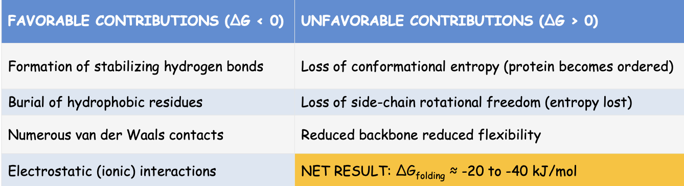
Protein Folding: A Delicate Thermodynamic Balance
what are 4 favorable and unfavorable contributions
This small net ΔG is a FEATURE, not a flaw:
Allows conformational flexibility
Enables regulation and function and allows controlled unfolding when needed
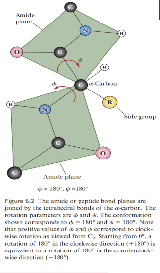
The Peptide Bond: Fundamental
what restricts rotation?
what is the peptide plane
what configuration pre dominates?
where does the backbone flexibility come from?
The Peptide Bond Is Rigid and Planar
C=O ↔ C–N resonance gives ~40% double-bond character → restricts rotation
Six atoms lie in a single plane: Cα–C–O–N–H–Cα (the peptide plane)
Trans configuration predominates (less steric clash)
Backbone flexibility comes only from rotation at φ (Cα–N) and ψ (Cα–C) bonds

Encoding of Folding Information in Primary Structure
describe analogy in the image
what does Φ (phi) represent
what does ψ (psi) represent
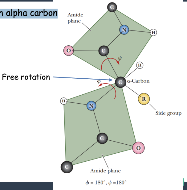
Analogy: rigid peptide planes connected by hinges at each alpha carbon
The conformation shown here is Φ = 180o and ψ = 180o
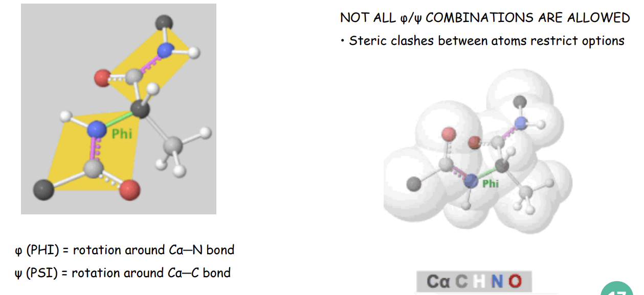
The angle about the Cα-N bond is denoted Φ (phi)
The angle about the Cα-C bond is denoted ψ (psi)
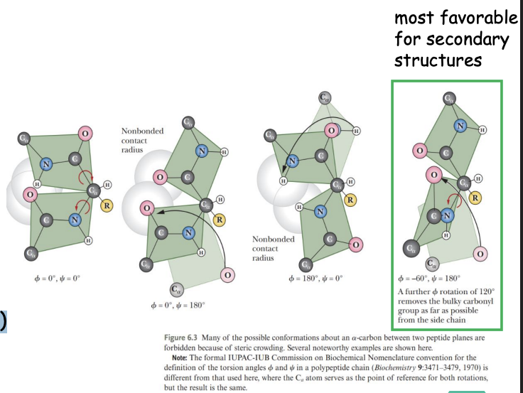
Some values of Φ and ψ are more likely than others
Defining φ (Phi) and ψ (Psi) Angles
Steric Constraints on φ/ψ Angles
what do most φ/ψ combinations cause?
what percentage of f φ/ψ space sterically allowed?
what does this restriction explain?
Most φ/ψ combinations cause steric clashes between backbone atoms
Only ~20% of φ/ψ space is sterically allowed
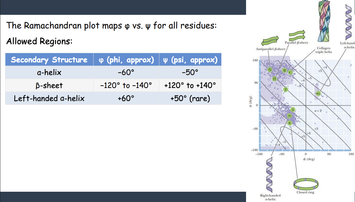
This restriction is why proteins adopt specific, repeatable secondary structures (α-helix, β-sheet)
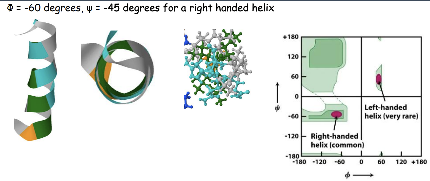
The Ramachandran Plot: A Map of Allowed Conformations
Secondary Structures: Local Folding Patterns
3 main types of folding patters?
function?
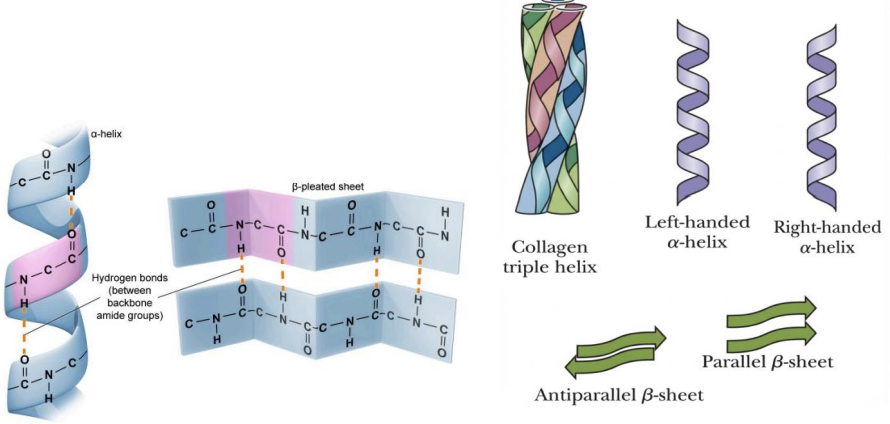
DEFINITION: Regular, repeating conformations stabilized by BACKBONE hydrogen bonds. Main types:
1. α-HELIX
Most common helix
2. β-SHEET
Extended strands
Inter-strand H-bonds
3. β-TURN
Reverses chain direction
4 residues
a-Helix Architecture
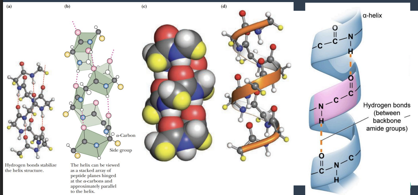
The α-Helix: Most Common Secondary Structure
which is the most common helix?
what do side chains do?
core structure?
H bond structure?
name the 6 parameters and their values
Right-handed helix (most common)
Side chains (R) project OUTWARD
Core is tightly packed (van der Waals contact)
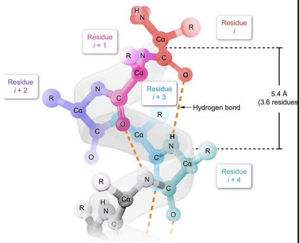
H-bonds are nearly parallel to helix axis
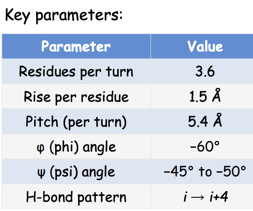
α-Helix: The i → i+4 Hydrogen Bonding Pattern
what residues do H bonds form with?
Each backbone C=O forms H-bond with N-H, four residues ahead:
Residue 1 (C=O) ──H-bond──→ Residue 5 (N-H)
Residue 2 (C=O) ──H-bond──→ Residue 6 (N-H)
Residue 3 (C=O) ──H-bond──→ Residue 7 (N-H)
... and so on
The α-Helix Dipole
what bonds points toward the N terminus and C terminus
what does the create?
what binds at the matching end?
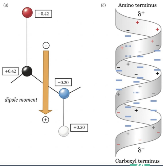
The α-Helix Has a Dipole
All N–H bonds point toward the N-terminus; all C=O bonds point toward the C-terminus
Creates a net dipole: N-terminus is δ⁺, C-terminus is δ⁻
Charged ligands bind at the matching end (e.g., negative ligands near the δ⁺ N-terminus)
α-Helix Architecture
Φ and ψ degrees for right handed helix?
Helices May be Polar, Nonpolar or Amphiphilic
what kind of faces do α-helices have?
what reveals this segregation?
3 functional roles?
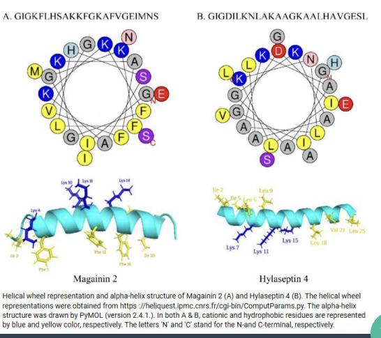
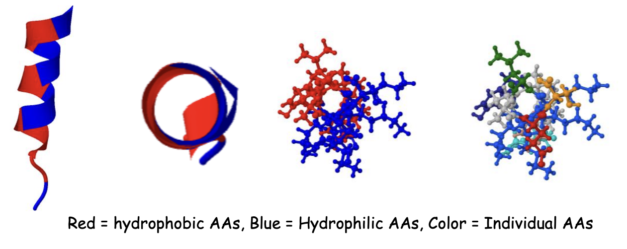
Many α-helices have distinct faces: one hydrophobic, one hydrophilic
A helical wheel (end-on view) reveals this segregation
Functional roles:
Membrane-spanning helices
Surface helices on globular proteins
Antimicrobial peptides that disrupt membranes
Another Amphipathic Helix
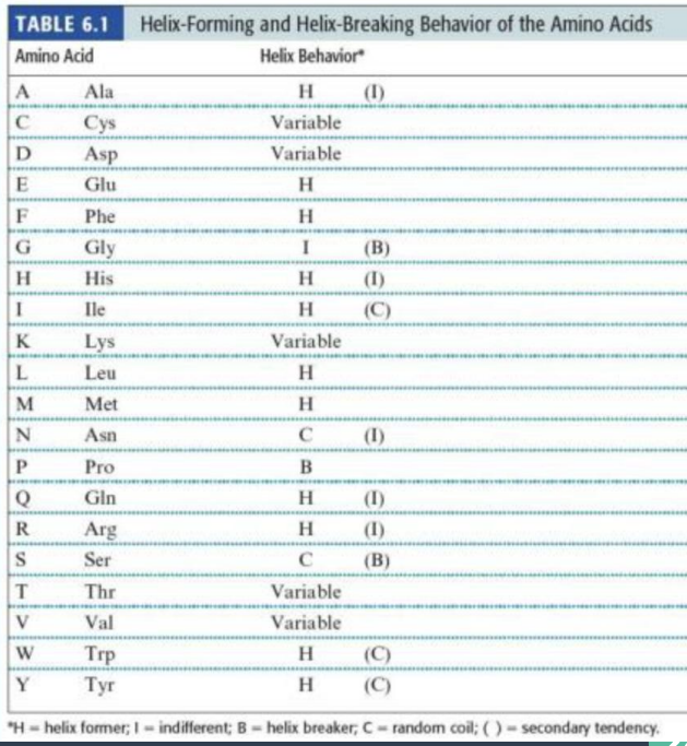
AAs can be classified as helix -formers or breakers
name 5 helix former AA
2 helix breakers AA
context dependent
HELIX FORMERS (favor helix):
Alanine (Ala) - small, fits well
Leucine (Leu) - hydrophobic, good packing
Methionine (Met) - flexible hydrophobic
Glutamate (Glu) - can form salt bridges
Lysine (Lys) - can form salt bridges
HELIX BREAKERS (disfavor helix):
Proline (Pro) - RIGID, causes kink, no N -H for H -bond
Glycine (Gly) - TOO FLEXIBLE, destabilizes helix
CONTEXT -DEPENDENT:
Most other amino acids
Depends on neighboring residues and environment
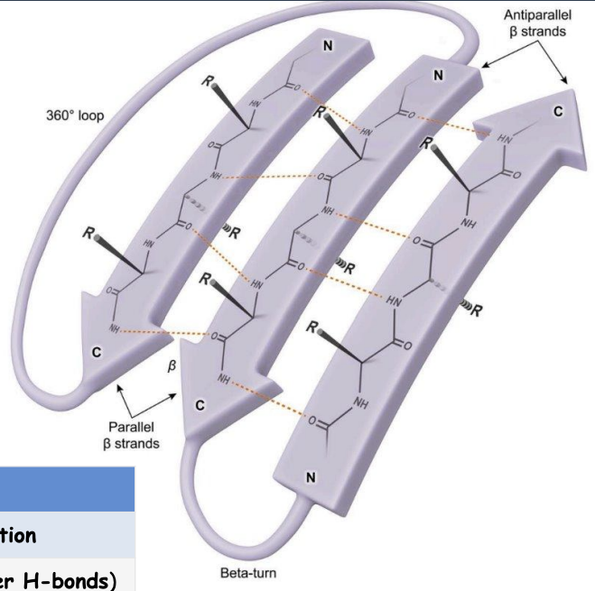
The B-Pleated Sheet
describe structure: what is it composed of?, how are strands connected?, what does it form?
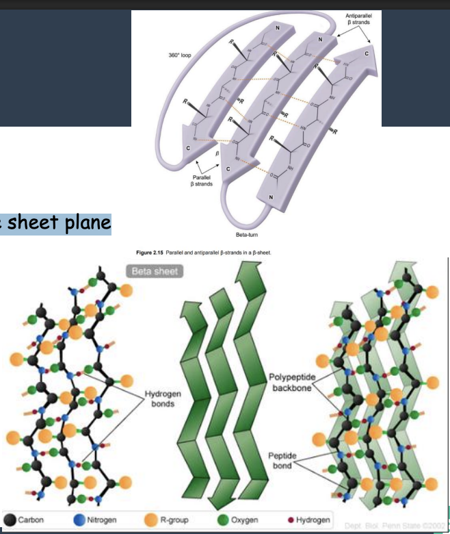
what are the 2 arrangements?
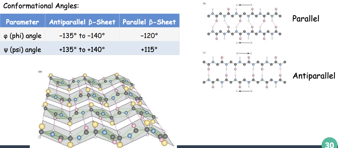
STRUCTURE:
Composed of β-STRANDS in extended conformation
Strands connected by INTER-STRAND H-bonds
Forms a pleated (corrugated) surface
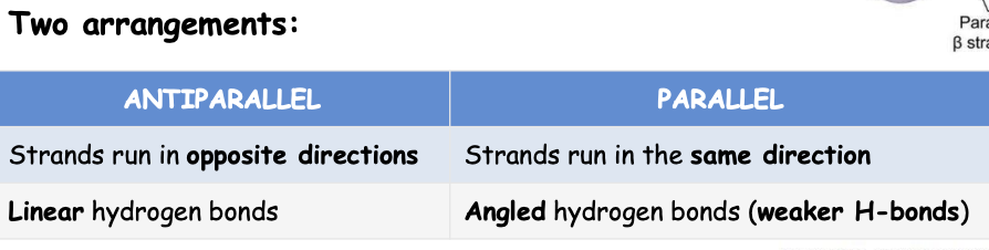
β-Sheet: Key Parameters
phi and psi angles in anti parallel and parallel B sheet
The b-Pleated Sheet: Side Chain Arrangement
where are the side chains located on the sheet plane?
what do the side chain create?
what does it allow?
SIDE CHAIN ARRANGEMENT:
Side chains alternate ABOVE and BELOW the sheet plane
Creates a "pleated" appearance
Allows for amphipathic sheets: - One face hydrophobic - Other face hydrophilic
β-Turns: Reversing Chain Direction
function?
what are the short turns (4 residues) stabilized by?
where do adjacent strands connect?
what is it enriched with?
where is it commonly found? and involved it what?
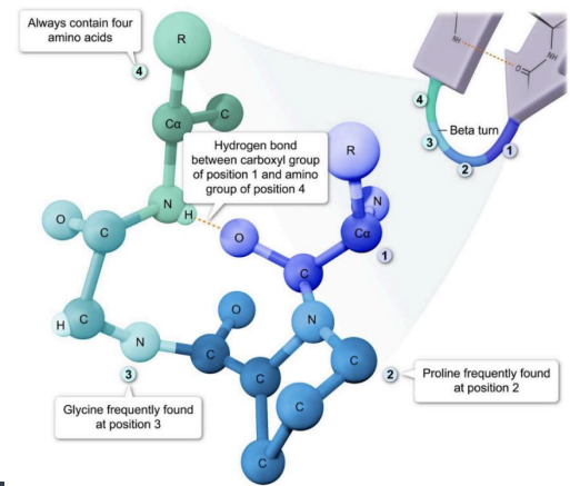
FUNCTION: Allow polypeptide chain to reverse direction
Short turns (4 residues) stabilized by an i → i+3 hydrogen bond
Connect adjacent strands in antiparallel β-strands
Enriched in Proline (rigid, φ-angle constraint) and Glycine (small, flexible it fits tight turns)
Commonly found on the protein surface, exposed to solvent and often involved in protein–protein interactions
Secondary Structure Summary
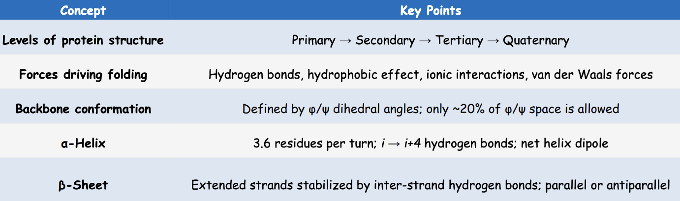
levels of protein structure
forces driving folding
backbone conformation
a-helix
B-Sheet