Exam 2 Hearing Science
1/100
There's no tags or description
Looks like no tags are added yet.
Name | Mastery | Learn | Test | Matching | Spaced | Call with Kai |
|---|
No analytics yet
Send a link to your students to track their progress
101 Terms
2 components of the auditory system
peripheral auditory system (outer, middle, inner ear)
central auditory system (auditory brainstem and auditory forebrain)
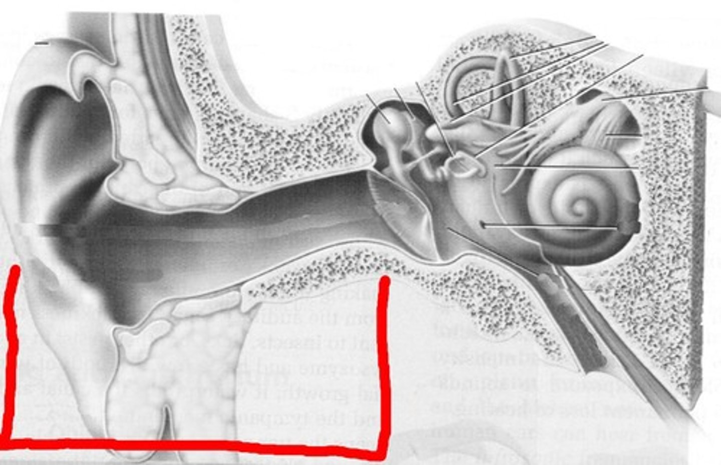
3 components of the peripheral auditory system
Outer ear, middle ear, and inner ear
Anatomy of the outer ear
pinna and external ear canal
Physiology of the outer ear (4 functions)
i. Collect sound
ii. Sound pressure gain (amply middle frequencies 1-5 kHz)
iii. Sound localization
iv. Protection of the tympanic membrane
Sound pressure gain
peaked around 2.5 kHz
primary contribution from the concha and the outer ear
This gain is crucial for hearing sensitivity in the speech frequency range.
What is sound localization?
The process by which the location of sound is determined.
What cues are used for localizing sound?
Intensity and phase (time) difference.
pinna anatomy
visable, cartiligenous part of the ear
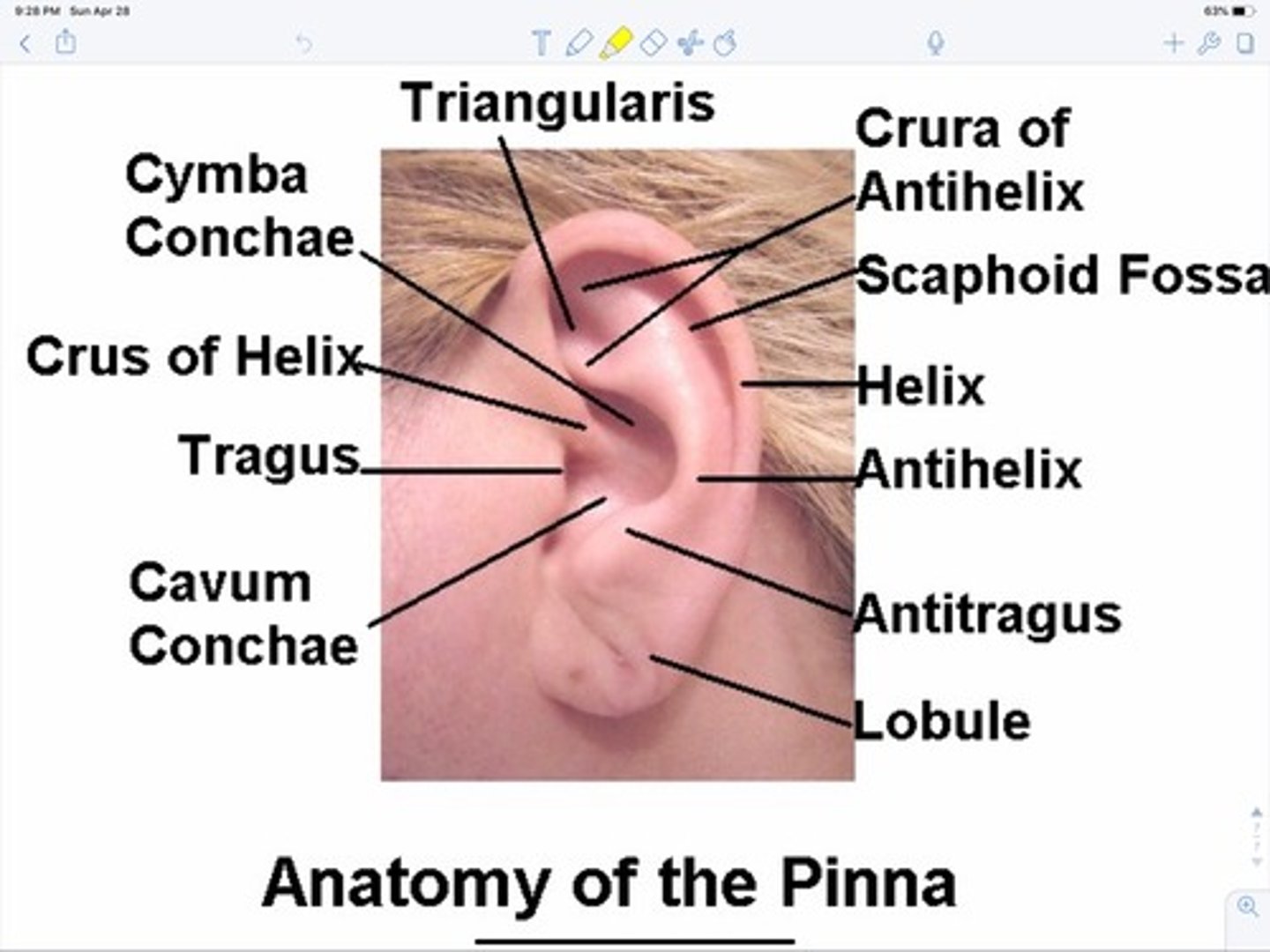
pinna physiology
sound localization in the midplane
acts as a funnel to direct sound waves towards the ear canal
external ear canal anatomy
a tube leading to the eardrum
external ear canal physiology
Provides an acoustic resonance effect, leading to sound pressure gain (peaking around 2.5 kHz)
middle ear anatomy
tympanic membrane
tympanic muscles
auditory ossicles
eustachian tube
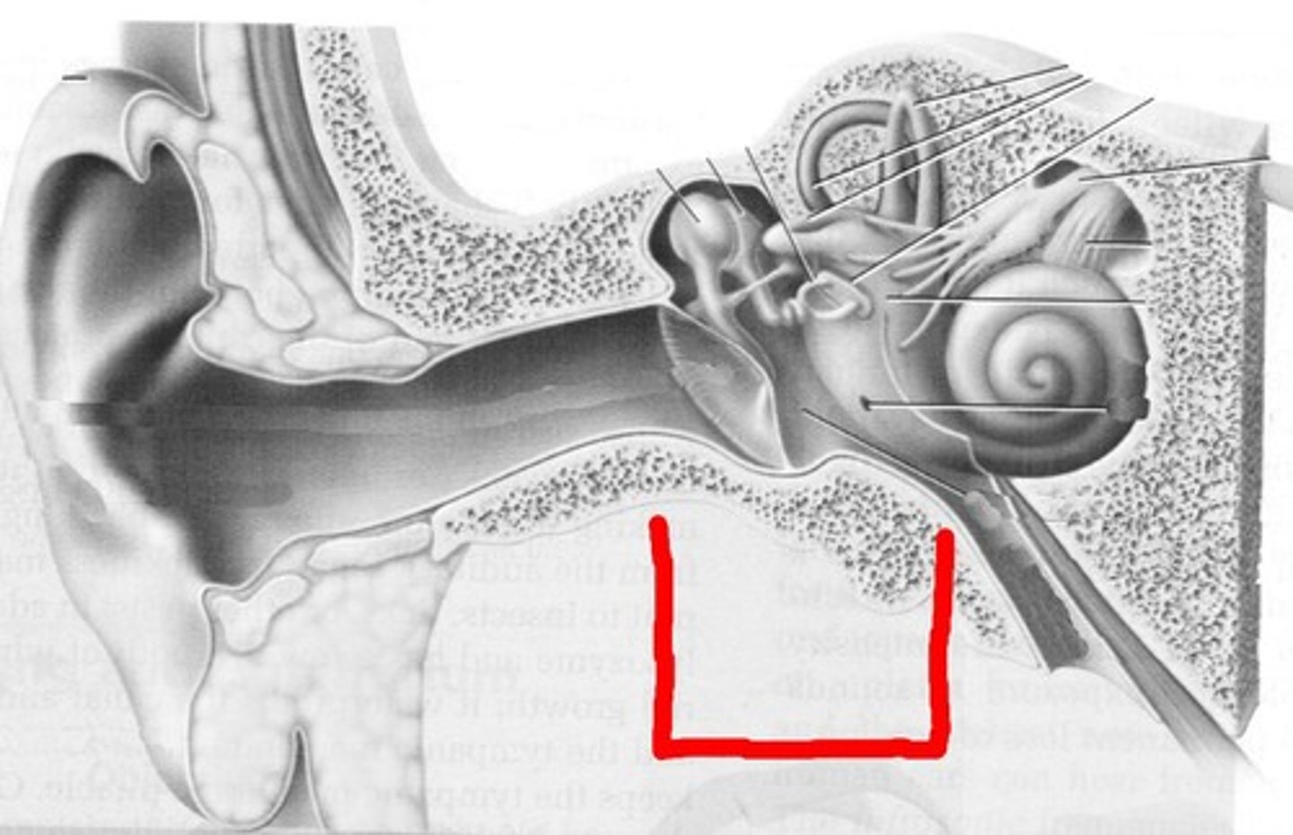
Middle Ear Physiology
sound transmission and protection of hearing
tympanic membrane anatomy
eardrum
tympanic membrane physiology
Transduces acoustic energy signals (air molecule vibration) into mechanical motion.
auditory ossicles anatomy
malleus, incus, stapes
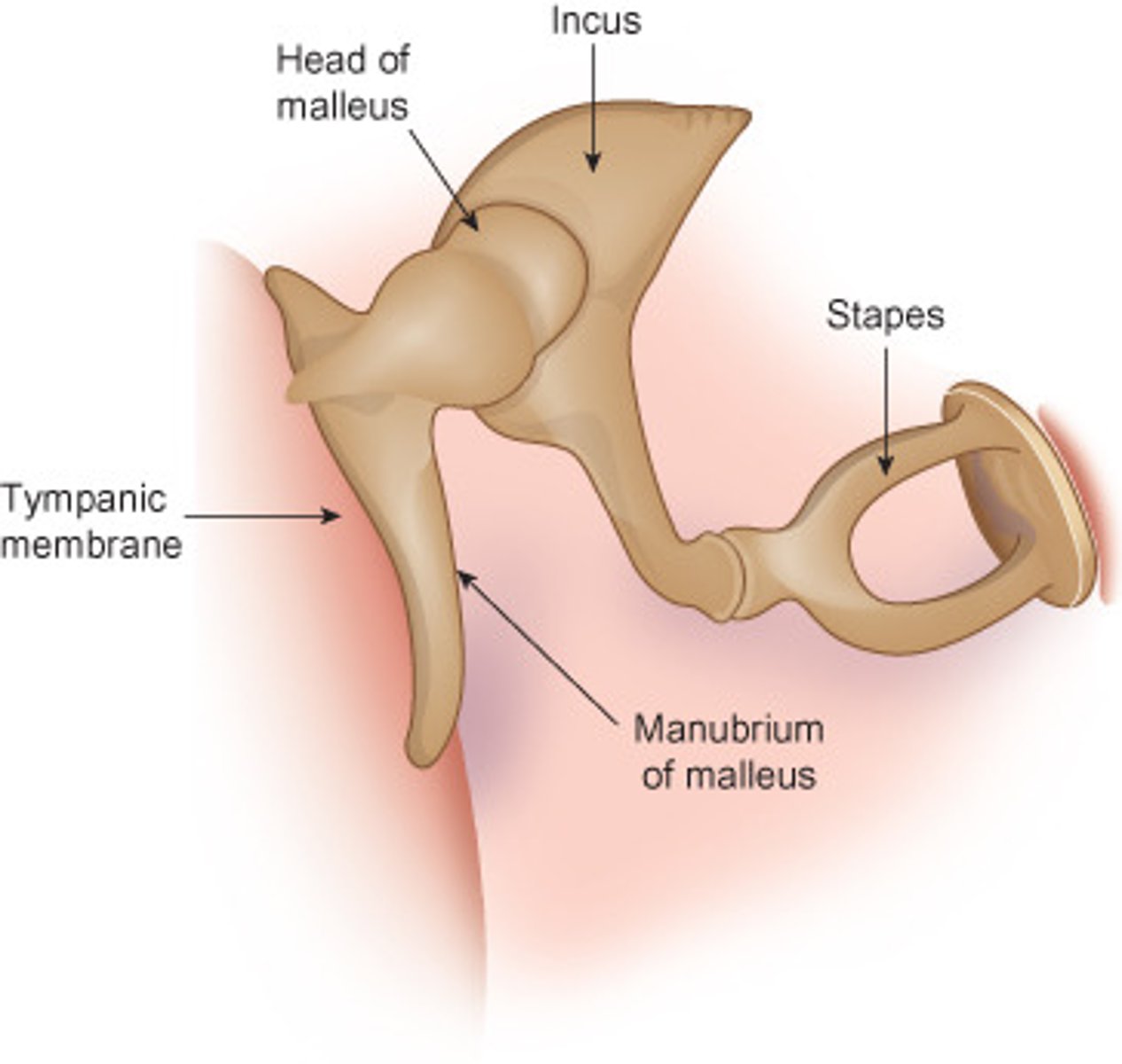
auditory ossicles physiology
Transduces the mechanical motion of the TM to fluid motion in the cochlea
Amplifies sound intensity across frequencies
At what frequency do the auditory ossicles provide peak amplification?
1000 Hz
How do the auditory ossicles transduce sound?
through transducing the mechanical motion of the ossicles to fluid motion in the cochlea (from the stapes moving in and out of the oval window)
Sound transfer functions of the middle ear
sound intensity amplified across frequencies with peaked amplification at 1000 Hz
Problem of sound transmission - What happens when vibration of sound wave transfers from the tympanic membrane in the air to fluid-filled cochlea?
Most of the acoustic energy will be reflected because the difference of acoustic impedance between the two sound media.
Solutions to the sound transmission problem
impedance mismatch problem - increase pressure/force at the oval window
sound mismatch issue
During the change between the ear drum and the fluid motion of the cochlea, the two mediums cause impedance mismatching. The sounds are mostly reflected (bouncing off an oval window).
Three mechanisms for impedance mismatch problem
area ratio, lever system, buckling of eardrum
Area Ratio (hydraulic affect)
Sound vibrates the large eardrum, but this force is concentrated onto the much smaller stapes footplate, greatly increasing pressure (approx. 18x gain)
lever system
The malleus and incus act as a lever, multiplying the force by about 2.1 times.
Buckling of ear drum
buckling motion increases the force transmitted to the middle ear's ossicles, effectively amplifying the sound pressure
acoustic reflexes
stapedius and tensor tympani (middle ear muscles) contract to lower sound transmission in the middle ear after receiving intense sounds
acoustic reflexes purpose
protects inner ear from intense sounds
tensor tympani physiology
contracts and increases tension on the tympanic membrane with intense sounds
stapedius physiology
contracts and works with the tensor tympani during high-intensity sounds to limit the motion of the ossicles and protect the inner ear.
two tympanic muscles
stapedius and tensor tympani
Eustachian tube anatomy
Connects the middle ear to the nasopharynx.
Eustachian tube physiology
Equalizes air pressure between the middle ear and the atmosphere.
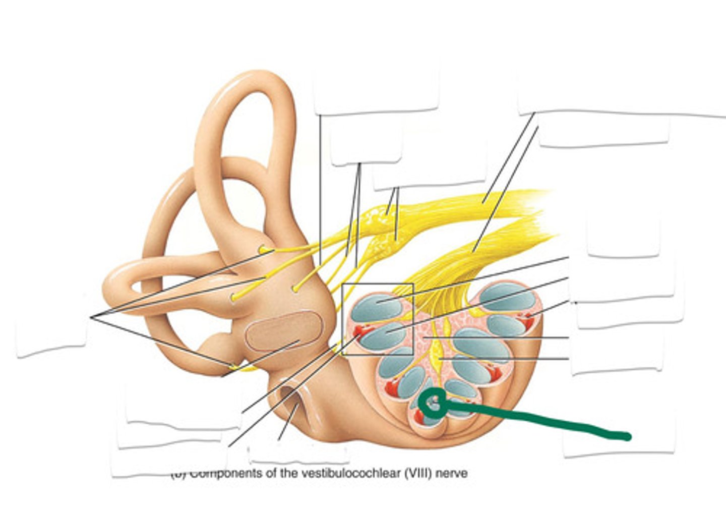
2 inner ear structures
vestibular apparatus and cochlea
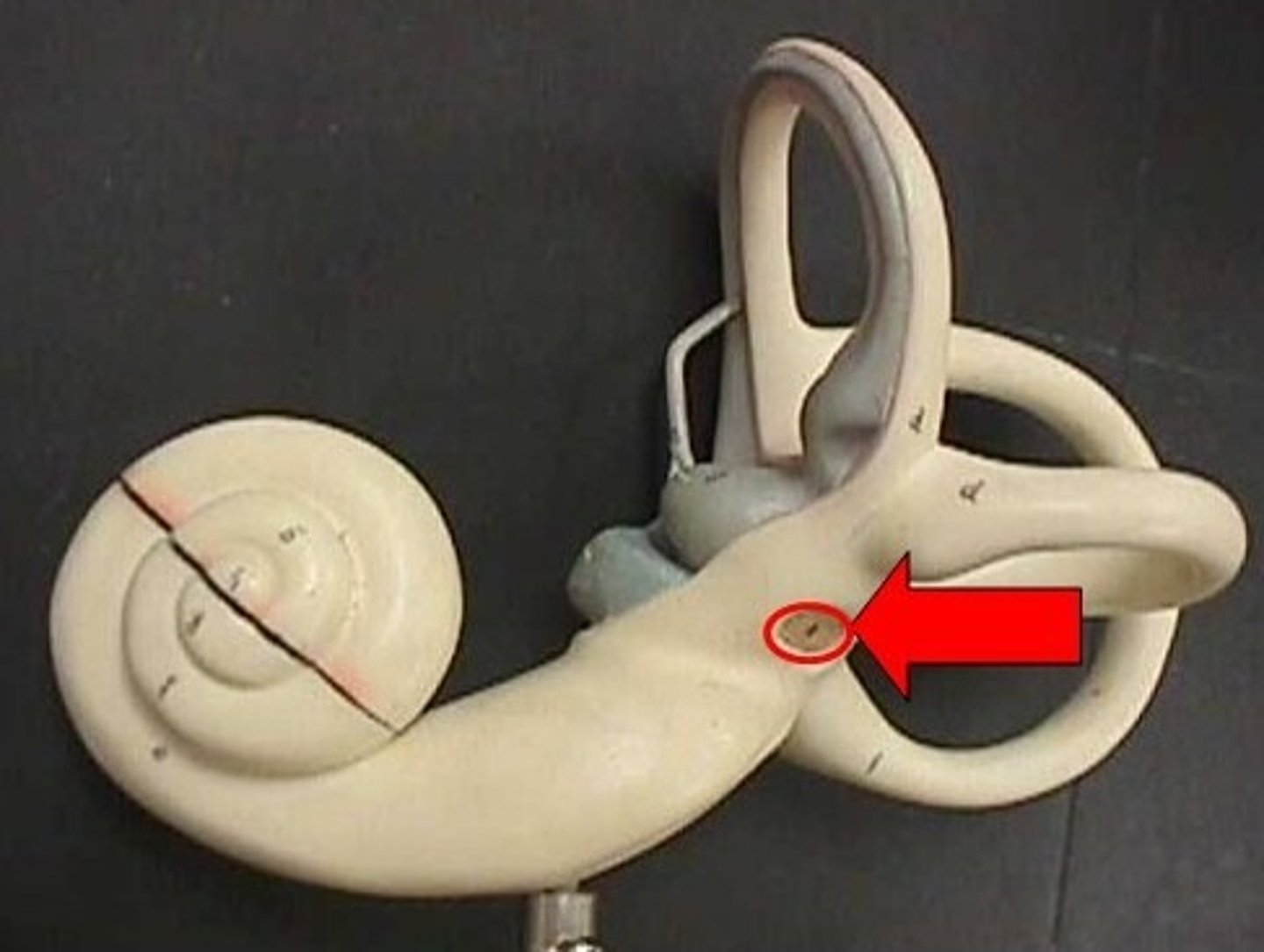
Where is the cochlea located?
Embedded in the temporal bone, medial to the middle ear cavity.
What is the shape of the cochlea?
Coiled shape with about 2.5 turns around a bony, hollow core called modiolus.
What are the dimensions of the cochlea?
35 mm long, 1 cm wide at the base, and 5 mm wide at the apex.
modiolus of cochlea
contains the auditory nerve and cell bodies of auditory neurons, which forms spiral ganglion surrounded by a ledge of bone (osseous spiral lamina)
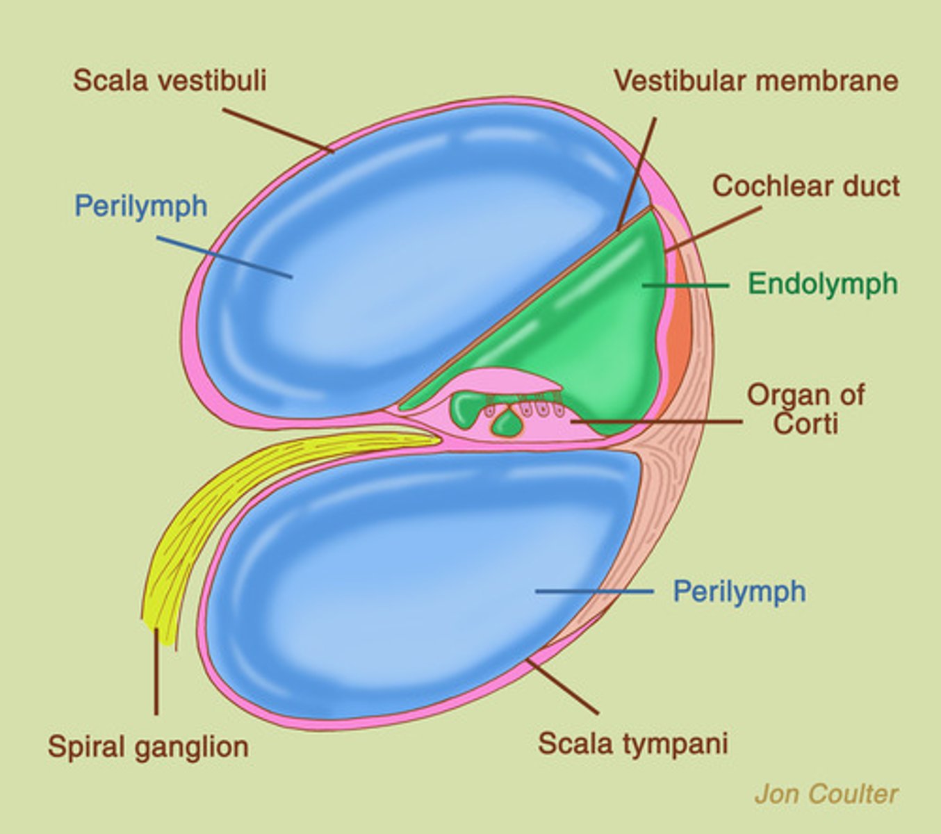
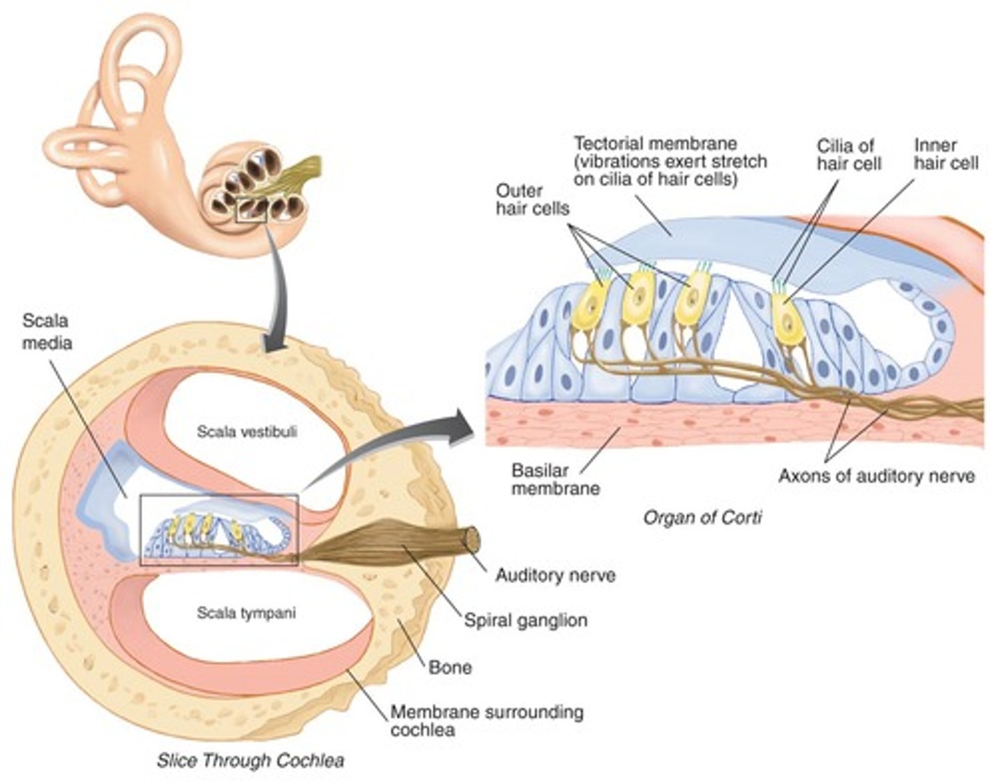
The Three Scalae of the Cochlea
scala vestibuli, scala media (also called cochlear duct), scala tympani
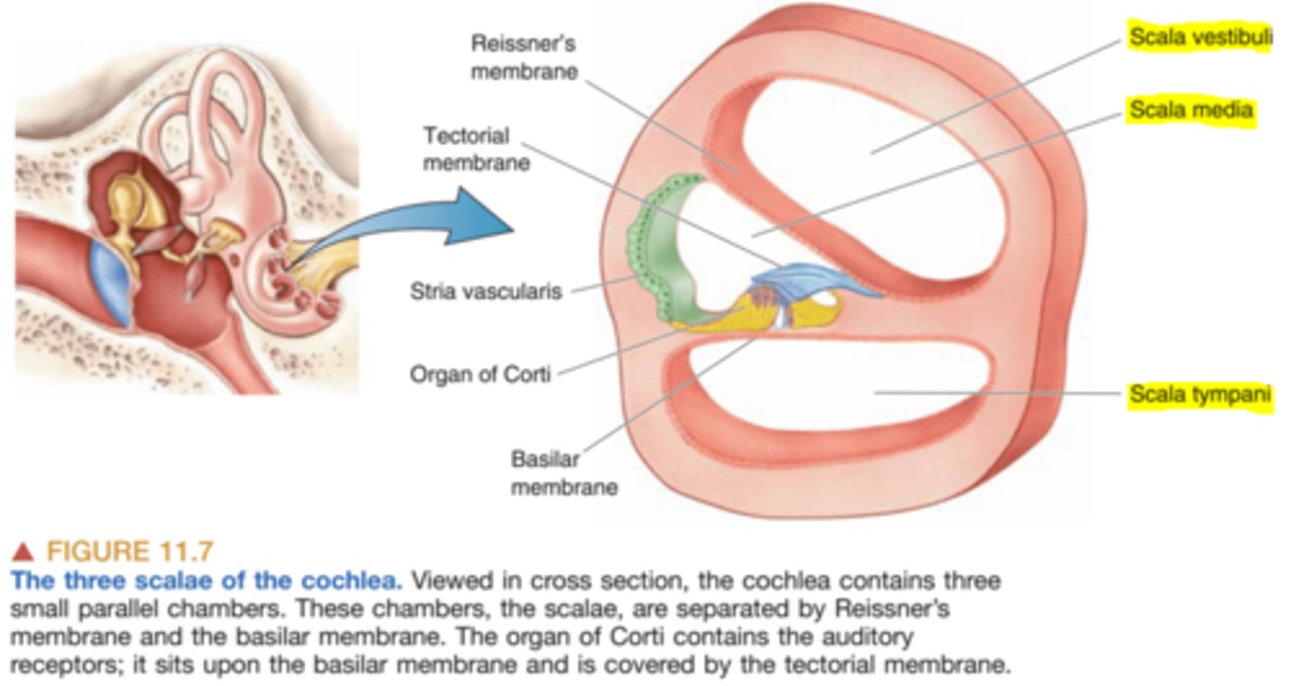
Two membranes to separate scalae in cochlea
Reissner's membrane and basilar membrane
Helicoterma
joint opening at the apex of the cochlea that connects scala vestibuli and scala tympani
Helicotrema function
allow fluid (perilymph) to move between the scala vestibuli and scala tympani, helping relieve pressure at low-frequency sounds.
scala vestibuli anatomy
Filled with perilymph and connected to the oval window
scala vestibuli physiology
Transmitting sound vibrations from the oval window to the rest of the cochlea
scala media (cochlear duct) anatomy
Filled with endolymph and contains the Organ of Corti
scala media (cochlear duct) physiology
converts sound waves into electrical signals to the brain
scala tympani anatomy
filled with perilymph and connected to the round window

scala tympani physiology
role in transmitting sound vibration to the auditory nerve via a fluid called perilymph
perilymph
fluid inside of cochlea
basilar membrane
A structure that runs the length of the cochlea in the inner ear and holds the auditory receptors, called hair cells.
BM base
Narrow and stiff; resonates high frequencies (4-5 kHz)
BM apex
wide and flaccid; resonates low frequencies (below 1 kHz)
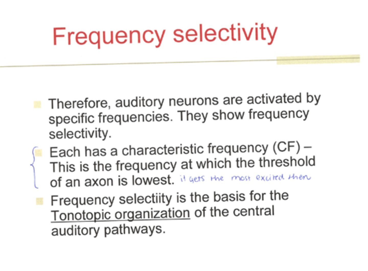
tonotopic organization
an arrangement in which neurons that respond to different frequencies and are organized anatomically in order of frequency
Basilar membrane function
Detects and separates different sound frequencies by vibrating at specific locations along its length, allowing the cochlea to encode pitch
Reissner's membrane anatomy
thin membrane inside the cochlea that separates the fluid-filled scala vestibuli from the scala media
Reissner's membrane function
maintain the separation of fluids (perilymph and endolymph), which is essential for proper inner-ear electrical balance and hearing
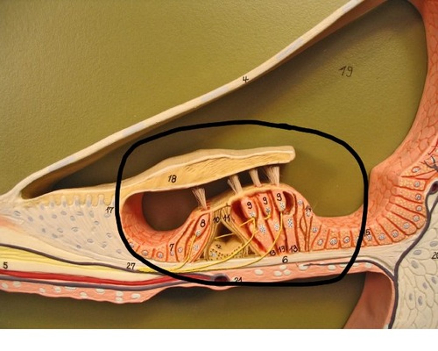
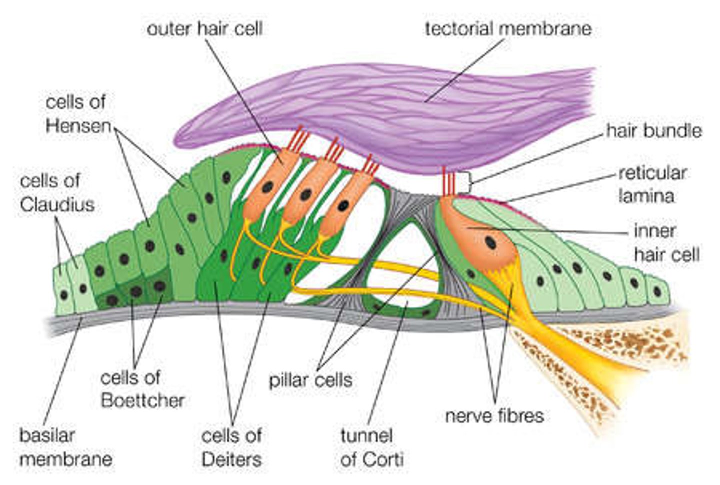
organ of corti anatomy
basilar membrane on the bottom, structure holding up outer and inner hair cells, outer hair cells with stereocilia, Reisner's/tectorial membrane on top
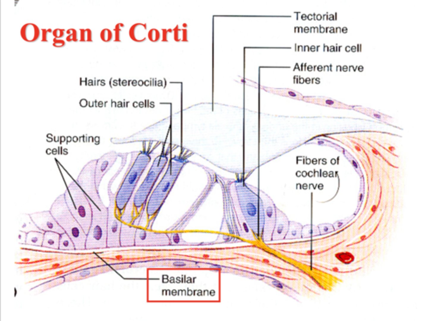
Outer Hair Cells (OHC)
Enhance the vibration of the basilar membrane, providing sensitivity and frequency selectivity (Cochlear Amplifier)
Inner Hair Cells (IHC)
The actual sensory transducers; convert mechanical motion into electrical signals.
True or False: We have more outer hair cells than inner hair cells
true
hair cells are also called
auditory receptors
supporting cells
- pilor and hensen cells
- structural and metabolic support to outer and inner hair cells
Stereocilia
hairlike extensions on the tips of hair cells in the cochlea that initiate the release of neurotransmitters when they are flexed
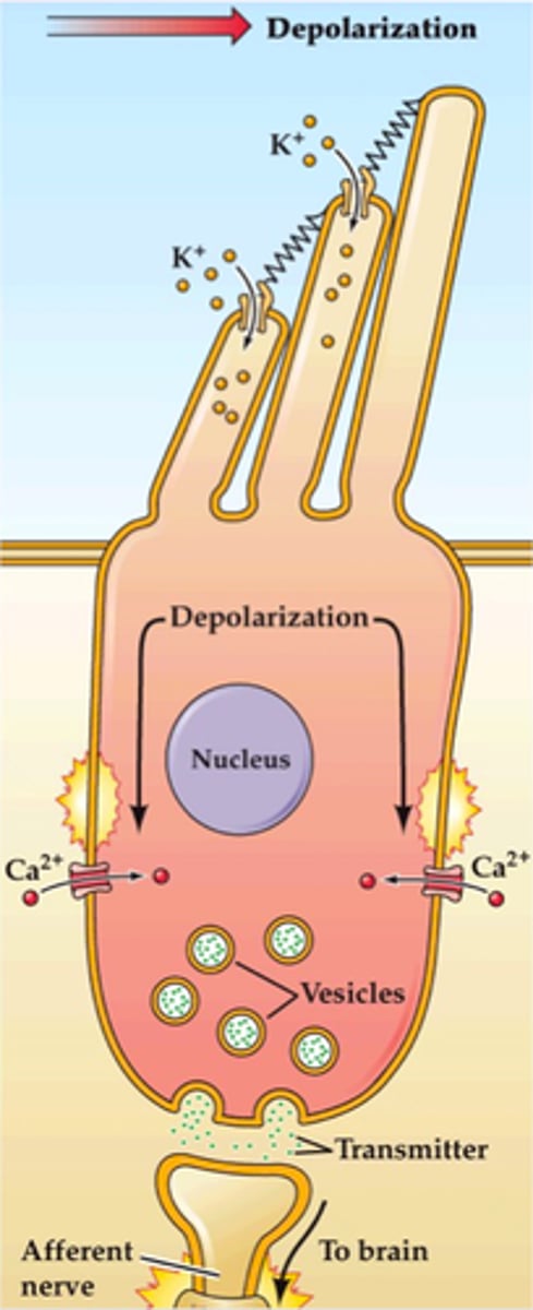
The stereocila need to be ______ to open the channel up
flat
Sound waves cause the ________ to vibrate.
basilar membrane
Traveling wave of the basilar membrane
displacement of BM caused by the flow of perilymph in the scala vestibuli and scala tympani
hair cell response to BM displacement
stereocillia bend, Mechanically gated K⁺ channels open, the hair cells are depolarized, which opens up voltage-gated Ca²⁺ channels, allowing for electrical waves to be sent to the brain and interpreted as sound
General sound transmission in cochlea
Flow of the fluid in the cochlea produced by the vibration of the stapes results in the displacement of basilar membrane.
frequency selectivity
The auditory system's ability to respond differentially to different frequencies and bands of frequencies
tuning curve
A graph that relates neural activity to a continuous range of stimulus properties
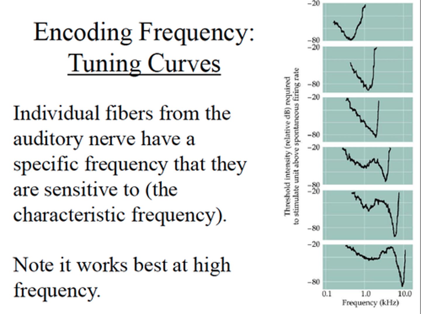
Characteristic frequency
The frequency of sound that a particular auditory nerve fiber is most sensitive to. It's the frequency that requires the least amount of sound energy for that region of the ear to respond.
Intensity Resolution
the ability to differentiate different sound levels
non-linear intensity resolution
Amplifying or dampening sounds to protect the ear
compression at characteristic frequency
A trick your inner ear uses to turn loud sounds way down, especially the exact sound frequency that the ear listens to, so that loud noises don't hurt you and soft noises are still heard.
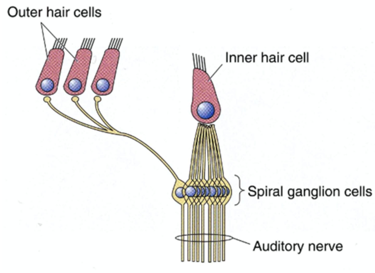
Auditory Nerve
a direct synaptic connection between the hair cells of the cochlea and the cochlear nucleus
How many fibers are in each human ear?
30,000
Type I spiral ganglion cells
95% of spiral ganglion cells, many are connected to one IHC (20 fibers to one IHC)
Type II spiral ganglion cells
10% of spiral ganglion cells, connected to OHC, one to many (one fiber to 10 OHC)
spontaneous firing rate
baseline electrical activity (spikes/second) of auditory nerve fibers
High Spontaneous Rate (HSR)
Active even without sound; high sensitivity, low threshold.
low spontaneous rate (LSR)
Requires more intense sound; low sensitivity, high threshold.
Intensity Resolution
Auditory nerve firing rate increases with sound intensity
Intensity resolution threshold
The lowest sound level that causes a nerve fiber to start responding
Intensity resolution saturation
The highest sound level where the neuron's firing rates stops increasing
Intensity Resolution Dynamic Range
Range of sound intensities between threshold and saturation
20-50dB: Fiber can accurately represent sound intensity changes
frequency selectivity
AN fibers are sharply tuned to a specific characteristic frequency (CF).
Phase Locking
AN firing is synchronized to a specific phase of the stimulus waveform (up to 4-5 kHz)
central auditory system contains
auditory brainstem and auditory forebrain
Auditory brainstem contains
Cochlear nuclei (CN), Superior Olivary Complex (SOC), nuclei of Lateral lemniscus (NLL), Inferior Collicus (IC)
Auditory forebrain contains
Medial geniculate body (MGB) and Auditory Cortex
Cochlear Nuclei divisions
Anteroventral (AVCN), Posteroventral (PVCN), and Dorsal (DCN)
Cochlear Nuclei function
first relay center for auditory nerve fibers (complex response patterns)
superior olivary complex
Receives bilateral inputs and localizes sound
nuclei of lateral lemniscus (NLL)
Helps with processing timing and temporal patterns
inferior colliculus (IC)
Combines the analysis of complex sound and the direction in space simultaneously
medial geniculate body (MGB)
The thalamic relay station. Processes and relays specific, detailed auditory information to the auditory cortex.
Auditory cortex divisions
primary auditory cortex and secondary auditory cortex
Auditory cortex tonotopic organization
systematic mapping of sound frequency (pitch) along a physical structure in the auditory system. Frequency of mapping is maintained
AC detection of complex features
Neurons respond to specific patterns, such as frequency-modulation (FM) detectors and temporal-modulation detectors.