Anatomy Exam 2 - Ali Alroalle
1/62
There's no tags or description
Looks like no tags are added yet.
Name | Mastery | Learn | Test | Matching | Spaced | Call with Kai |
|---|
No analytics yet
Send a link to your students to track their progress
63 Terms
Match each erector spinae muscle group with the parts it includes.
Includes cervical, thoracic, and lumbar parts
Includes cervical and thoracic parts
Includes capitis, cervical, and thoracic parts
Match each of the options above to the items below.
A. Iliocostalis group
B. Longissimus group
C. Spinalis group
A. Iliocostalis group - 1
B. Longissimus group - 3
C. Spinalis group - 2
Match each extrinsic muscle of the eye with the action they perform.
Pulls the eye inferiorly and medially
Adducts the eye
Abducts the eye
Match each of the options above to the items below.
A. Medial rectus
B. Lateral rectus
C. Inferior rectus
A. Medial rectus - 2
B. Lateral rectus - 3
C. Inferior rectus - 1
If you contract only your left sternocleidomastoid muscle, it rotates your head to the _____ side of the body.
Right
Match each muscle of the anterior neck with its function.
Elevates the hyoid bone
Depresses the hyoid bone
Depresses and fixes hyoid during opening of mouth
Depresses mandible; elevates hyoid bone
Match each of the options above to the items below.
A. Digastric
B. Geniohyoid
C. Sternohyoid
D. Omohyoid
A. Digastric - 4
B. Geniohyoid - 1
C. Sternohyoid - 2
D. Omohyoid - 3
Which of the following muscles are in the superficial layer of the urogenital triangle?
Check All That Apply
Puborectalis
Bulbospongiosus
Ischiocavernosus
Superficial transverse perineal muscle
Bulbospongiosus
Ischiocavernosus
Superficial transverse perineal muscle
The muscle that has its superior attachment on the mastoid process of the temporal bone and has a bilateral action of flexing the neck is the?
sternocleidomastoid.
middle scalene.
splenius capitis.
sternocleidomastoid.
The collection of muscles in the pelvic floor that function to support the pelvic viscera and act as a sphincter at the anorectal junction, urethra, and vagina is the?
levator ani.
coccygeus.
gluteus maximus.
external anal sphincter.
levator ani.
Match each muscle of the pharynx with its function.
Elevates pharynx and larynx
Opens auditory tube when swallowing
Elevates soft palate when swallowing
Constricts pharynx in sequence
Match each of the options above to the items below.
A. Levator veli palatini
B. Tensor veli palatini
C. Middle constrictor
D. Palatopharyngeus
A. Levator veli palatini - 3
B. Tensor veli palatini - 2
C. Middle constrictor - 4
D. Palatopharyngeus - 1
Which of the following is a function of the erector spinae muscles?
Check All That Apply
To move the upper limbs
To maintain posture
To rotate the neck
To extend the vertebral column
To maintain posture
To extend the vertebral column
The muscle that primarily functions to pull the eye superiorly, and secondarily functions to move the eye medially, is the _____ _____ muscle.
Superior rectus
From superficial to deep, place the muscles of the abdominal wall in the correct order.
External oblique
Internal oblique
Transverse abdominis
External oblique
Internal oblique
Transverse abdominis
Which of the following are functions of the abdominal wall muscle?
Check All That Apply
Flexion of the vertebral column
Extension of the vertebral column
Compression of the abdominal wall
Rotation of the hips
Flexion of the vertebral column
Compression of the abdominal wall
Which muscle is the most powerful of the masticatory muscles, and functions to elevate and protract the mandible?
Masseter
Temporalis
Pterygoid
Mandibular
Masseter
Match each muscle of the tongue with its function.
Depresses and retracts tongue
Protracts tongue
Elevates posterior part of tongue
Elevates and retracts tongue
Match each of the options above to the items below.
A. Genioglossus
B. Styloglossus
C. Hyoglossus
D. Palatoglossus
A. Genioglossus - 2
B. Styloglossus - 4
C. Hyoglossus - 1
D. Palatoglossus - 3
Match each muscle of the pelvic floor with its function.
Ejects urine; stiffens penis
Voluntarily constricts urethra
Forms pelvic floor
Match each of the options above to the items below.
A. Coccygeus
B. Bulbospongiosus
C. External urethral sphincter
A. Coccygeus - 3
B. Bulbospongiosus - 1
C. External urethral sphincter - 2
The primary protractor of the scapula is the?
trapezius.
pectoralis major.
serratus anterior.
rhomboid major.
serratus anterior.
Which muscles flex the arm at the glenohumeral joint?
Check All That Apply
Supraspinatus
Teres minor
Pectoralis major
Deltoid (anterior fibers)
Latissimus dorsi
Coracobrachialis
Biceps brachii
Pectoralis major
Deltoid (anterior fibers)
Coracobrachialis
Biceps brachii
Match each muscle of the leg with its action.
Plantar flexes and inverts the foot
Flexes the leg
Extends toes 2–5
Medially rotates the tibia
Match each of the options above to the items below.
A. Extensor digitorum longus
B. Gastrocnemius
C. Tibialis posterior
D. Popliteus
A. Extensor digitorum longus - 3
B. Gastrocnemius - 2
C. Tibialis posterior - 1
D. Popliteus - 4
True or false. The quadriceps femoris is the prime mover of knee extension.
True
The agonist of forearm flexion is the?
brachialis.
brachioradialis.
triceps brachii.
supinator.
brachialis.
Which of the following muscles abduct the hand at the wrist?
Check All That Apply
Extensor carpi radialis brevis
Extensor carpi radialis longus
Adductor pollicis
Flexor carpi radialis
Extensor carpi radialis brevis
Extensor carpi radialis longus
Flexor carpi radialis
True or false. In general, the muscles of the anterior compartment of the forearm tend to extend the wrist and fingers, while muscles in the posterior compartment of the forearm tend to flex the wrist and fingers.
False
Which of the following muscles flex the thigh?
Check All That Apply
Gracilis
Iliacus
Sartorius
Pectineus
Rectus femoris
Psoas major
Iliacus
Sartorius
Rectus femoris
Psoas major
The abductor pollicis brevis functions to abduct the _____ .
Thumb
Which of the following thigh muscles has its distal attachment in the head of the fibula?
Adductor magnus
Gracilis
Sartorius
Biceps femoris
Biceps femoris
Match each muscle with its action on the scapula.
Protracts scapula, depresses scapula
Elevates scapula, retracts scapula
Prime mover in scapula protraction, superiorly rotates scapula
Match each of the options above to the items below.
A. Pectoralis minor
B. Serratus anterior
C. Rhomboid major
A. Pectoralis minor - 1
B. Serratus anterior - 3
C. Rhomboid major - 2
Match each muscle that moves the arm with their action(s).
Flexes arm
Agonist of arm extension, adducts arm, medially rotates arm
Medially rotates arm
Agonist of arm flexion, adducts arm, medially rotates arm
Extends arm, adducts arm
Match each of the options above to the items below.
A. Latissimus dorsi
B. Pectoralis major
C. Triceps brachii (long head)
D. Biceps brachii
E. Subscapularis
A. Latissimus dorsi - 2
B. Pectoralis major - 4
C. Triceps brachii (long head) - 5
D. Biceps brachii - 1
E. Subscapularis - 3
The large, three-headed muscle that is the prime extensor of the forearm at the elbow joint is the _____ _____ .
Triceps brachii
Which muscles are located in the posterior compartment of the forearm?
Check All That Apply
Extensor carpi radialis longus
Extensor carpi radialis brevis
Flexor pollicis longus
Extensor carpi ulnaris
Extensor digitorum
Flexor digitorum
Extensor carpi radialis longus
Extensor carpi radialis brevis
Extensor carpi ulnaris
Extensor digitorum
The muscles that move the pectoral girdle attach to the scapula and the _____ .
Clavicle
Which muscle encircles the opening of the mouth?
Corrugator supercilii
Levator labii
Risorius
Orbicularis oris
Orbicularis oris
The muscles that have their origin on the mandible and you would use to stick out your tongue are the left and right _____ muscles.
Genioglossus
True or false. During normal, relaxed exhalation, the internal intercostals contract to assist with this process.
False
Which of the following muscles that move the head and neck have their superior attachment in the inferior nuchal line of the occipital bone?
Check All That Apply
Scalene muscles
Rectus capitis posterior major
Rectus capitis posterior minor
Sternocleidomastoid
Rectus capitis posterior major
Rectus capitis posterior minor
Which of the following muscles elevate the ribs during inhalation?
Check All That Apply
Serratus posterior superior
External intercostalis
Transversus thoracis
Internal intercostals
Serratus posterior superior
External intercostalis
Which of the following muscles extends the metatarsophalangeal joint of the great toe?
Flexor hallucis brevis
Extensor hallucis brevis
Lumbricals
Plantaris
Extensor hallucis brevis
The rectus capitis posterior major and minor muscles function to?
protract the shoulders.
rotate the head.
extend the neck.
depress the chin.
extend the neck.
If you palpate around your eyebrows, you are feeling the _____, which marks the termination of the frontal region.
occipital protuberance
superciliary arches
zygomatic arch
mastoid process
superciliary arches
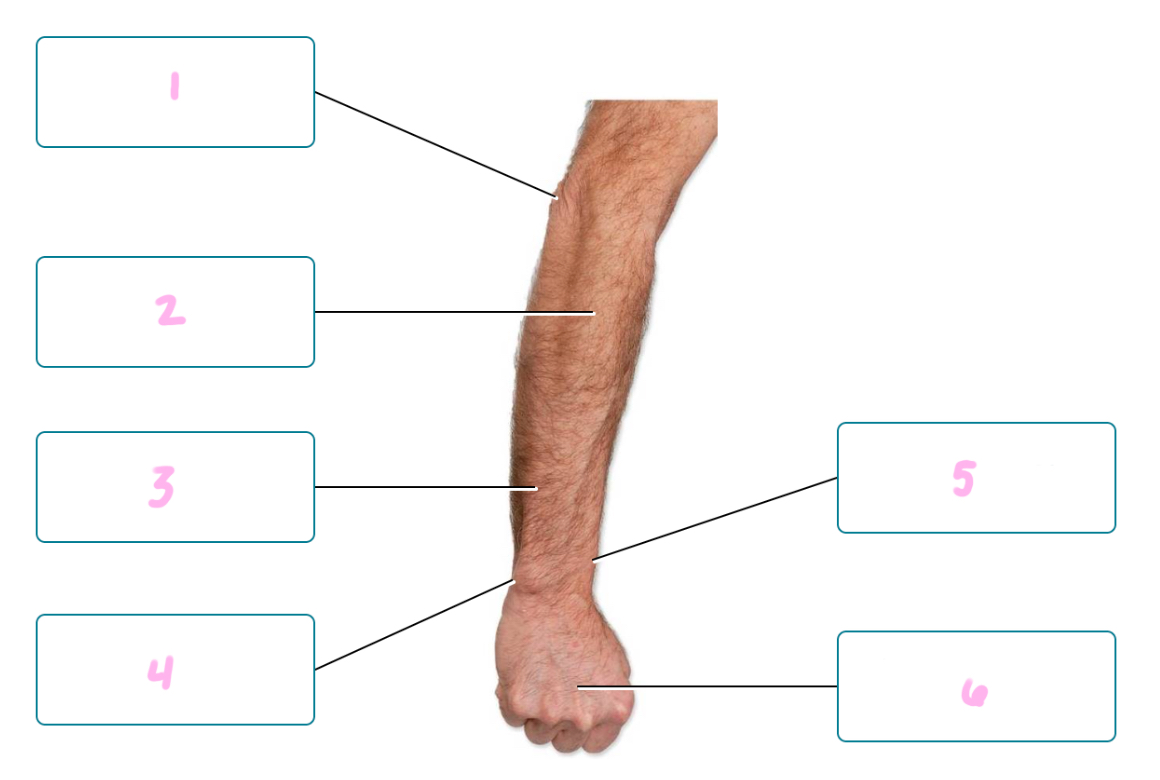
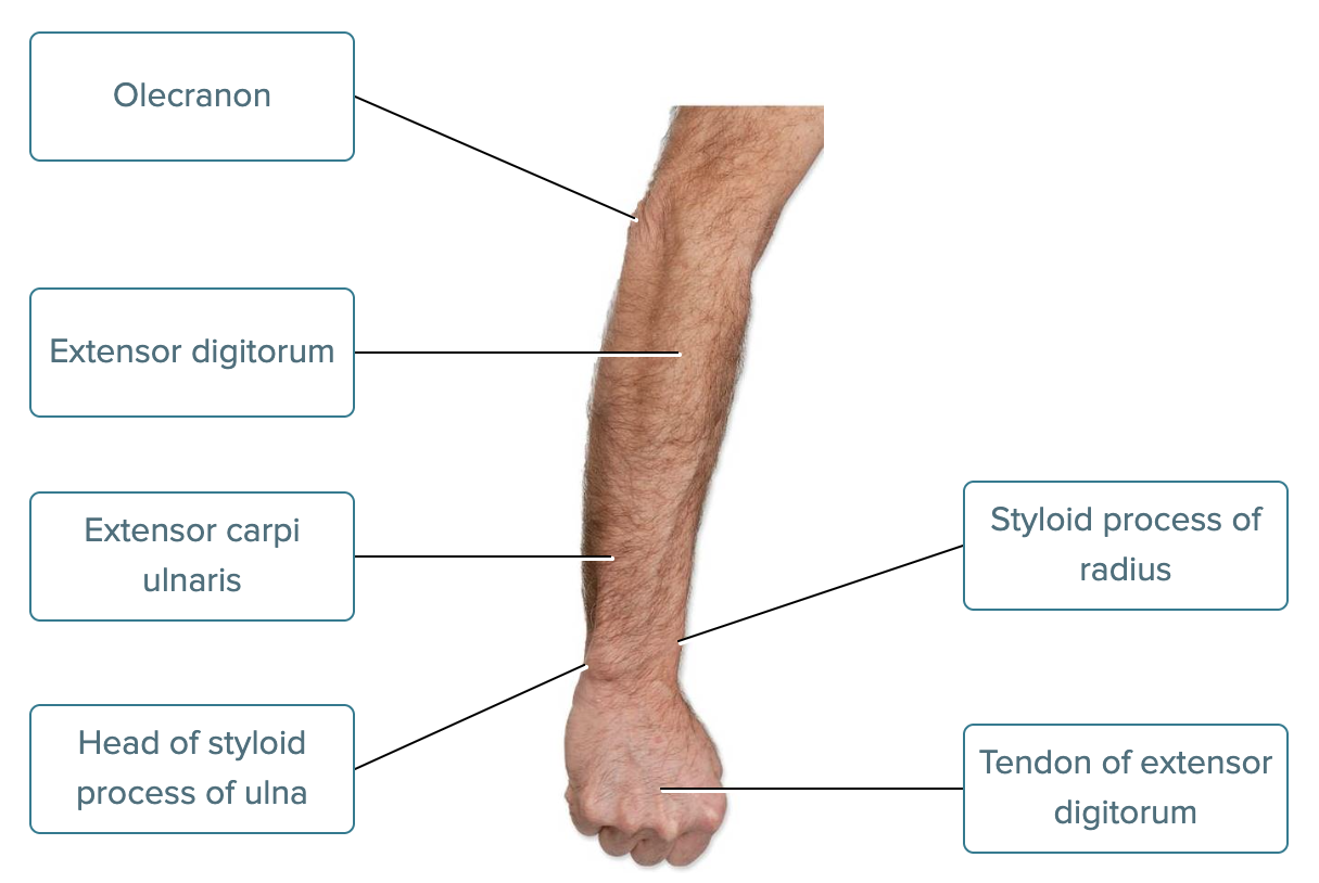
Label the structures in this posterior view of the right forearm.
Styloid process of radius
Extensor digitorum
Tendon of extensor digitorum
Extensor carpi ulnaris
Olecranon
Head of styloid process of ulna
1 - Olecranon
2 - Extensor digitorum
3 - Extensor carpi ulnaris
4 - Head of styloid process of ulna
5 - Styloid process of radius
6 - Tendon of extensor digitorum
Match each region of the cranium and face with its description.
Includes the forehead
Includes the eyes
Includes the ear
Posterior part of the cranium
Includes the sides of the skull
Includes the nose
Includes the cheek
Match each of the options above to the items below.
A. Frontal region
B. Temporal region
C. Occipital region
D. Auricular region
E. Orbital region
F. Nasal region
G. Buccal region
A. Frontal region - 1
B. Temporal region - 5
C. Occipital region - 4
D. Auricular region - 3
E. Orbital region - 2
F. Nasal region - 6
G. Buccal region - 7
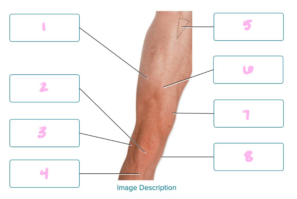
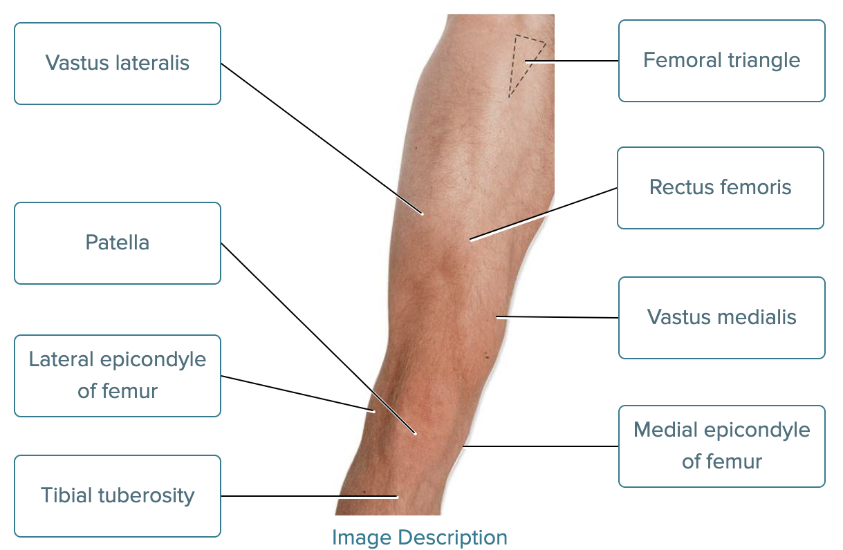
Label the structures in an anterior view of the right thigh.
Lateral epicondyle of femur
Vastus lateralis
Medial epicondyle of femur
Femoral triangle
Rectus femoris
Patella
Tibial tuberosity
Vastus medialis
1 - Vastus lateralis
2 - Patella
3 - Lateral epicondyle of femur
4 - Tibial tuberosity
5 - Femoral triangle
6 - Rectus femoris
7 - Vastus medialis
8 - Medial epicondyle of femur
Match each technique that health care professionals use when examining surface anatomy with its description.
Tapping firmly on body sites to detect vibrations
Directly observe the structure of surface features
Feeling to locate anatomic structures under the skin
Listen to sounds emitted from organs
Match each of the options above to the items below.
A. Visual inspection - 2
B. Palpation - 3
C. Percussion - 1
D. Auscultation - 4
A. Visual inspection
B. Palpation
C. Percussion
D. Auscultation
Another term for the _____ system is the afferent nervous system.
Sensory
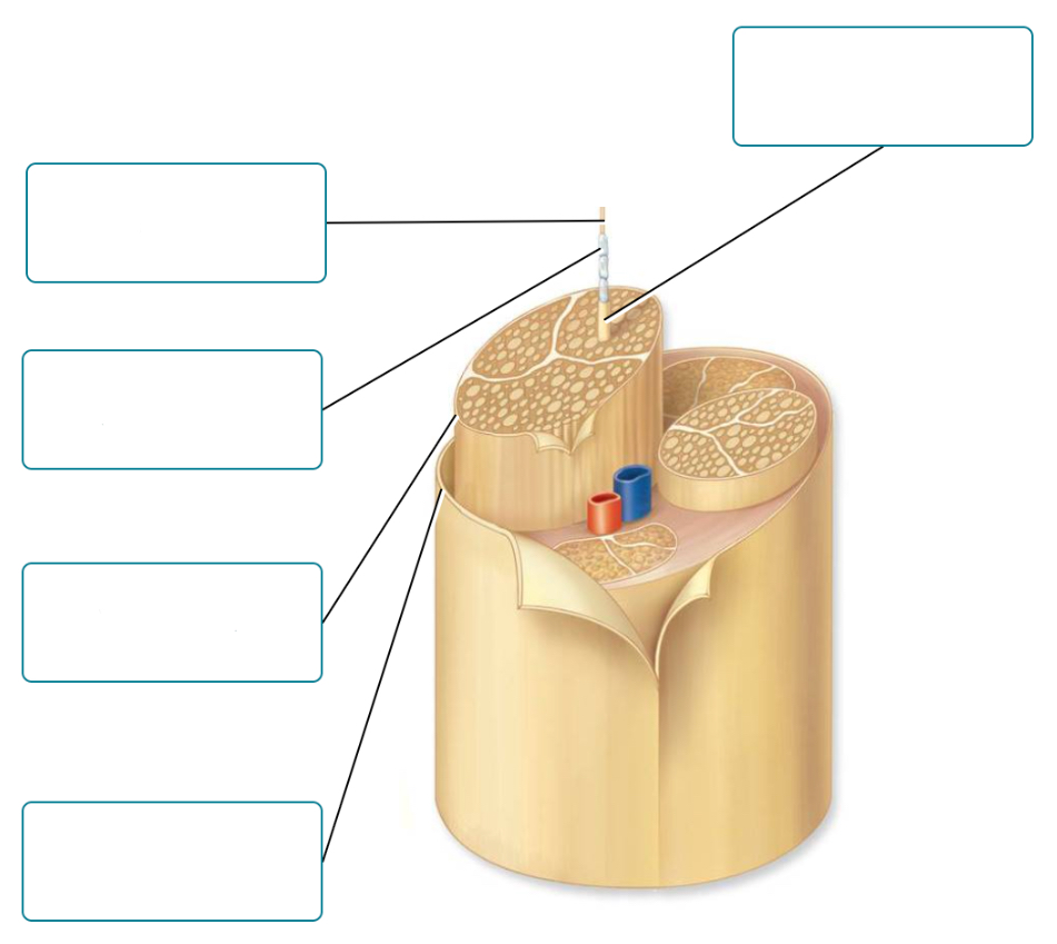
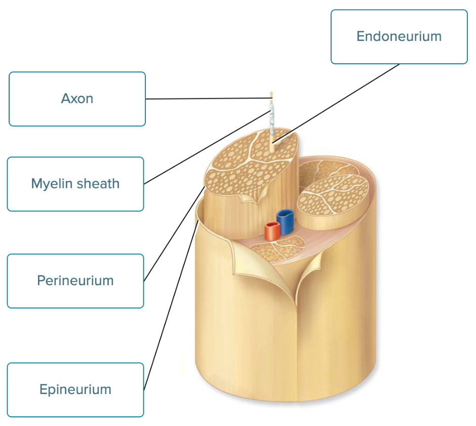
Label the structures of a nerve.
Epineurium
Axon
Perineurium
Endoneurium
Myelin sheath
1 - Axon
2 - Myelin sheath
3 - Perineurium
4 - Epineurium
5 - Endoneurium
Which of the following are true of chemical synapses?
Check All That Apply
Chemical synapses are the most numerous type of synapse in the human body.
Chemical synapses are found primarily in smooth muscle cells.
Chemical synapses are unidirectional (one way).
Chemical synapses allow two-way signaling.
Chemical synapses are the most numerous type of synapse in the human body.
Chemical synapses are unidirectional (one way).
While waiting for your food at your favorite restaurant, you see your favorite dish being carried to the table next to yours. The sight and smell of the food causes your salivary glands to produce saliva. What type of neural circuit caused this single reaction from the multiple stimuli you experienced?
Converging circuit
Reverbrating circuit
Parallel-after discharge circuit
Diverging circuit
Converging circuit
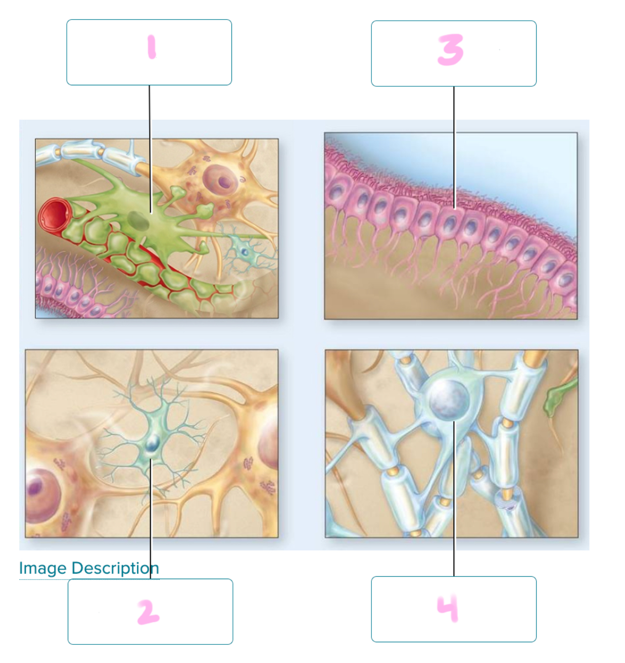
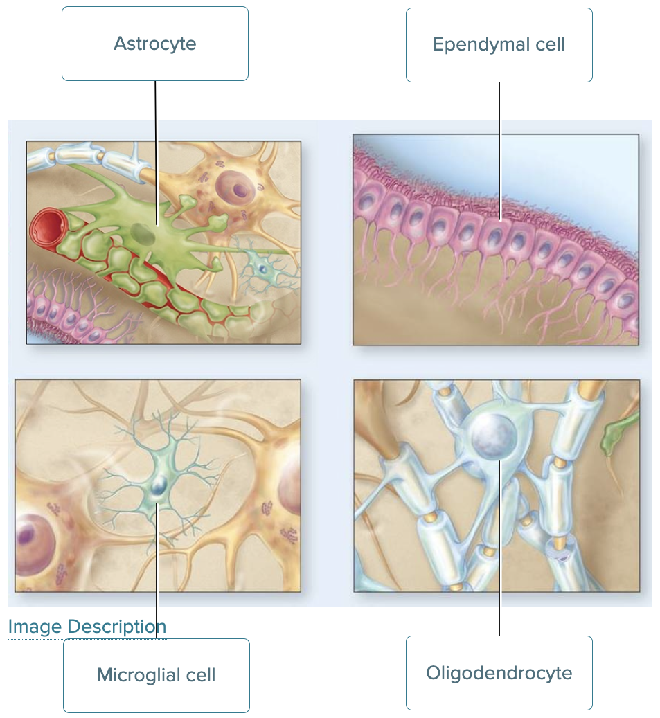
Identify each glial cellsof the CNS.
Oligodendrocyte
Ependymal cell
Microglial cell
Astrocyte
1 - Astrocyte
2 - Microglial cell
3 - Ependymal cell
4 - Oligodendrocyte
Which of the following are a function of the nervous system?
Check All That Apply
Processing and evaluating information
Collecting information
Responding to information
Processing and evaluating information
Collecting information
Responding to information
The _____ nervous system controls the contraction of skeletal muscles.
Somatic
In a nerve, groups of axons are wrapped into separate bundles called _____ .
Fascicles
True or false. Ganglia are clusters of neuron cell bodies located within the CNS.
False
A typical synapse in the CNS consists of a presynaptic neuron and a postsynaptic neuron, separated by a narrow space called the?
presynaptic space.
postsynaptic space.
synaptic cleft.
synaptic membrane.
synaptic cleft.
One cause of hydrocephalus is a malfunction of the structures that normally drain CSF from the subarachnoid space into the venous blood. In other words, there is a problem with the?
arachnoid villi.
choroid plexuses.
cerebral aqueduct and central canal.
median and lateral apertures.
arachnoid villi.
Which of the following brain structures are composed of white matter?
Check All That Apply
Cerebral cortex
Corpus callosum
Cerebral nuclei
Septum pellucidum
Internal capsule
Corpus callosum
Septum pellucidum
Internal capsule
What is the CN VII cranial nerve name?
Facial
What is the CN VIII cranial nerve name?
Vestibulocochlear
What is the CN IX cranial nerve name?
Glossopharyngeal
What is the CN X cranial nerve name?
Vagus
What is the CN XI cranial nerve name?
Accessory
What is the CN XII cranial nerve name?
Hypoglossal
Match each brain vesicle that has developed by the fifth week of development with the structure that they will eventually form.
Forms the thalamus
Forms the medulla oblongata
Does not form a secondary vesicle
Forms the cerebellum
Forms the cerebrum
Match each of the options above to the items below.
A. Telencephalon
B. Diencephalon
C. Mesencephalon
D. Metencephalon
E. Myencephalon
A. Telencephalon - 5
B. Diencephalon - 1
C. Mesencephalon - 3
D. Metencephalon - 4
E. Myencephalon - 2
During the fifth week of embryonic development, ____ brain vesicles form.
Five
Place in order the circulation of cerebrospinal fluid.
CSF flows from the third ventricle through the cerebral aqueduct into the fourth ventricle
CSF flows into the arachnoid villi and drains into the dural venous sinuses
CSF is produced by the choroid plexus in the ventricles
CSF flows through the subarachnoid space and removes waste products
CSF flows through the paired lateral apertures or the single medial aperture and into the central canal of the spinal cord
1 - CSF is produced by the choroid plexus in the ventricles
2 - CSF flows from the third ventricle through the cerebral aqueduct into the fourth ventricle
3 - CSF flows through the paired lateral apertures or the single medial aperture and into the central canal of the spinal cord
4 - CSF flows through the subarachnoid space and removes waste products
5 - CSF flows into the arachnoid villi and drains into the dural venous sinuses