Exam 4: Hand Complex
1/75
Earn XP
Description and Tags
Extrinsic and intrinsic hand muscles
Name | Mastery | Learn | Test | Matching | Spaced | Call with Kai |
|---|
No analytics yet
Send a link to your students to track their progress
76 Terms
The hand complex enables us to?
grasp, pinch, manipulate objects
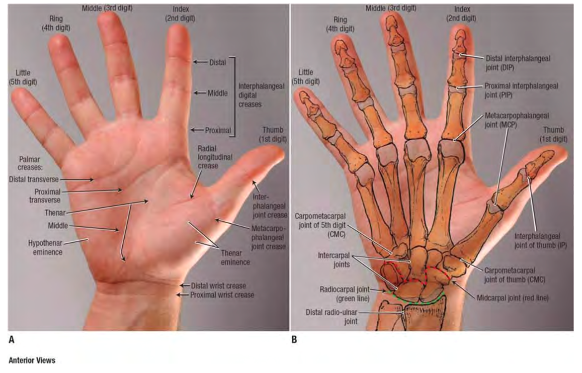
What are the bones of the hand?
5 metacarpals: Fingers/Digits 1-5 (named by digit; ex: 1st metacarpal = thumb metacarpal)
5 proximal phalanx (P1)
4 middle phalanx (P2): absent in thumb
5 distal phalanx (P3)
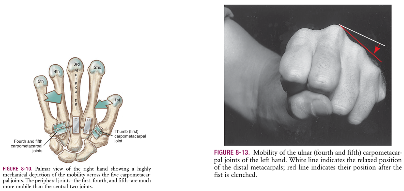
What are the joints of the hand?
Carpometacarpal (CMC) joints
Metacarpophalangeal (MCP)
Proximal interphalangeal (PIP)
Distal interphalangeal (DIP)
Thumb only: Interphalangeal (IP)
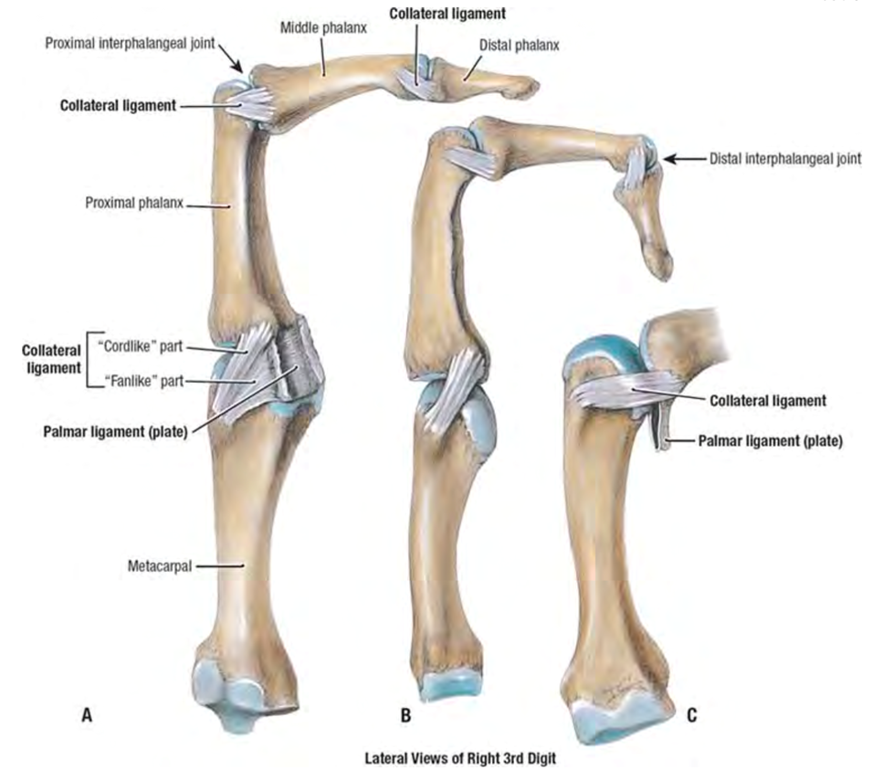
Primary Stabilizers
Collateral ligaments
True ligament: cord-strong ligament
Accessory component: fan-like weaker ligament
Volar plate: strong fibrocartilage plate
Palmar surface anatomy: Identify the…
Distal wrist & palmar crease
Thenar muscle mass
Joint creases for MCP, PIP, DIP, thumb IP joints
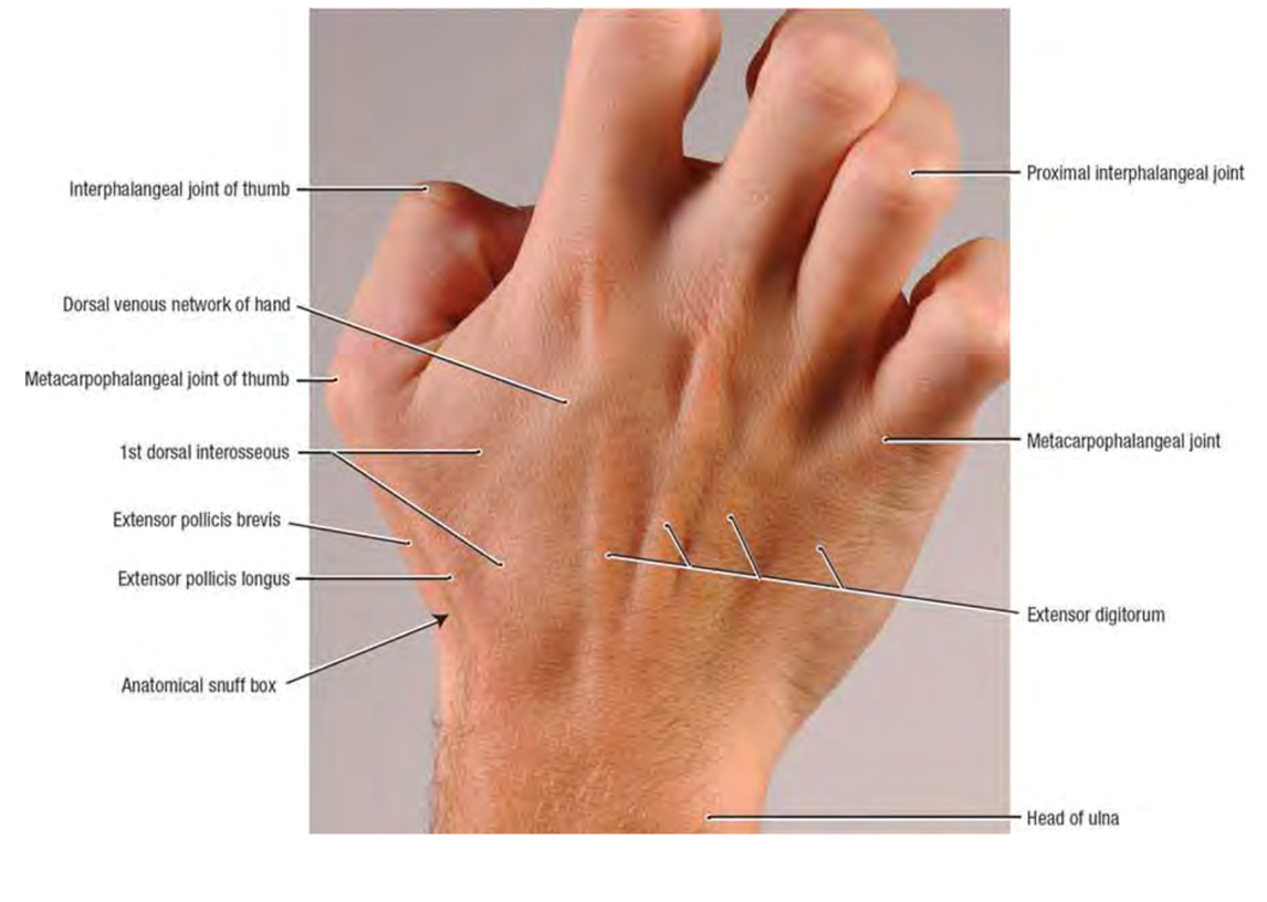
Dorsal surface anatomy: Identify the…
Extensor digitorum (EDC) tendons
1st, 2nd, 3rd extensor compartments
Metacarpophalangeal (MCP) joints
Proximal interphalangeal (PIP) joints
Distal interphalangeal (DIP) joints
1st dorsal interosseous muscle
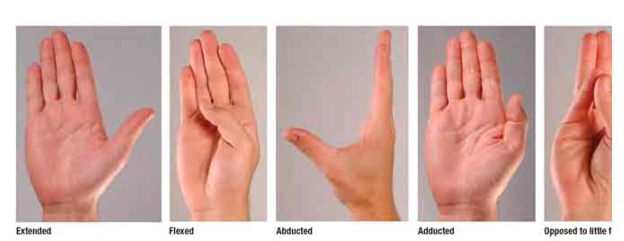
What are the 4 movements of the fingers?
Flexion
Extension
Abduction
Adduction
What are the 6 movements of the thumb?
Flexion (composite flexion = full flexion of all thumb joints)
Extension
Radial & palmar abduction
Adduction
Opposition
How does joint stability demands in the thumb joints differ from the finger joints?
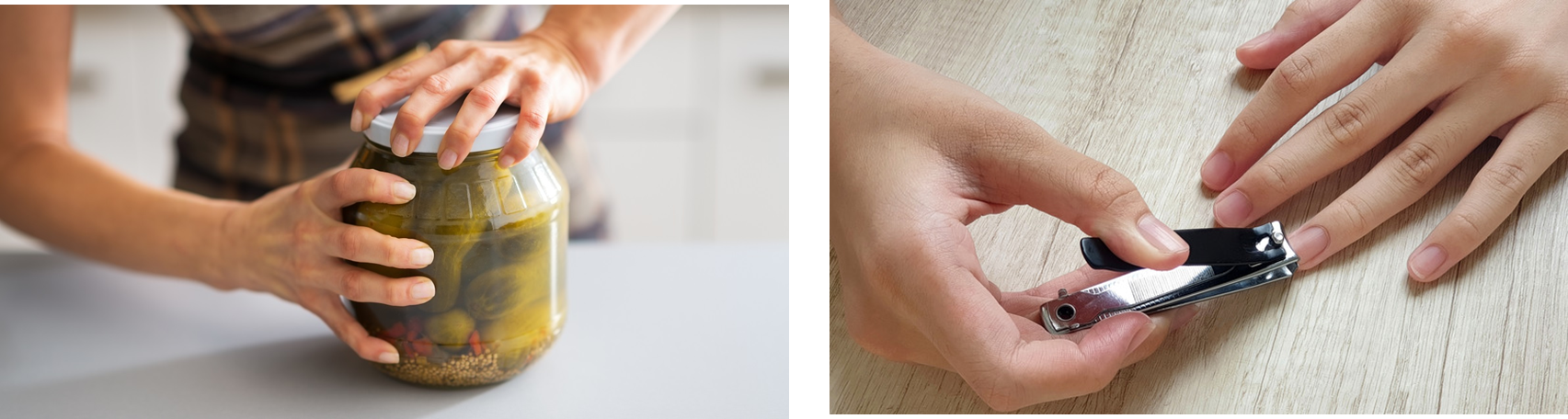
Thumb: power & precision (mobility)
Fingers: stability more important to grasp
Finger joint: 2nd - 5th Carpometacarpal (CMC) joints
Inconsistent classification, so debate on plane VS saddle joint
4th/5th CMC joints more mobile than 2nd/3rd CMC joints
Finger joint: Metacarpophalangeal (MCP) joint
Condyloid joint (lacks axial rotation)
2 degrees of freedom: Flexion/extension & abduction/adduction
Finger joint: Proximal interphalangeal (PIP) & Distal interphalangeal (DIP) joint
Hinge joint: Flexion/extension
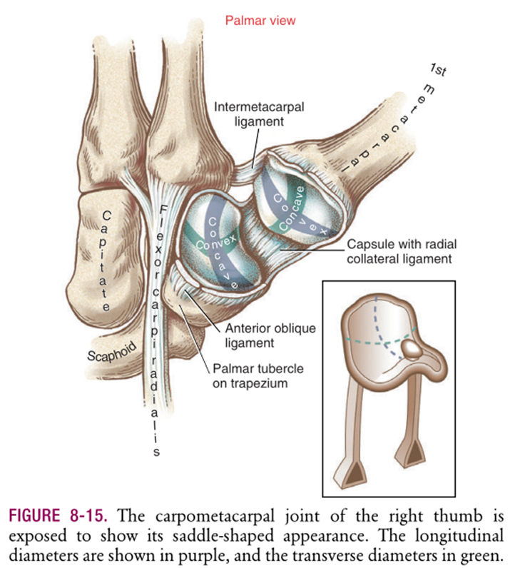
Thumb joint: 1st Carpometacarpal (CMC) joint - Saddle joint
2 degrees of freedom
Flexion/extension
Abduction/adduction
Thumb joint: 1st Metacarpophalangeal (MCP) joint - Condyloid joint
Functions more like “hinge” joint w/ very limited side-to-side motion
Interphalangeal (IP) thumb joint is what kind of joint?
Hinge joint
Carpometacarpal (CMC) joints
1st/4th/5th CMC joints more mobile than 2nd/3rd CMC joints
Allows opposition motion
The thumb CMC joint is a special joint located at the…
Articulation between 1st metacarpal & trapezium
Biomechanics:
Most mobile, complex of the CMC joints due to saddle-shape
Loose joint capsule to accommodate large motion
Relies on ligaments & tendons for stability
1 lb force @ tip of thumb = 12 lbs at base
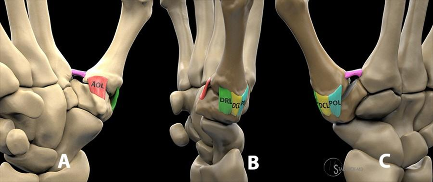
(2) Ligaments of the unstable thumb CMC joint
Volar Anterior Oblique Ligament (AOL): Variable anatomy; curtain-like structure covering volar joint surface
Dorsal Deltoid Ligament: Primary stabilizers of thumb CMC joint
Has 3 stout bands (fan-shaped like deltoid): dorsoradial, dorsal central, posterior oblique ligaments
Originate from dorsal tubercle of triquetrum & insert onto the dorsal base of 1st metacarpal
Thumb CMC joint osteoarthritis: Pathophysiology
Degenerative attenuation of stabilizing ligaments, followed by increased mechanical stress of the CMC joint
40% & 25% of incidence in women & men 75+
Causes debilitating pain & weakness in pinching & loading activities
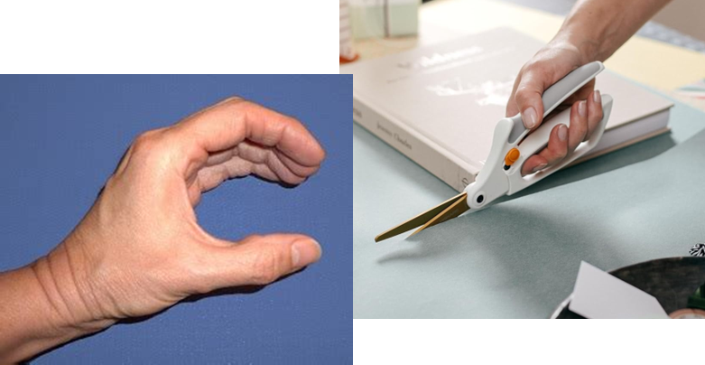
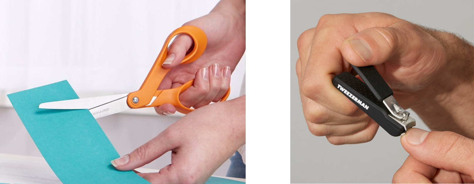
Thumb ergonomics: Since CMC OA comes from stress on ligaments & mechanical load on the joint surface, should patients with CMC OA be positioned in its close-packed (maximizing congruency/ligament tautness) or loose-packed (minimizing compression) joint position?
Loose-packed position to minimize further compression/not stretch out ligament any further. At mid-range; least congruent
EX: natural resting position of thumb (not fully abducted in either direction is the position of comfort), using ergonomic scissors w/ larger grips ✂
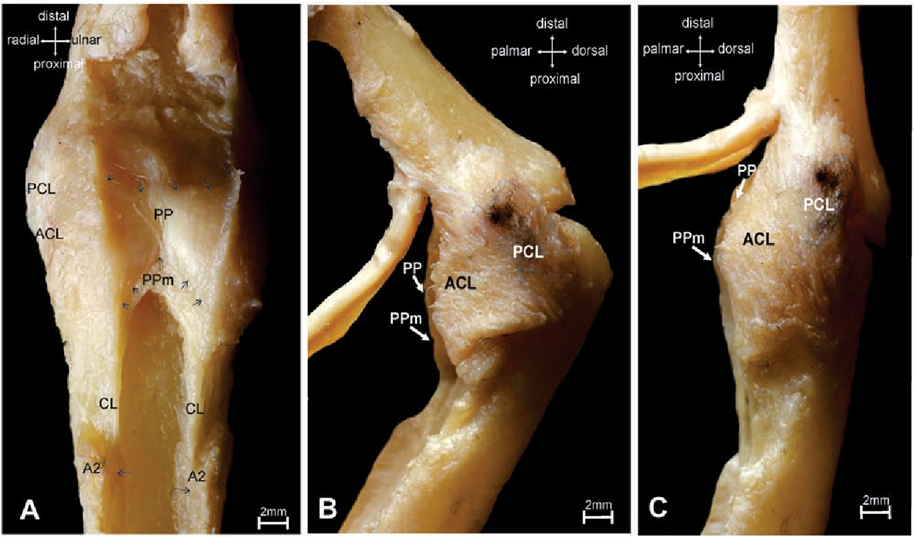
Hand Joint Stabilizer: Collateral ligaments
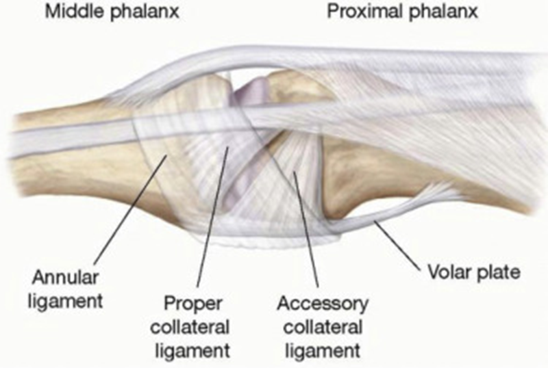
Located on radial & ulnar sides of the joint to provide lateral (side-to-side) stability
2 components
Proper collateral ligament: “cord” like
Accessory collateral ligament: “fan” shaped
Hand joint stabilizers are shared ligamentous structures between the…?
MCP, PIP, DIP, IP joints of the fingers & thumb
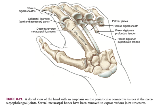
Hand Joint Stabilizer: Volar plate

Prevents hyperextension
Strong fibrocartilage on volar aspect of joint, spanning across joint space
Proximally reinforced by check-rein ligaments on either (lateral) side
Forms “floor” of finger joints
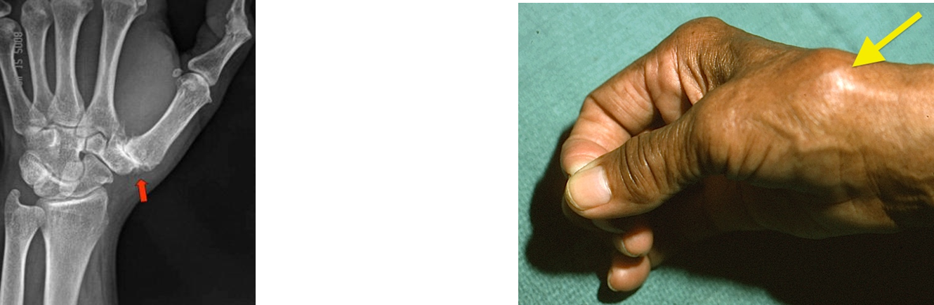
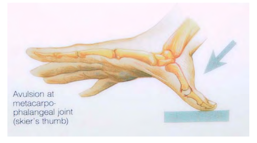
Skier’s thumb is a tear of the…?
Ulnar collateral ligament (UCL) of thumb MCP joint
MOI: Fall/direct trauma to thumb in valgus stress
Chronic tear of thumb MCP UCL ligament = Gamekeeper’s thumb
Finger MCP Joints (2nd - 5th MCP joints)
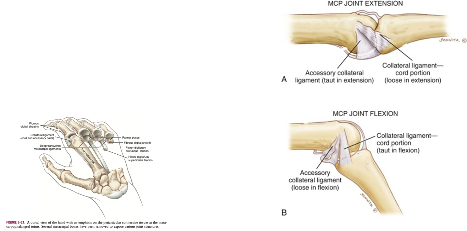
Collateral ligaments
Proper collateral ligament: taut in flexion
Accessory collateral ligament: taut in extension
Deep transverse metacarpal ligaments: Strong bands connecting volar plates of adjacent finger MCP joints
Closed & open pack finger joint positions
Closed-pack position: Fully congruent joint surfaces held tightly together by max tension in joint ligaments.
MP joints: flexion
IP joints: extension
*IMPORTANT DISTINCTION: In IP joints, proper collateral ligaments are taut in all motions. Volar plates are taut in extension, making extension the closed-pack position.
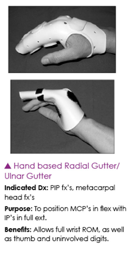
To prevent contracture fraction in fracture splinting, the…
MCPs positioned in 70* flexion
PIP & DIP joints in full extension
also called “Intrinsic plus/safe position”
Extrinsic hand muscles are finger & thumb muscles that originate in the forearm…
OUTSIDE of the hand
Extrinsic hand muscles: Flexors of Anterior forearm
Intermediate layer: Flexor digitorum superficialis (FDS)
Deep layer: FDP + Flexor pollicis longus (FPL)
Extrinsic hand muscles: Extensors (Posterior forearm)
Superficial layer: ED, EDM
Deep layer: APL, EPB/L, EI
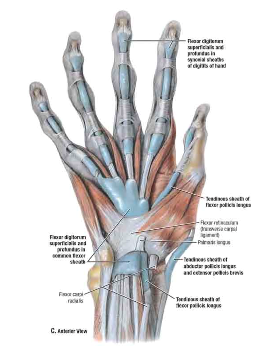
The flexor retinaculum (transverse carpal ligament) forms the roof of the carpal tunnel, enclosing 9 tendons:
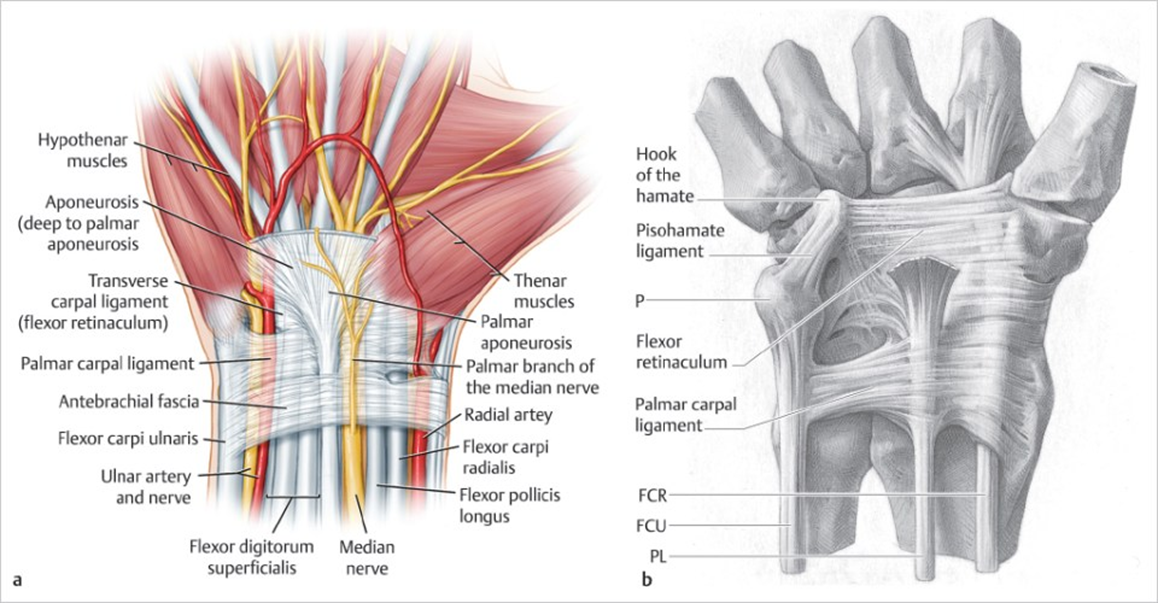
FPL: 1 tendon
FDS & FDP: 4 tendons each
Finger flexors (digits 2-5): Flexor digitorum superficialis (FDS)
Action: Digit 2-5 flexion at PIP joint
Origins:
Humeroulnar head: medial epicondyle and coronoid process of ulna
Radial head: oblique line of radius
Insertion: Shaft of middle phalanges
Nerve innervation: Median nerve
Finger flexors (digits 2-5): Flexor digitorum profundus (FDS)
Action: Digit 2-5 flexion at DIP joint
Origin: proximal ¾ of anteromedial surface of ulna & interosseous membrane
Insertion: Base of distal phalanges
Nerve innervation:
Digits 2&3: AIN of Median nerve
Digits 4&5: Ulnar nerve
Flexor digitorum superficialis (FDS) vs. Flexor digitorum profundus (FDP) Anatomy
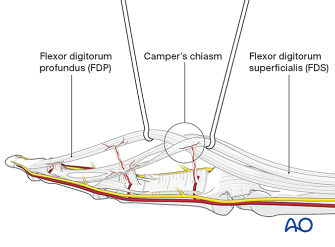
Runs superficial to the FDP: at its insertion, the FDS splits into two tendon slips wrapping itself around the FDP to allow the FDP to pass through = Camper’s chiasm
FDP doesn’t divide & cont. to insert into distal phalanx
FDS = PIP flexion
FDP = DIP flexion
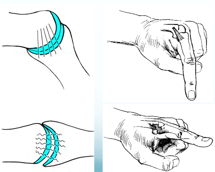
Isolate the actions of FDS by flexing the targeted digit…
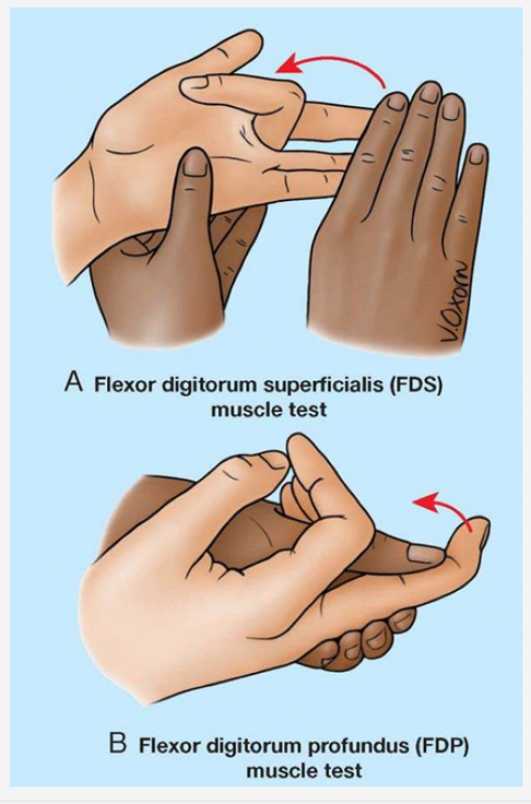
while holding rest of digits in extension
FDS has common muscle belly
Isolate the actions of FDP by holding the targeted digit…
just proximal to DIP joint
only allow DIP to flex, while keeping PIP in extension
Thumb flexor - Flexor pollicis longus (FPL) Overview
Part of deep layer of anterior forearm muscles
Action: thumb flexion @ IP & MCP joints
Origin: Anterior surface of radius & adjust interosseous membrane + interosseous membrane
Insertion: base of distal phalanx of thumb
AIN nerve innervation (median n.)
Flexor Tendon Pulleys
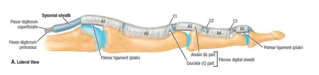
Function: Prevent bowstringing of the flexors
Fingers: 5 annular pulleys (A1-A5) and 3 cruciate pulleys
Annular pulleys much stronger than cruciate pulleys
A2 pulley = strongest
Thumb: 2 annular pulleys (A1, A2) & 1 oblique pulley
A1 pulley at MCP joint
Oblique pulley in middle of proximal phalanx: most critical against bowstringing
Trigger finger/thumb
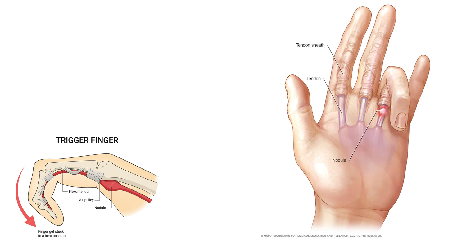
Pathophysiology: Inflamed nodule forms on thickened flexor tendon sheath. Nodule becomes trapped proximal to A1 pulley structure when the digit’s trying to extend from full flexion.
MOI: overuse/repetitive gripping
Finger Extensors: Superficial VS Deep Layers
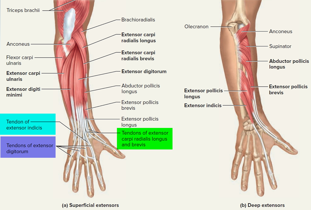
Superficial layer: EDC, EDM
Deep layer: Extensor indicis (EI)
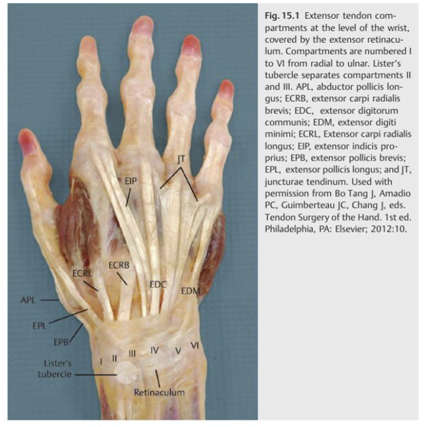
6 fibro-osseous tunnels form compartments, a thick fibrous band securing the extensor tendons
1st compartment: APL & EBP
2nd compartment: ECRB/L
3rd compartment: EPL
4th compartment: EDC & EDI
5th compartment: EDM
6th compartment: ECU
Finger Extensor: Extensor indicis (EI)
Action: Digit 2 extension (index/pointer finger)
Origin: Distal 3rd of ulna & interosseous membrane
Insertion: Digit 2 extensor expansion
Nerve: PIN - cont. of deep branch of radial n.
Dorsal extensor compartment: 4th
Unique: Tendons linked to each other in dorsal hand, w/ juncturae tendinae: CT band
Finger Extensor: Extensor digitorum communis (EDC)
Action: Digit 2-5 extension
Origin: Lateral epicondyle
Insertion: Digit 2-5 extensor expansion
Nerve: PIN - cont. of deep branch of radial n.
Dorsal extensor compartment: 4th
Unique: Tendons linked to each other in dorsal hand, w/ juncturae tendinae: CT band
Finger Extensor: Extensor digiti minimi (EDM)
Action: Digit 5 extension
Origin: Lateral epicondyle
Insertion: Digit 5 extensor expansion
Nerve: PIN (cont. of deep branch of radial n.)
Dorsal extensor compartment: 5th
Unique: Tendons linked to each other in dorsal hand, w/ juncturae tendinae: CT band
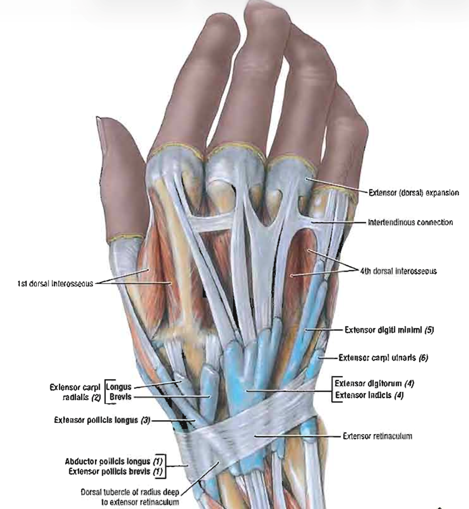
Finger Extensor: Juncturae tendinae (JT)
CT band linking adjacent tendons of EDC proximal to MCP joints
highly variable in structure
assist in centralizing extensor tendon
Finger extension: Why are we able to independently extend the index and small fingers but not the middle and ring fingers?
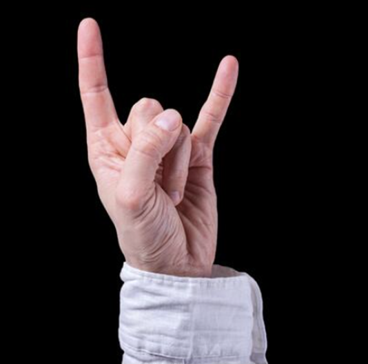
Because they each have their own individual extensor!
Extensor expansion is a…?
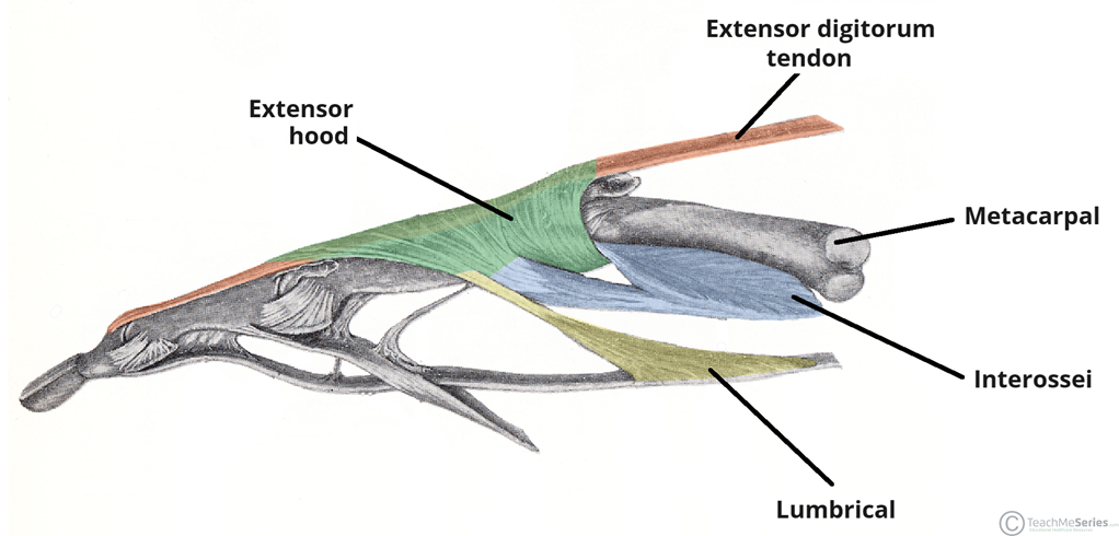
Complex web of CT structure that EDC tendon & intrinsic hand muscles insert into
Facilitates finger extension!
Deep layer of Thumb Extensors are the…?
APL, EPB/L (3)
Assist w/ wrist radial deviation
Form border of anatomical snuffbox
In dorsal forearm
Anatomical Snuffbox 📐
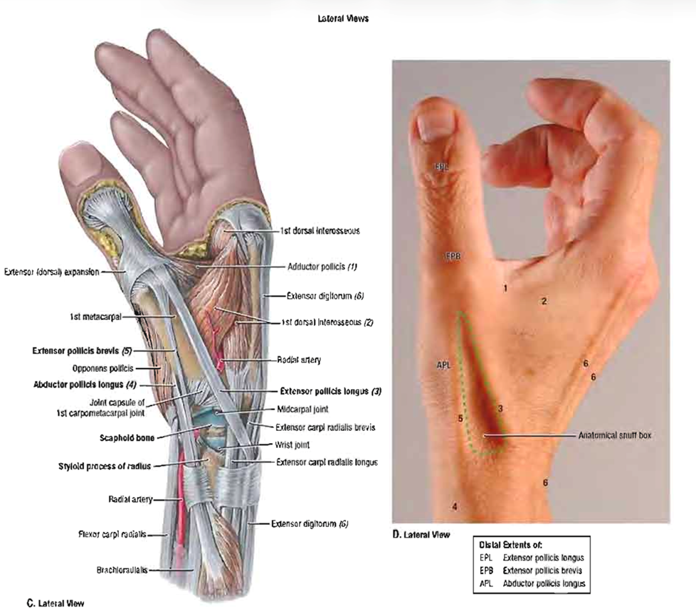
Radial border: 1st dorsal compartment (triangle) - Shared compartment between APL & EPB
Ulnar border: EPL
Floor: Scaphoid - Pain w/ pressure in this location = scaphoid fracture
Thumb Extensor: Abductor Pollicis Longus (APL)
Action: Thumb abduction & extension @ CMC
Origin: Posterior proximal halves of ulnar, radius, interosseous membrane
Insertion: base of 1st metacarpal
Nerve: PIN - cont. of deep branch of radial n.
1st Compartment
Thumb Extensor: Extensor pollicis brevis (EPB)
Action: Thumb extension @ CMC & MCP
Origin: Posterior distal third of radius & interosseous membrane
Insertion: base of thumb proximal phalanx
Nerve: PIN - cont. of deep branch of radial n.
1st Compartment
Thumb Extensor: Extensor pollicis longus (EPL)
Action: Thumb extension at ALL thumb joints
Origin: Posterior middle third of ulna & interosseous membrane
Insertion: base of thumb distal phalanx
Nerve: PIN (cont. of deep branch of radial n.)
Compartment: 3rd
Thumb extensor isolated function, from proximal to distal:
APL: thumb CMC abduction/extension
EPB: thumb MP extension
EPL: thumb IP extension

De Quervain’s Tenosynovitis is Stenosing tenosynovitis = Degenerative thickening of the tendon sheath & retinaculum of tendons in 1st dorsal extensor compartment (APL + EPB)
Which motions would become painful for the patient?
Grasping, lifting, or twisting; due to pain at the radial side of the wrist, triggered by thumb movement & wrist deviation.
Intrinsic hand muscles: Vital for fine-motor hand control, divided into 4 groups (within the hand)
Perform fine motor skills
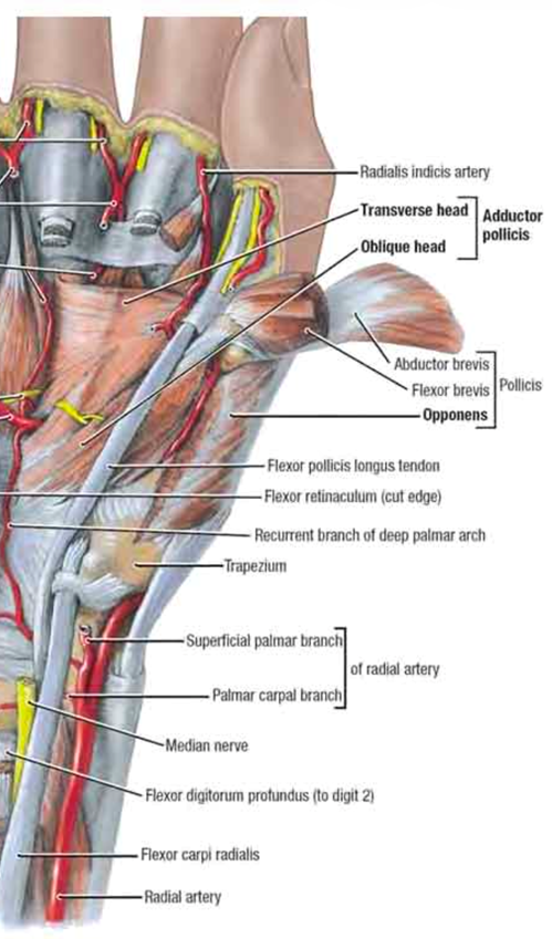
Muscles of thenar eminence (meaty part of thumb): ABP, flexor pollicis brevis, opponens pollicis
Muscles of hypothenar eminence (bulk on pinky side): Flexor digiti minimi, abductor digit minimi, opponens digiti minimi
Adductor pollicis (thumb adductor)
Lumbricals & interossei
These fill the space between our thumb metacarpals
Thenar & hypothenar “AFO” muscles function to…
synergically to perform opposition
What are the thenar muscles? “AFO”
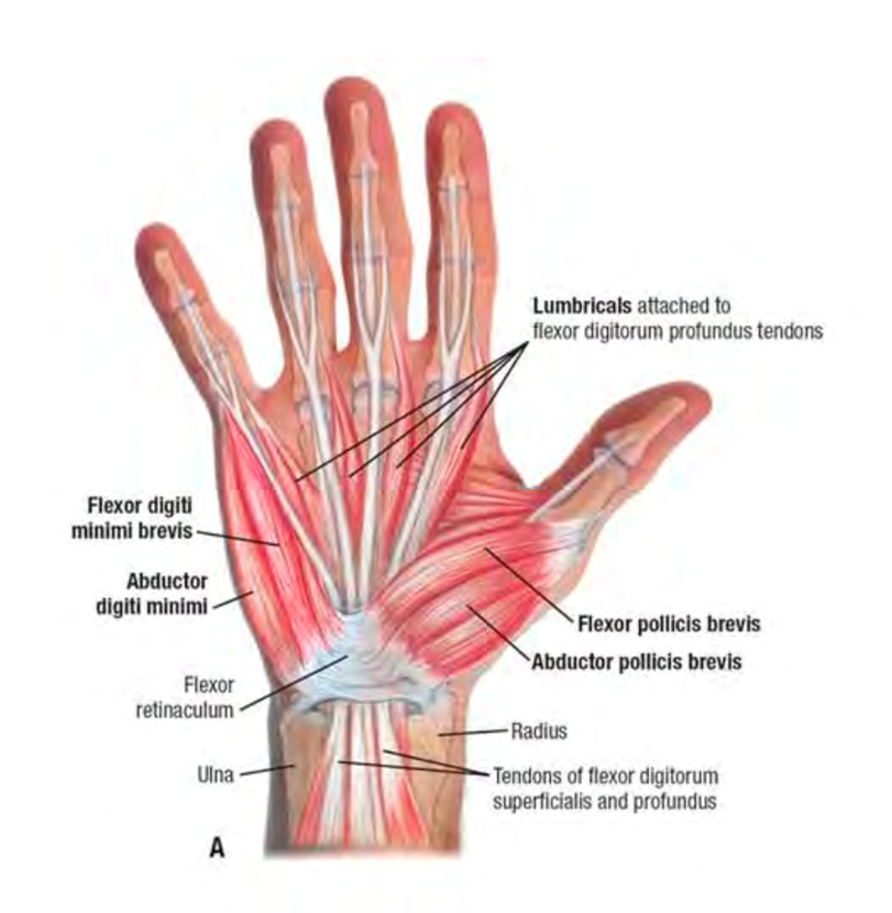
Abductor pollicis brevis
Flexor pollicis brevis
Opponens pollicis (deeper to other 2 thenar muscles)
What are the hypothenar muscles? “AFO”
Abductor digiti minimi
Flexor digiti minimi brevis
Opponens digiti minimi (deeper to other 2 hypothenar muscles)
Thenar Muscles: Abductor pollicis brevis (APB)
Action: Thumb abduction
Origin: Flexor retinaculum & tubercles of scaphoid & trapezium
Insertion: Lateral side of base of thumb proximal phalanx
Nerve: Recurrent branch of median n. (pass thru carpal tunnel)
MOST powerful muscle of thenar muscle
Crosses thumb MCP joint
Thenar Muscles: Flexor pollicis brevis (FPB)
Action: Thumb MP & CMC flexion
Origin: Flexor retinaculum & tubercles of scaphoid & trapezium (same as OP)
Insertion: Lateral side of base of thumb proximal phalanx
Nerve: Recurrent branch of median n. (pass thru carpal tunnel)
Has 2 heads: superficial & deep
Crosses thumb MCP joint
Thenar Muscles: Opponens pollicis (OP)
Action: Thumb opposition
Origin: Flexor retinaculum & tubercles of scaphoid & trapezium (same as FPB)
Insertion: Lateral side of 1st metacarpal (Doesn’t cross thumb MCP joint b/c it ends here)
Nerve: Recurrent branch of median n. (pass thru carpal tunnel)
Hypothenar Muscles: Abductor digiti minimi
Action: 5th digit abduction
Origin: Pisiform
Insertion: Medial side of base of 5th digit proximal phalanx
Nerve: Deep branch of ulnar n.
Hypothenar Muscles: Flexor digiti minimi brevis
Action: 5th digit proximal phalanx flexion
Origin: Flexor retinaculum & hook of hamate
Insertion: Medial side of base of 5th digit proximal phalanx
Nerve: Deep branch of ulnar n.
Hypothenar Muscles: Opponens digiti minimi
Action: 5th digit opposition
Origin: Flexor retinaculum & hook of hamate
Insertion: Medial border of 5th metacarpal
Nerve: Deep branch of ulnar n.
Combined action of the thenar & hypothenar muscles is?
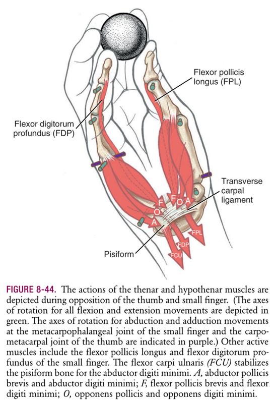
Thenar muscles: Position thumb in varying amounts of opposition to facilitate grasping. Combines CMC abduction, flexion, medial rotation
Hypothenar muscles: Raise & “cup” ulnar border of hand
Adductor pollicis: Overview
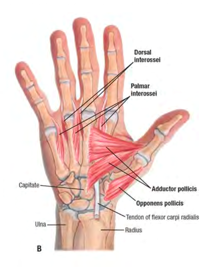
Action: Thumb adduction
2 Origins
Oblique head: base of 2nd & 3rd metacarpal, capitate, adjacent carpals
Transverse head: anterior surface of 3rd metacarpal shaft
Insertion: Medial base of thumb proximal phalanx
Nerve: Deep branch of ulnar n. (like hypothenar muscles)
Ape Hand deformity in Carpal Tunnel Syndrome: Median n. compression @ carpal tunnel → atrophy of thenar muscles
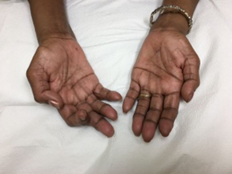
What do you notice about the resting hand posture of the patient w/ this deformity?
Atrophy in thenar muscles, thumb is completely flat in same plane as palm. Can’t bring thumb into palmar abduction
What accounts for this posture? Which muscles are working & overcompensating?
Lack of participation from thumb thenar muscles
Thumb adductor pollicis muscle overworked, keeping thumb close to palm (APM deformity)
Palmar & dorsal interossei
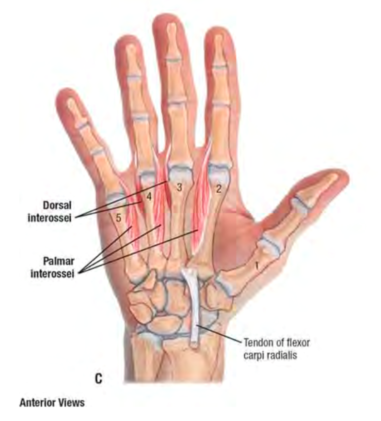
Palmar: Unipennate, palmar surface of 2nd, 4th, 5th metacarpals
Dorsal: Bipennate, adjacent sides of 2 metacarpals
ALL insert into bases of proximal phalanges & extensor expansion
Deep branch of ulnar n. innervated
Actions of the interossei are?
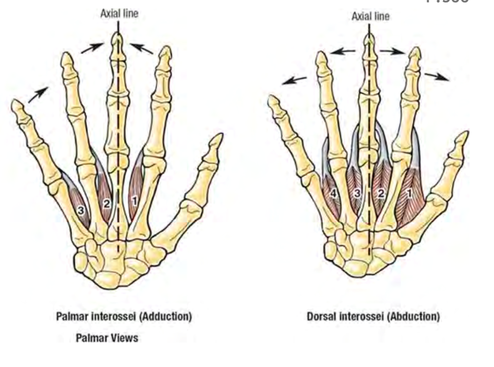
PAD: Palmar interossei ADduct
DAB: Dorsal interossei ABduct
4 Lumbricals “earth worm” numbered from radial to ulnar:
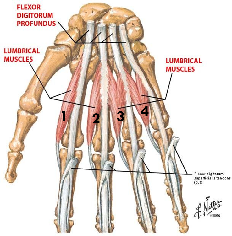
Lumbricals 1-2 = unipennate (median n. innervation)
Lumbricals 3-4 = bipennate (deep branch of ulnar n.)
Lumbricals 1-2
Action: Digits 2-3, MCP flexion & IP extension
Origins: lateral 2 FDP tendons
Insertion: Extensor expansion (lateral side)
Nerve: Median n. (same as FDP origin)
Structure: Unipennate
Lumbricals 3-4
Action: Digits 4-5, MCP flexion & IP extension
Origins: Medial 3 FDP tendons
Insertion: Extensor expansion (lateral side)
Nerve: Deep branch of ulnar n. (same as FDP origin)
Structure: Bipennate
Lumbricals & interossei work together to perform?
Finger MCP flexion & IP extension
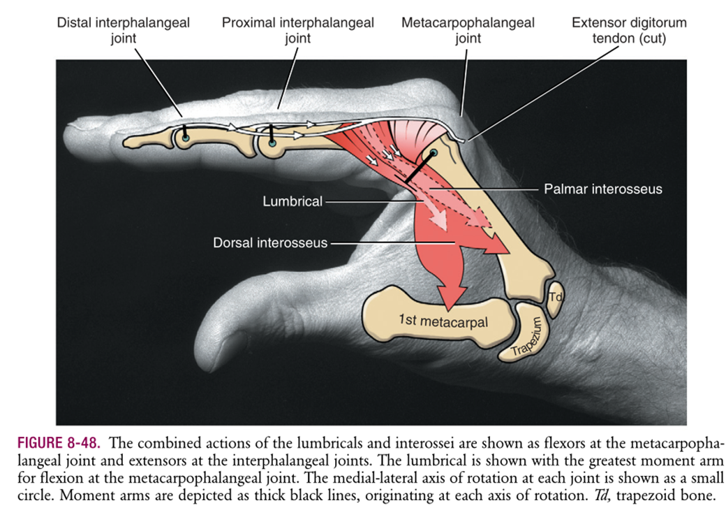
Intrinsic plus position is what combined action of the intrinsic muscles:
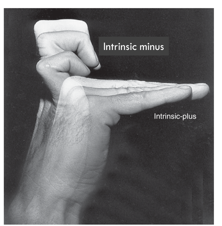
MP flexion + IP extension
Intrinsic minus position is the combined action of the extrinsic muscles WITHOUT action of the intrinsics
MP extension + IP flexion
MCPs are hyperextended instead of being in neutral extension because EDC muscle acts unopposed on the MCP joints
Clinical: Ulnar Claw
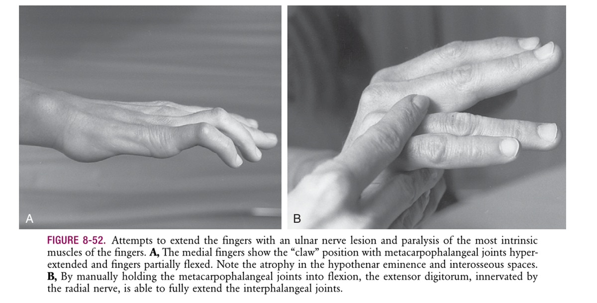
Intrinsic muscles affected by ulnar n. lesion @ wrist level
Affected hand can’t extend digits 4+5 b/c paralyzed lumbricals/interossei = extensor digitorum cause hyperextension @ MCP joints, while intact FDP pulls IP joints into flexion
If you had carpal tunnel syndrome, would the hypothenar or thenar muscles be intact?
Hypothenar muscles since they’re innervated by the ulnar nerve, not median nerve (thenar muscles).