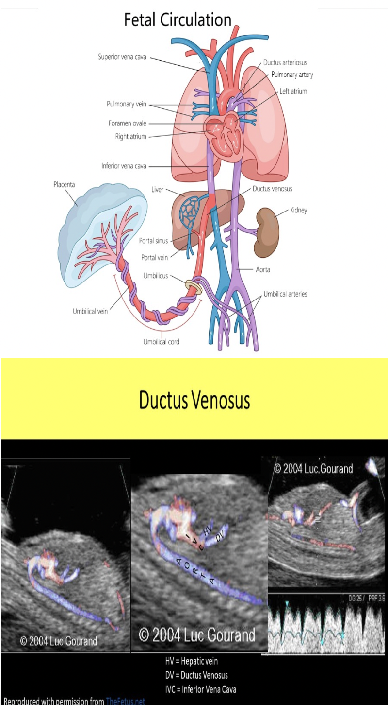
2nd/3rd Trimester Anatomy
1/154
There's no tags or description
Looks like no tags are added yet.
Name | Mastery | Learn | Test | Matching | Spaced | Call with Kai |
|---|
No analytics yet
Send a link to your students to track their progress
155 Terms
The fetal umbilical arteries branch from the:
A. fetal internal iliac arteries
B. maternal aorta
C. maternal internal iliac arteries
D. fetal aorta
A. fetal internal iliac arteries
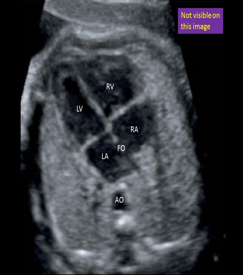
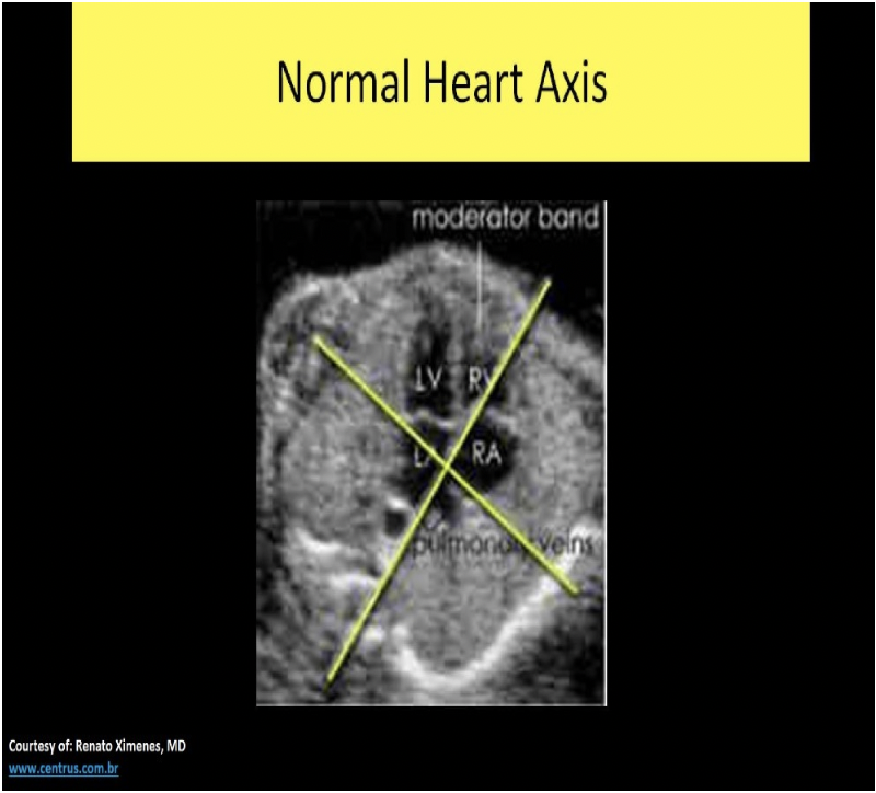
Locate the left ventricle. The structure may or may not be visible on this image

Locate the superior vena cava
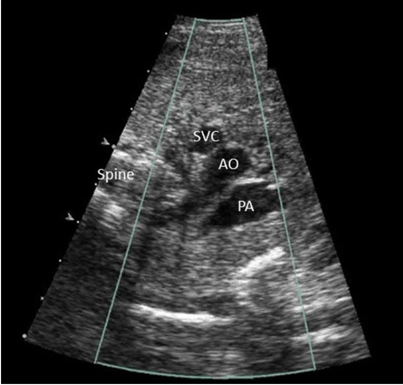
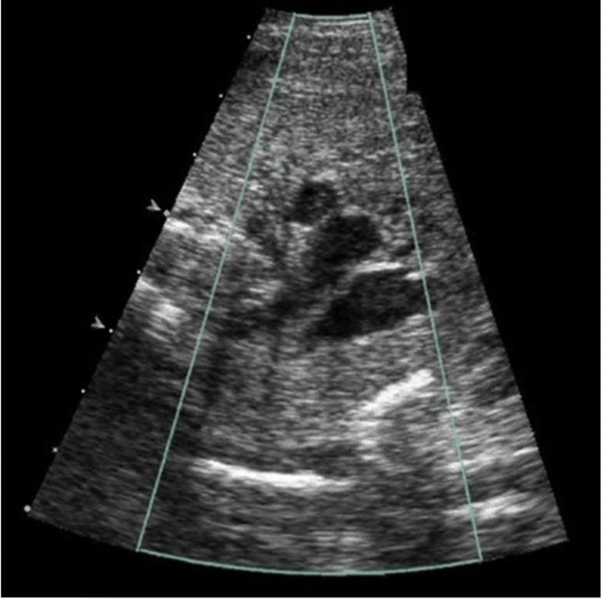
The attached image is a _____ plane used to evaluate which great vessel?
A. coronal, IVC
B. sagittal, aorta
C. sagittal, IVC
D. coronal, aorta
B. sagittal, aorta
Note the sagittal image of the spine and the parallel course of the vessel. The Aorta originates from the superior portion of the heart and courses posteriorly through the abdomen adjacent to the spine.
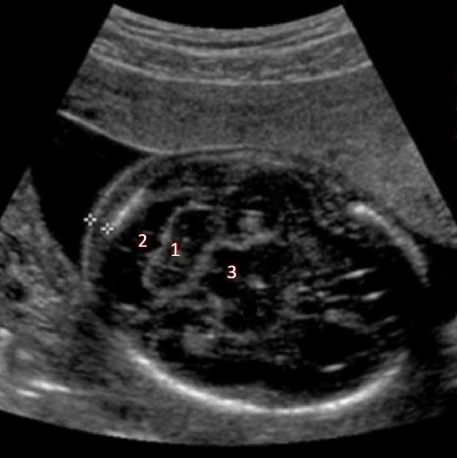
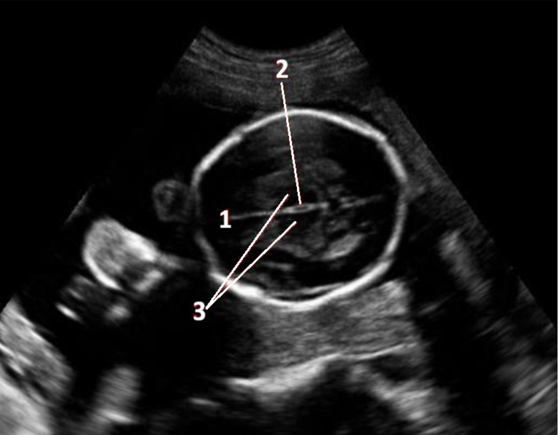
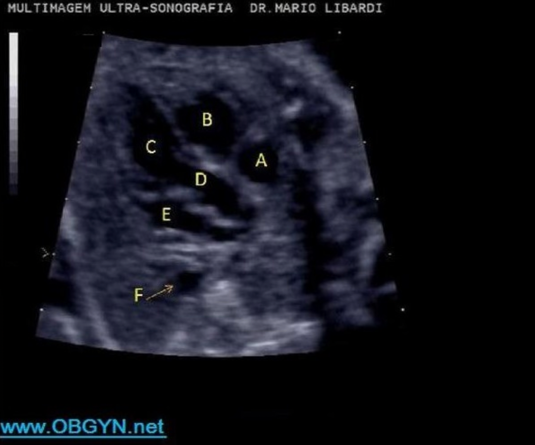
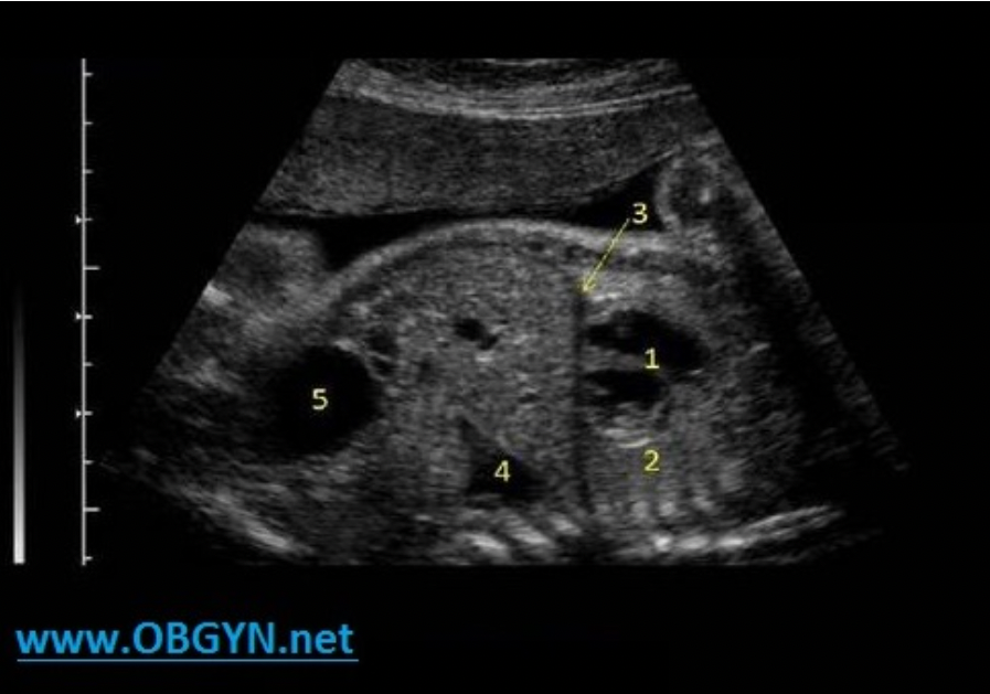
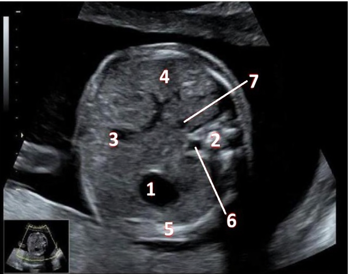
What fetal structure is indicated by #3?
A. caudate nucleus
B. thalamus
C. cisterna magna
D. cerebellum
B. thalamus
cerebellum
cisterna magna
thalamus
The fetal testicles normally descend into the scrotum before weeks gestation.
A. 16
B. 22
C. 34
D. 26
C. 34
The fetal testicles normally descend into the scrotum between 26 and 34 weeks gestation. If one or both fail to descend, cryptorchidism is present.
The visualization of a normal stomach indicates a normal _____ is present
A. small intestine
B. esophagus
C. palate
D. colon
B. esophagus

What fetal measurement can be assessed in this view?
A. HC
B. cerebellum diameter
C. BPD
D. lateral ventricle diameter
B. cerebellum diameter
Chromosomal defects may be present in a pregnancy where the amnion and chorion have not completely fused by ____ gestational age.
A. 8 weeks
B. 12 weeks
C. 16 weeks
D. 20 weeks
C. 16 weeks
At the start of the 12th week, the amnion and chorion begin to fuse and should be complete by week 16. If the amnion and chorion are not fused by 16 weeks of gestation there may be associated fetal structural and/or chromosomal abnormalities. (60%)
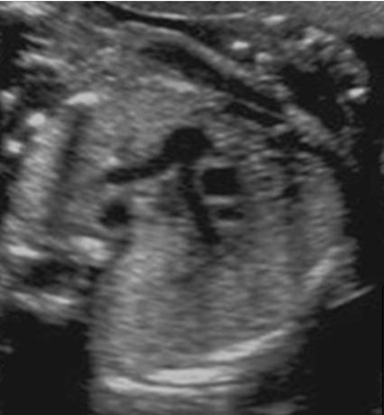
Locate the right pulmonary artery
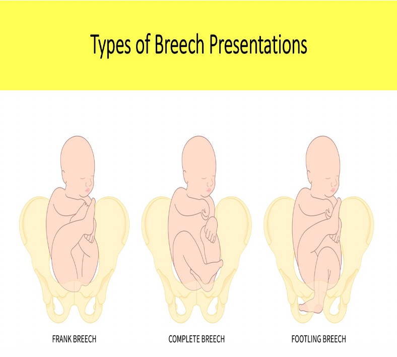
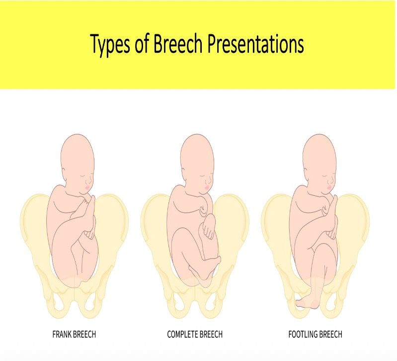
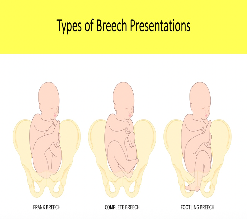
Which type of breech position is defined as the fetus presenting butt first and sitting cross legged with knees bent?
A. Frank
B. Complete
C. Partial
D. Footling
B. Complete
Frank breech = butt down legs straight up in front of abdomen
Complete breech = butt down legs folded, knees bent, ankles crossed
Footling breech = feet down, baby "standing" on cervix
The fetal stomach should be routinely visualized by week _____
A. 10
B. 12
C. 16
D. 14
D. 14
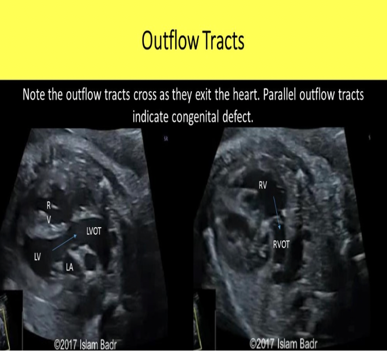
The proximal pulmonary artery and aorta demonstrate a normal appearance when:
A. the diameter of the aorta is at least 2X the diameter of the pulmonary artery
B. the aorta crosses anterior to the pulmonary artery as they both exit the heart
C. the pulmonary artery crosses anterior to the aorta as they both exit the heart
D. they exit the heart parallel to one another
C. the pulmonary artery crosses anterior to the aorta as they both exit the heart
The proximal pulmonary artery and aorta demonstrate a normal appearance when the pulmonary artery crosses anterior to the aorta as they both exit the heart. The PA is the same as or slightly larger in diameter than the aorta in the 2nd and 3rd trimester fetus. Parallel great vessels OR the aorta crosses anterior to the pulmonary artery = transposition of the great vessels
The cisterna magna is enlarged if it measures more than:
A. 5mm
B. 7mm
C. 10mm
D. 12mm
C. 10mm
A cisterna magna with an AP dimension greater than 10mm can indicate dandy walker malformation or mega cisterna magna. If the AP measurement is less than 2mm, Arnold Chiari II malformation could be present.
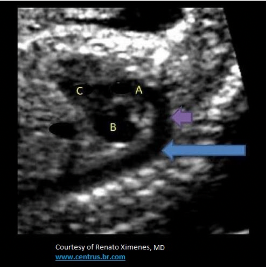
What fetal structure is indicated by #1?
A. falx cerebri
B. cisterna magna
C. caudate nucleus
D. corpus callosum
A. falx cerebri
falx cerebri
third ventricle
thalamus lobes
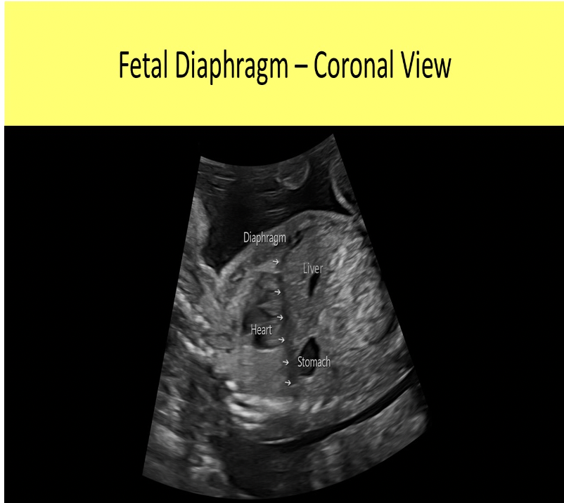
Which of the following describes the normal fetal diaphragm?
A. hyperechoic dome shaped structure with the top of the dome facing cephalad
B. hypoechoic cup shaped structure with the bottom of the cup facing caudal
C. hypoechoic dome shaped structure with the top of the dome facing cephalad
D. hyperechoic linear structure that separates the abdomen from the chest
C. hypoechoic dome shaped structure with the top of the dome facing cephalad
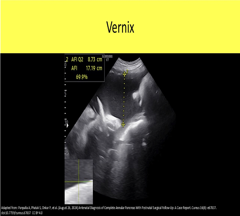
______ refers to a normal finding of sebum and epithelial cells that cause white free-floating debris within the amniotic fluid late in pregnancy.
A. vernix
B. meconium
C. Chadwick's sign
D. pseudomyxomatous peritonei
A. vernix
Vernix refers to a normal finding of sebum and epithelial cells that cause white free-floating debris within the amniotic fluid late in pregnancy. Meconium refers to an abnormal finding of fetal waste, sebum and epithelial cells, that cause hypochoic free-floating debris within the amniotic fluid late in pregnancy. Normally meconium is visualized in the fetal colon in near term pregnancies. If the fetus has a bowel movement, it is a sign of fetal distress.
The _____ is located between the right atrium and ventricle, while the _____ is located between the left atrium and ventrible
A. tricuspid valve, mitral valve
B. tricuspid valve, pulmonary valve
C. mitral valve, tricuspid valve
D. mitral valve, aortic valve
A. tricuspid valve, mitral valve
The tricuspid valve is located between the RA and RV. The mitral valve is located between the LA and LV. The aortic valve is located between the LV and aorta. The pulmonary valve is located between the RV and pulmonary artery.
Dizygotic twins:
A. are always the same sex.
B. are the less common than monozygotic twins.
C. are formed by the union of 2 sperm and one egg.
D. will usually demonstrate two chorions, two amnions and two placentas.
D. will usually demonstrate two chorions, two amnions and two placentas.
A dizygotic gestation occurs when 2 OVA are fertilized by 2 SPERM.
It is the most common type of twins. They always have 2 chorions, 2 amnions, 2 placentas (can appear as one large placenta); The babies can be of the same sex or different.
Which cranial structure is considered abnormal if it increases in size between ultrasound exams performed at 20 and 28 weeks?
A. thalamus
B. lateral ventricle
C. cerebellum
D. choroid plexus
B. lateral ventricle
The lateral ventricles are usually consistent in size throughout pregnancy. An increase in size between serial exams can indicate a developing problem with CNS drainage.
Normal amniotic fluid levels are mandatory for proper development of the fetal:
A. bladder
B. digestive tract
C. kidneys
D. lungs
D. lungs
Normal amniotic fluid levels are mandatory for proper development of the fetal lungs. If oligohydramnios is present, this leads to pulmonary hypoplasia.

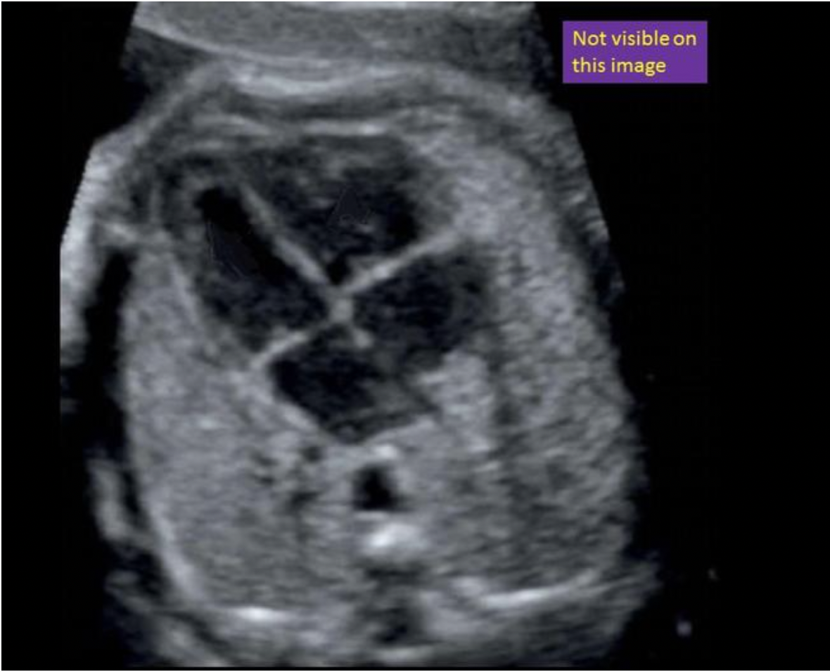
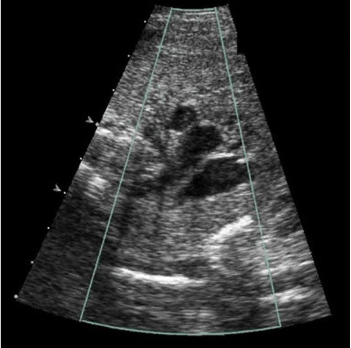
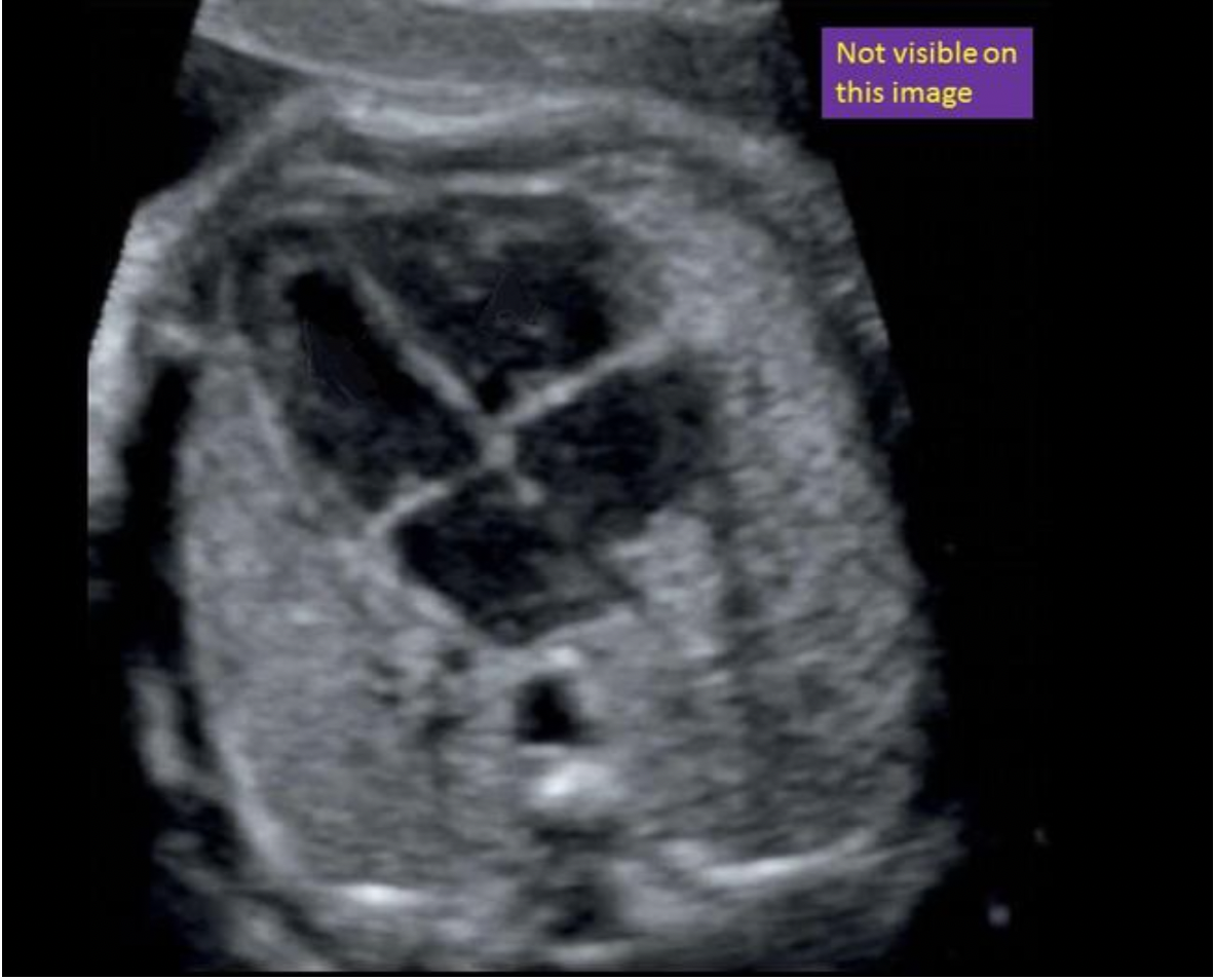
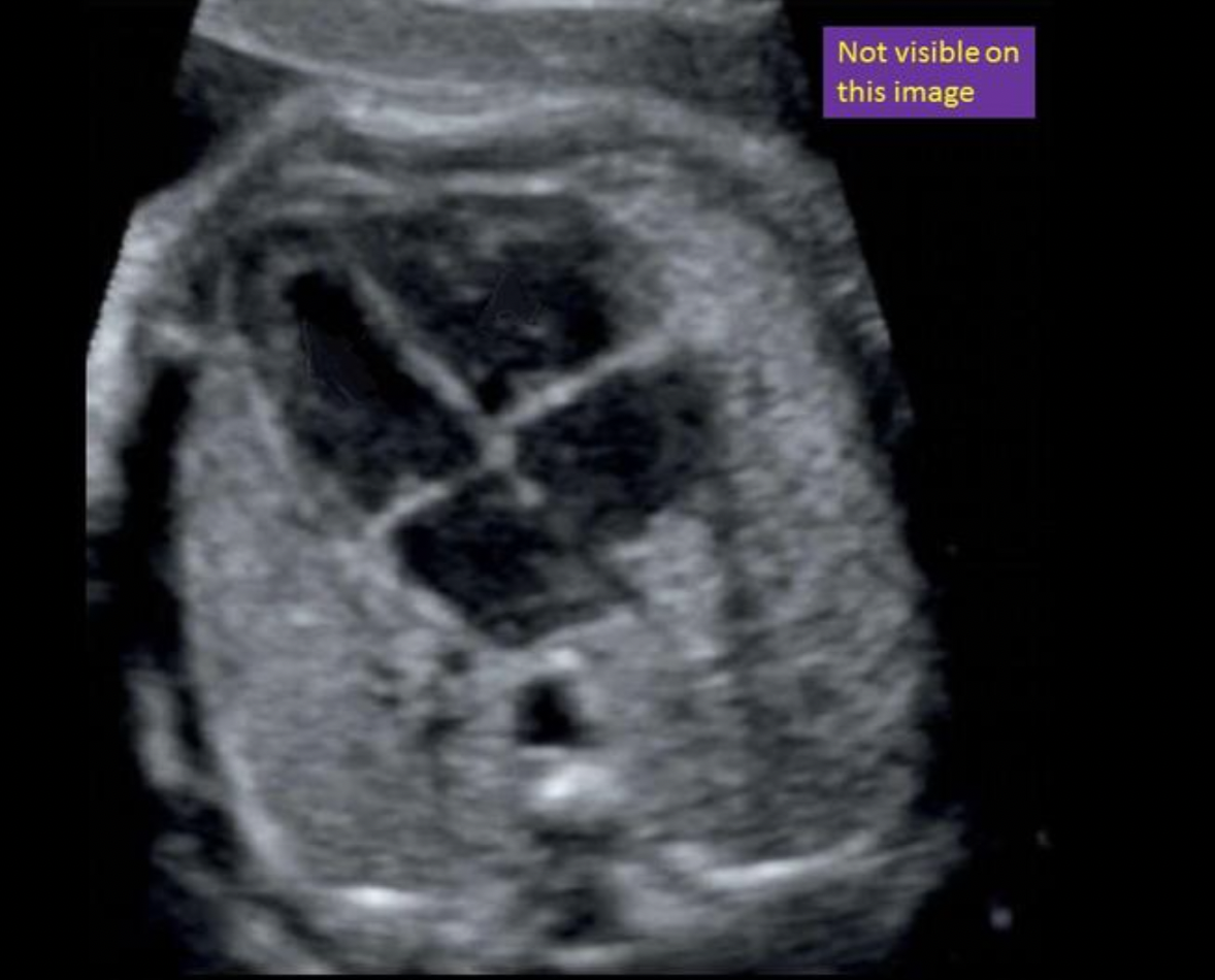
Locate the inferior vena cava. The structure may or may not be visible on this image
The IVC is not visible in this image. The inferior vena cava is located at a level inferior to the 4 chamber view. The IVC courses up the abdomen into the chest to attach to the inferior right atrium. It is best demonstrated in a sagittal view of the abdomen and chest.
What is the location of the placenta?
A. anterior
B. posterior
C. fundal
D. low lying
A. anterior
Note the placenta located on the anterior uterine wall
The proximal femoral epiphyseal plates should be visualized sonographically by what gestational age?
A. 13 wks
B. 18 wks
C. 30 wks
D. 35 wks
D. 35 wks
The proximal femoral epiphyseal plates can be visualized sonographically by 35wks and the distal epiphyseal plates by 33 weeks. Care should be taken not to include them in the femur length measurement.
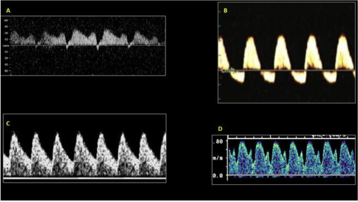
Which of the waveforms above is a normal ductus venosus waveform?
Image D
Image A is a Doppler evaluation of the ductus venosus that demonstrates increased diastolic resistance with atrial reversal.
This type of waveform indicates IUGR.
Image B is a Doppler evaluation of the umbilical artery that demonstrates high resistance flow with diastolic flow reversal. This type of waveform indicates IUGR.
Image C is a Doppler evaluation of the MCA that demonstrates low resistance flow with increased diastolic flow. This type of waveform indicates head sparing with IUGR.
Image D demonstrates Doppler evaluation of the ductus venosus that is normal
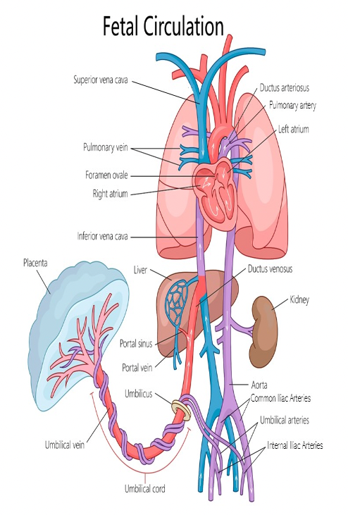
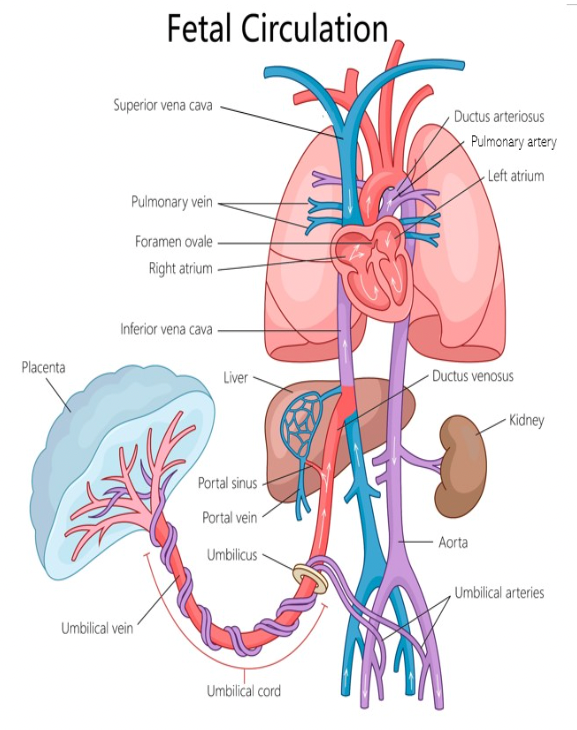
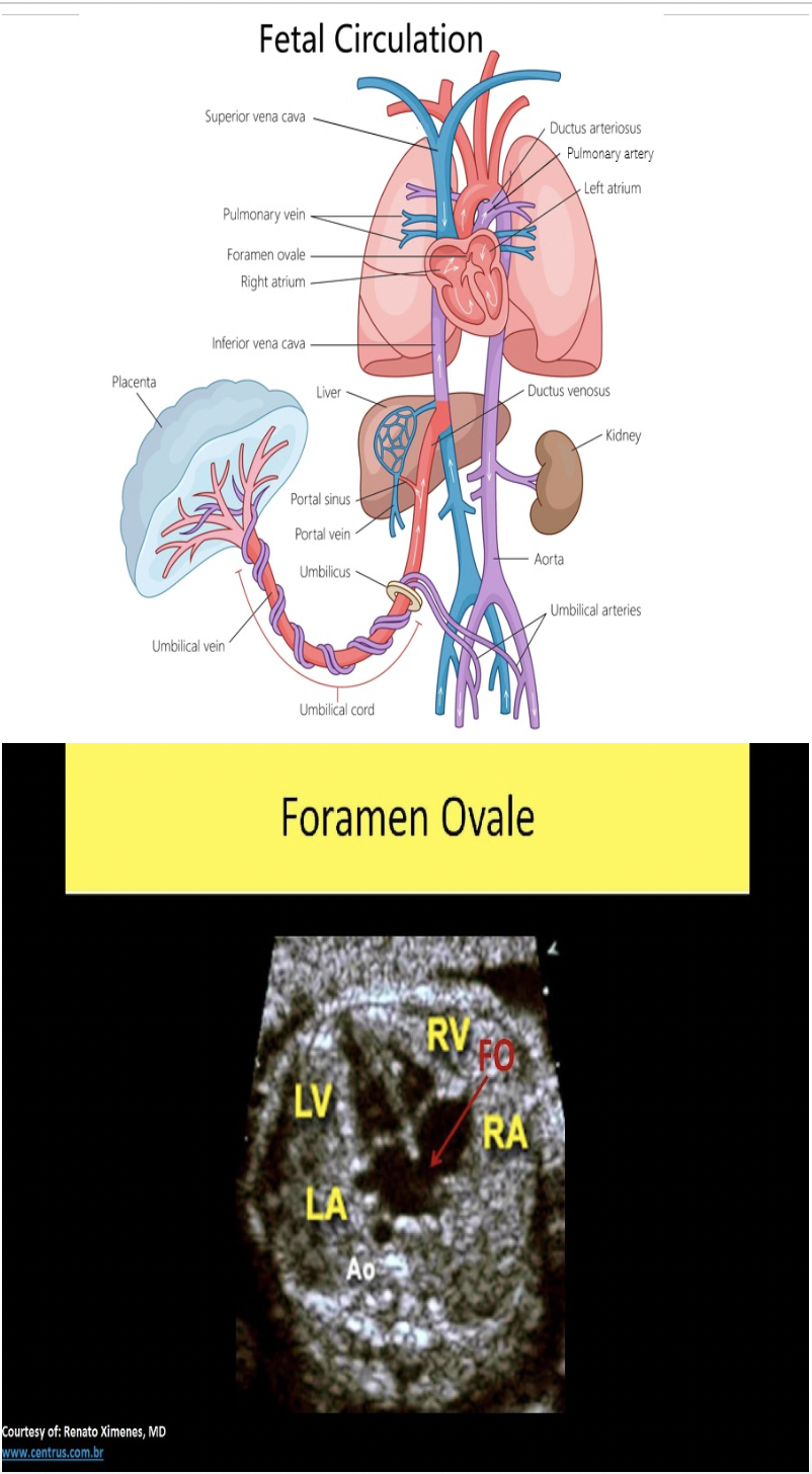
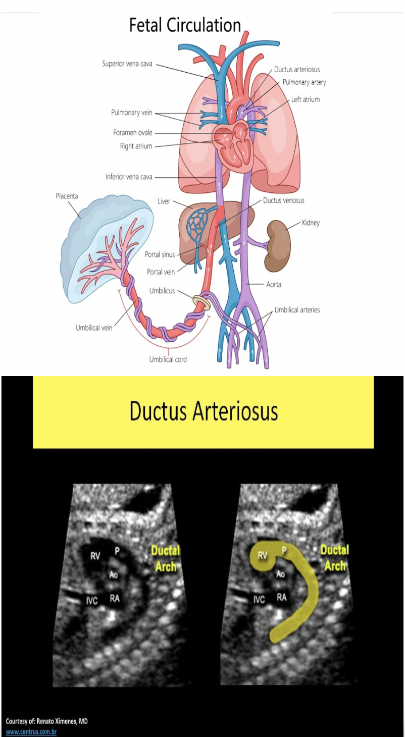
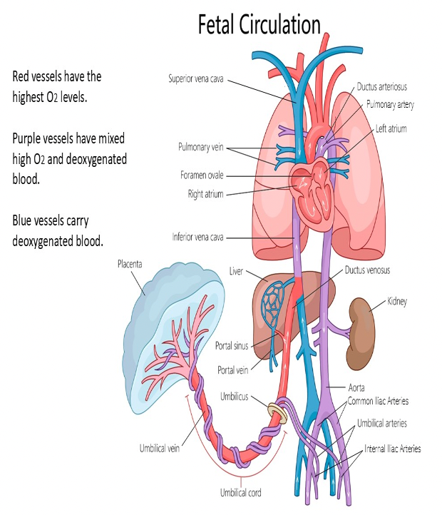
Which of the following terms describes the vessel that allows some umbilical blood to bypass the fetal liver in fetal circulation?
A. umbilical vein
B. ductus arteriosus
C. ductus venosus
D. foramen ovale
C. ductus venosus
The foramen ovale describes the opening in the interatrial septum that allows normal shunting between the atria. It functions to as a shunt for the fetal flow to bypass the fetal lungs. The foramen ovale normally closes very soon after birth. The ductus arteriosus describes the connecting vessel from the main pulmonary trunk to the descending aorta in fetal circulation. It functions as a shunt to bypass the fetal lungs. The ductus venosus describes the vessel that shunts blood flow around the fetal liver in fetal circulation. Both ductus connections also close shortly after birth.
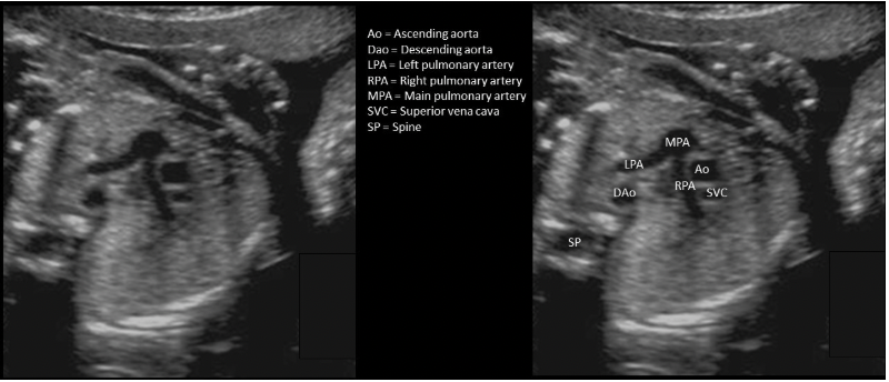
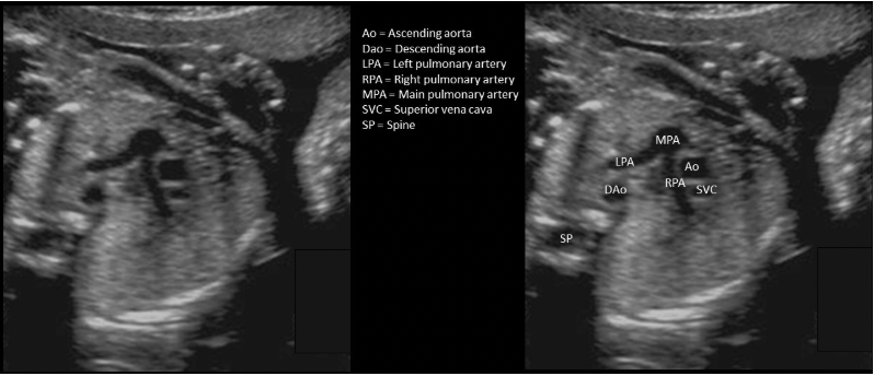
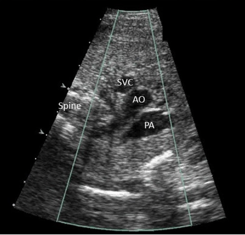
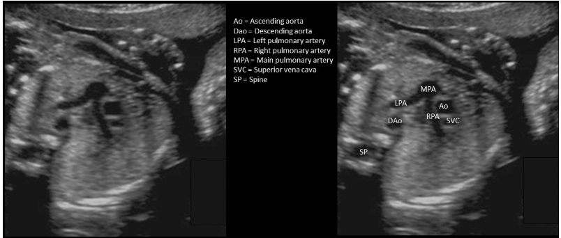
Click on the aorta on the three vessel view
Which of the following statements correctly describes the image displayed?
A. The image demonstrates a two vessel cord.
B. The image demonstrates a transverse view of the fetus at the level of the stomach.
C. The image demonstrates a normal cord with two arteries.
D. The image demonstrates the lumbar spine and pelvic bones
C. The image demonstrates a normal cord with two arteries.
The image demonstrates 2 arteries are present in the umbilical cord (3 vessel cord)
Average normal heart rate in a 2nd trimester fetus is
A. 100-140bpm
B. 160-200bpm
C. 80-120bpm
D. 120-160bpm
D. 120-160bpm
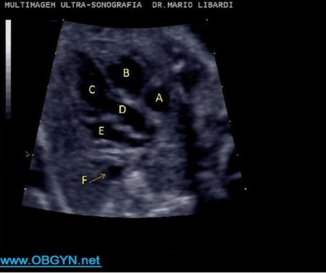
Letter A indicates which cardiac structure?
A. right ventricle
B. RVOT
C. left atrium
D. LVOT
D. right atrium
D. right atrium
right atrium
right ventricle
left ventricle
LVOT
left atrium
aorta
The normal direction of flow through the fetal foramen ovale is
A. umbilical vein to fetal IVC
B. left atrium to right atrium
C. pulmonary artery to aorta
D. right atrium to left atrium
D. right atrium to left atrium
The fetal lungs are not functional while in utero. This leads to increased resistance to incoming blood flow. In the fetus the right heart pressures exceed the left heart pressures causing flow thru the foramen ovale from right atrium to left atrium.
The normal fetal bladder should empty and refill every _____
A. 15-20 minutes
B. 30-45 minutes
C. 1-2 hours
D. 3-4 hours
B. 30-45 minutes
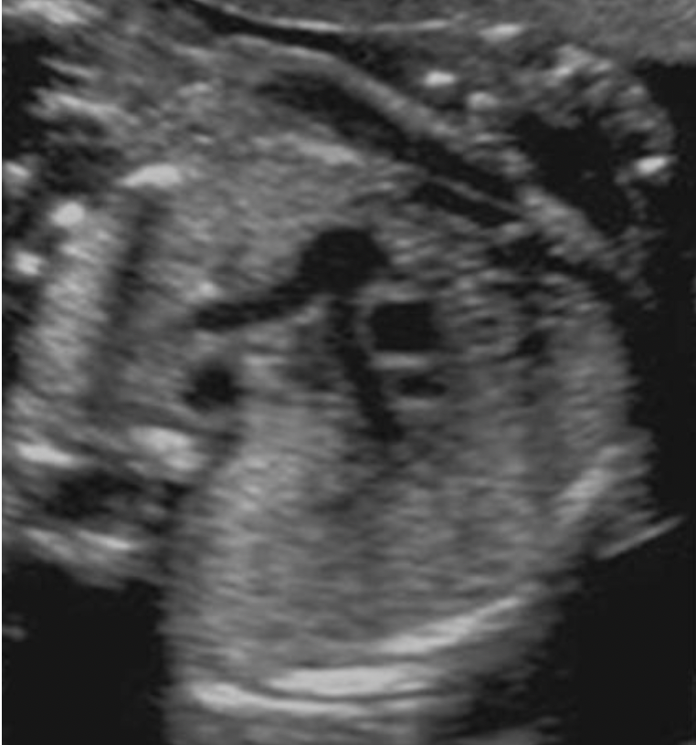
All of the following correctly describe a normal fetal spine, except:
A. tapering of the sacral spine
B. a skin dimple on the back marks the tip of the normal spinal cord
C. widening of the proximal spine
D. in the late 3rd trimester the spinal cord tip is at the level of L3
B. a skin dimple on the back marks the tip of the normal spinal cord
The neural tube usually closes by the 6th week of gestation. The vertebral column is formed by surrounding mesoderm tissues. The internal lumen of the tube forms the ventricles and central spinal canal. At 10 weeks the spinal cord is the same length as the central spinal canal. At 26 weeks the spinal cord tip should be at the level of S1. In the late 3rd trimester, the spinal cord tip is at the level of L3. The normal spine should demonstrate widening at the top of the cervical spine and tapering of the sacral spine. The skin normally covers the entire spine with a smooth contour. A skin dimple or small tuft of hair can indicate a spina bifida occulta.
What normal fetal structure produces alpha-fetoprotein?
A. liver
B. kidneys
C. pancreas
D. heart
A. liver
The fetal yolk sac and liver produce AFP that is released into the blood stream and dispersed into the maternal blood through the placenta.
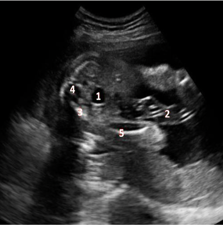
What fetal structure is indicated by #4?
A. sacral spine
B. cord
C. lumbar spine
D. pelvic bone
A. sacral spine
bladder
umbilical cord
pelvis
sacral spine
femur
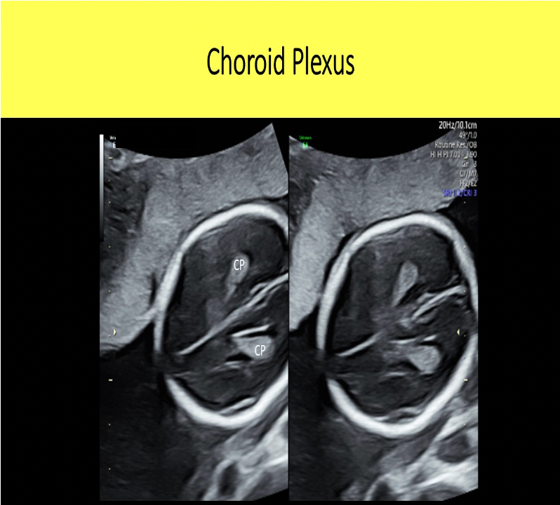
What is the function of the choroid plexus?
A. a choroid plexus is responsible for releasing multiple types of hormones that control fetal growth and metabolism.
B. The choroid plexus is responsible for production of cerebrospinal fluid.
C. The choroid plexus is responsible for releasing multiple types of hormones that control fetal lung maturation.
D. The choroid plexus is responsible for releasing multiple types of hormones that control fetal circulation and blood pressures.
B. The choroid plexus is responsible for production of cerebrospinal fluid.

The blue arrow indicates which structure?
A. aortic arch
B. main pulmonary artery
C. descending aorta
D. ascending aorta
C. descending aorta
What is the lateral bone of the forearm called?
A. fibula
B. humerus
C. radius
D. ulna
C. radius
When describing upper extremity anatomy it is assumed the patient is in anatomic position. The palms of the hands are turned forward which indicates the radius is on the lateral aspect of the arm.
Which type of breech position is defined as fetus with butt first with legs extended adjacent to fetal abdomen?
A. Complete
B. Frank
C. Partial
D. Footling
B. Frank
Frank breech = butt down legs straight up in front of abdomen
Complete breech = butt down legs folded, knees bent, ankles crossed
Footling breech = feet down, baby "standing" on cervix
Which cardiac chamber is normally located most anterior in the fetus?
A. right atrium
B. left atrium
C. right ventricle
D. left ventricle
C. right ventricle
The right ventricle is the most anterior chamber of the heart. The left atrium is the most posterior chamber of the heart.
Which normal cranial structures are identified within the lateral ventricles bilaterally?
A. cerebellar pedicles
B. lobes of the thalamus
C. choroid plexus
D. cavum septum pellucidum
C. choroid plexus
The lateral ventricles normally contain the choroid plexus.
#5 represents what fetal structure?
A. hydronephrosis
B. lung tissue
C. urinary bladder
D. stomach
C. urinary bladder
Heart
Lung
Diaphragm
Stomach
Bladder
The most common type of twins is ____
A. dizygotic
B. monozygotic
C. identical
D. monoamniotic
A. dizygotic
Dizygotic or fraternal twins are the most commonly occurring type of twins, more common in African Americans than Caucasians and more common in older women (>37yrs).
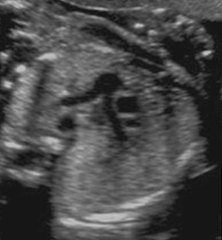
Locate the descending aorta. The structure may or may not be visible in the image

The ductus arteriosus is:
A. an opening between the right and left atrium to shunt flow away from the fetal lungs
B. a vessel that connects the umbilical vein to the IVC to allow flow to bypass the liver
C. a vessel that connects the pulmonary artery to the aorta to allow flow to bypass the fetal lungs
D. a vessel that connects the umbilical artery to the aorta to allow flow to bypass the fetal lungs
C. a vessel that connects the pulmonary artery to the aorta to allow flow to bypass the fetal lungs
The ductus arteriosus is a vessel that connects the pulmonary artery to the aorta to allow most flow to bypass the fetal lungs.

#2 represents what fetal structure?
A. diaphragm
B. lung tissue
C. liver
D. thymus
B. lung tissue
Heart
Lung
Diaphragm
Stomach
Bladder
In normal pregnancies, fetal age derived from biometric measurements should be within _____ of the average fetal parameter age for that measurement.
A. 10%
B. 15%
C. 5%
D. 7.5%
A. 10%
#4 represents what fetal structure?
A. urinary bladder
B. stomach
C. lung tissue
D. hydronephrosis
B. stomach
Heart
Lung
Diaphragm
Stomach
Bladder
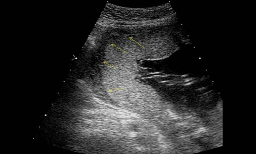
What are the arrows pointing to on the image?
A. hematoma formation within a placental abruption
B. placental lake formation posterior to the placenta
C. normal retroplacental space
D. amnion and chorion junction
C. normal retroplacental space
The image demonstrates a normal placenta and retroplacental space
The _____ empties blood into the right atrium and, while the _____ empties blood into the left atrium.
A. pulmonary veins, inferior vena cava
B. inferior vena cava, pulmonary veins
C. inferior vena cava, superior vena cava
D. tricuspid valve, mitral valve
B. inferior vena cava, pulmonary veins
The IVC and SVC empty blood into the right atrium, while the pulmonary veins empty blood into the left atrium.
The purple arrow indicates which structure?
A. main pulmonary artery
B. ascending aorta
C. aortic arch
D. desce
C. aortic arch
Which of the following fetal structures is not subject to changes in shape due to molding?
A. femur length
B. abdomen circumference
C. biparietal diameter
D. head circumference
A. femur length
Molding occurs when external uterine pressure causes changes to the shape of a fetal structure. The fetal cranium and abdomen are most susceptible to these changes, while the long bones are not affected. Oligohydramnios, premature rupture of membranes and low fetal position are all causes of molding.
During renal development, the kidneys normally migrate _____
A. from their origination point centrally in the abdomen to a more lateral location adjacent to the ribs.
B. from the pelvic region superiorly into the lower abdomen.
C. from the lower right side of the spine superiorly and separate once the fetus reaches 11 wks.
D. from the upper abdomen into an inferior retroperitoneal location.
B. from the pelvic region superiorly into the lower abdomen.
During renal development, the kidneys normally migrate from the pelvic region superiorly into the lower abdomen. A pelvic kidney occurs when the kidney fails to migrate superior/posterior into the proper location.
Which hormone stimulates the formation of the cervical mucous plug found with pregnancy?
A. Oxytocin
B. bhCG
C. Progesterone
D. Estrogen
C. Progesterone
Estrogen stimulates the cervix to produce mucous just prior to ovulation. Increasing progesterone levels with pregnancy cause the cervical mucous to "dry up" and the cervix to tighten leading to mucous plug formation.
Which of the following structures is considered the most reliable for determining fetal situs?
A. stomach and heart
B. liver and gall bladder
C. aorta and IVC
D. stomach and liver
C. aorta and IVC
Fetal situs: Document the position of the aorta and IVC to determine left/right side of the fetus - more reliable than noting stomach position. Other structures identified to assist in documenting fetal situs: stomach, liver, spleen, portal sinus, umbilical vein, gallbladder
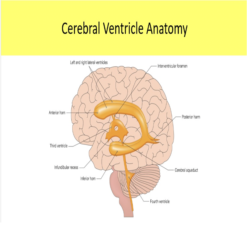
The aqueduct of Sylvius connects the _____ in the brain.
A. lateral ventricles and 4th ventricle
B. 3rd and 4th ventricles
C. 4th ventricle and cisterna magna
D. 3rd ventricle and lateral ventricles
B. 3rd and 4th ventricles
Letter F indicates which cardiac structure?
A. right atrium
B. aorta
C. LVOT
D. coronary sinus
E. left atrium
B. aorta
right atrium
right ventricle
left ventricle
LVOT
left atrium
aorta
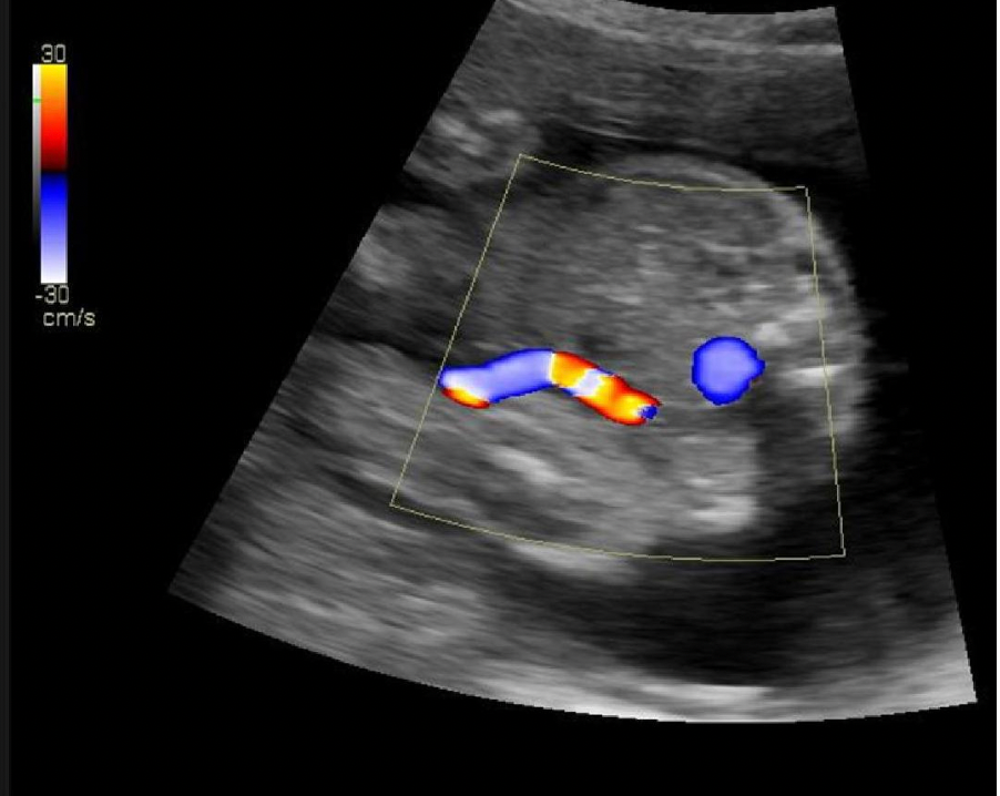
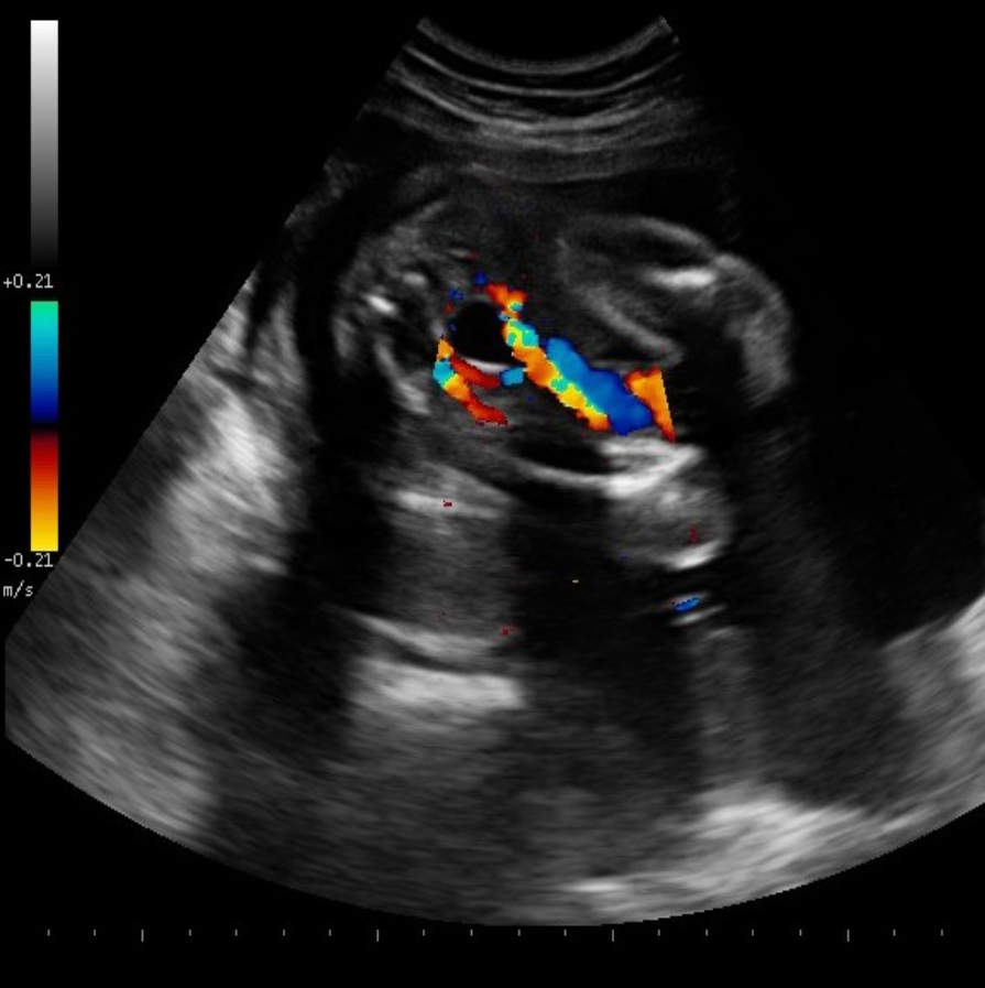
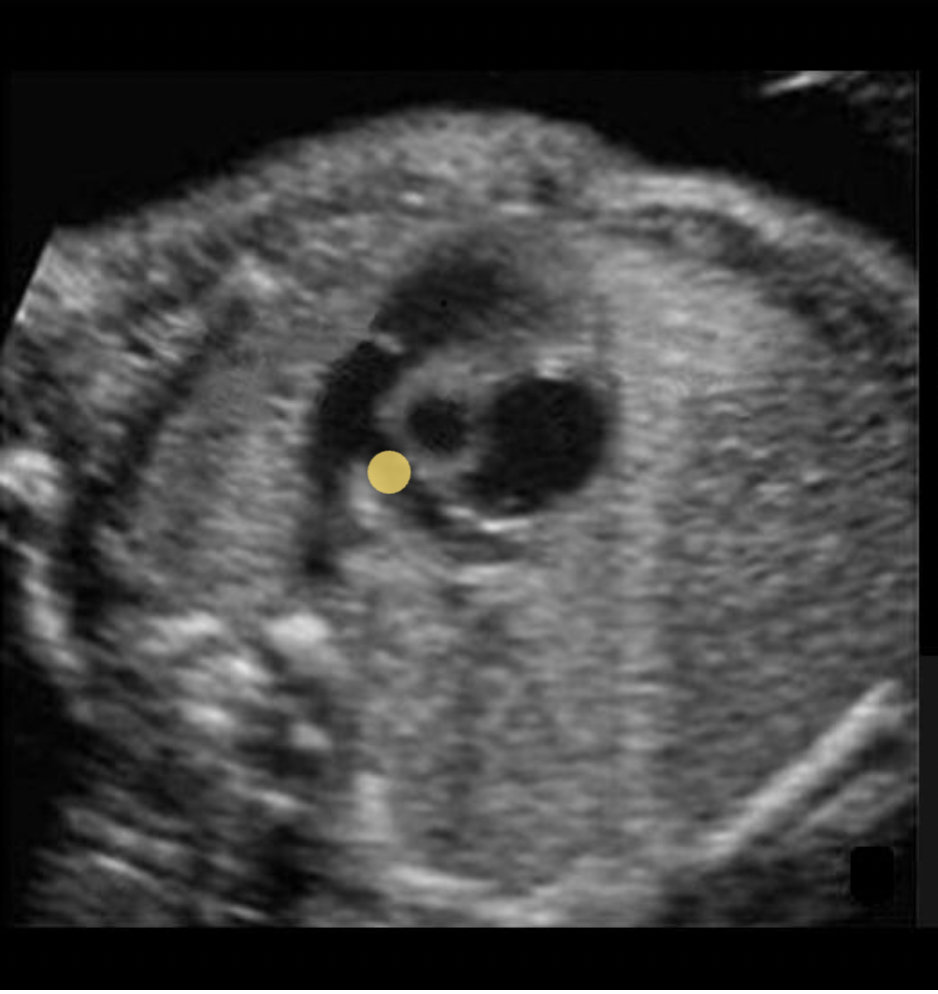
What is represented by the blue circle of color Doppler displayed on the image?
A. umbilical varix
B. inferior vena cava
C. umbilical artery
D. aorta
D. aorta
Note the liver and spine position on the image. The blue circle is to the left of the spine which indicates the aorta.
Locate the superior vena cava on the three vessel view
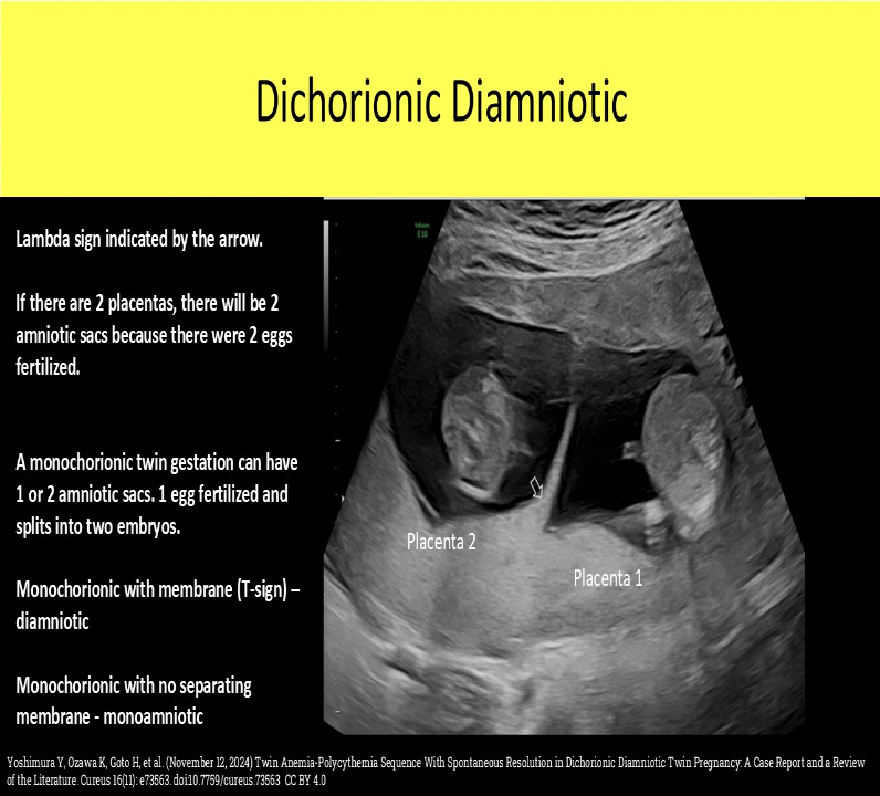
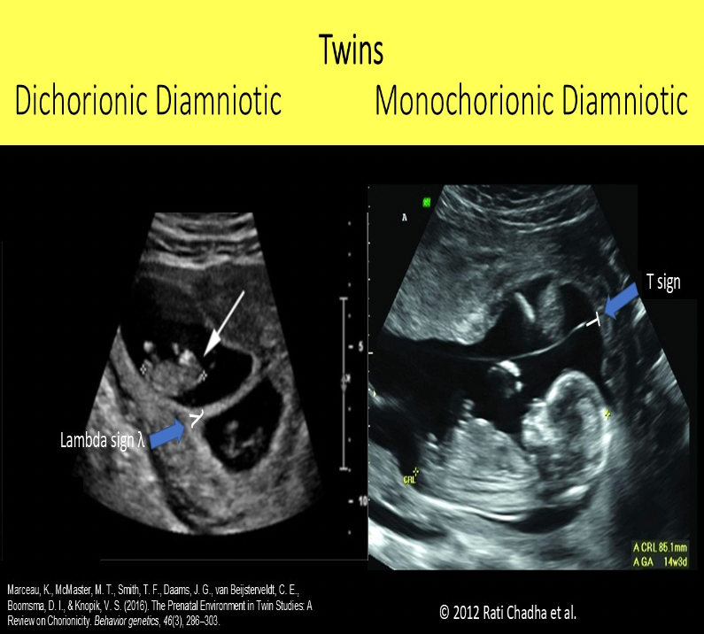
The lambda sign indicates indicates _____, while the T sign indicates _____
A. monochorionic monoamniotic twins, conjoined twins
B. dichorionic diamniotic twins, monochorionic diamniotic twins
C. dichorionic diamniotic twins, monochorionic monoamniotic twins
D. monochorionic diamniotic twins, dichorionic diamniotic twins
B. dichorionic diamniotic twins, monochorionic diamniotic twins
The lambda sign indicates dichorionic diamniotic twins. The thick membrane has a triangle of placental tissue at the base of the membrane. The T sign indicates monochorionic diamniotic twins. The membrane attaches to the uterine wall without placental tissue at the base.
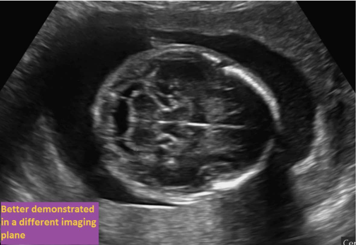
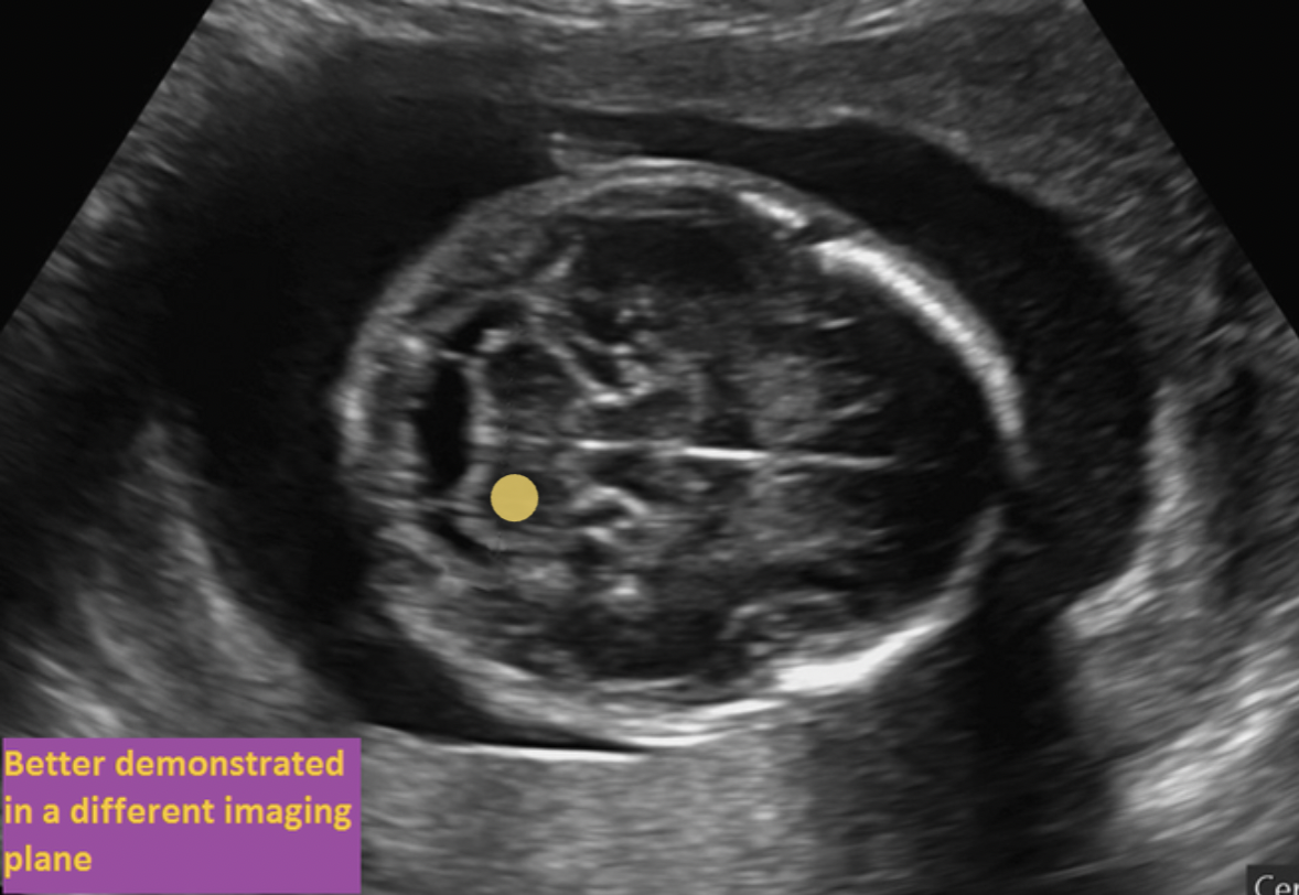
Where is the choroid plexus located on the image? If the structure is normally evaluated in a different imaging plane, select the purple box
The choroid plexus are located within the lateral ventricles of the brain. They would be visible in an axial plane that is superior to the current one.
Which cardiac view of the fetal heart is demonstrated on the image?
A. RVOT
B. Ductus arteriosus
C. LVOT
D. Ductus venosus
C. LVOT
Note the echogenic walls of the LVOT/aortic root extending superiorly in the chest and the adjacent mitral valve seen inferiorly to the proximal LVOT.
The normal heart occupies about ______ of the fetal chest
A. 60%
B. 33%
C. 25%
D. 55%
B. 33%
The normal heart occupies about 1/3 of the fetal chest

Letter D indicates which cardiac structure?
A. RVOT
B. left ventricle
C. right ventricle
D. LVOT
E. right atrium
D. LVOT
right atrium
right ventricle
left ventricle
LVOT
left atrium
aorta
Fetal stool is referred to as referred to as _____, while fetal urine is _____
A. meconium, amniotic fluid
B. Wharton jelly, amniotic fluid
C. vernix, meconium
D. meconium, vernix
A. meconium, amniotic fluid
Fetal stool is referred to meconium, while fetal urine is referred to as amniotic fluid. Vernix is the white particles floating in the amniotic fluid later in pregnancy (fetal hair and skin cells). Wharton jelly is inside the umbilical cord.

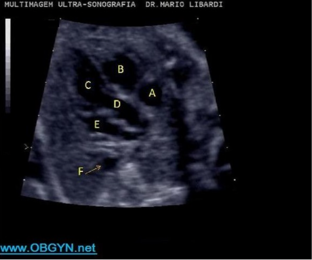
Letter B indicates which cardiac structure?
A. right atrium
B. left ventricle
C. right ventricle
D. left atrium
D. left atrium
ascending aorta
left atrium
left ventricle
The ratio of kidney diameter to the abdominal diameter should stay between ______ for the entire pregnancy
A. 0.3 - 0.4
B. 0.5 - 0.57
C. 0.33 - 0.44
D. 0.23 - 0.27
D. 0.23 - 0.27
The ratio of kidney diameter to the abdominal diameter should stay between 0.23 - 0.27 for the entire pregnancy. An increased kidney diameter will lead to an abnormally increased ratio. Mass formation or cystic disease are potential causes for increased kidney size.
What is the functional unit of the placenta?
A. lobule
B. lobe
C. cotyledon
D. nephron
C. cotyledon
The placenta has 15-30 cotyledons. They are functional lobules of placental tissue separated by septations of maternal tissue (basal layer).

What fetal structure is indicated by #1?
A. cord
B. bladder
C. lumbar spine
D. sacral spine
B. bladder
bladder
umbilical cord
pelvis
sacral spine
femur
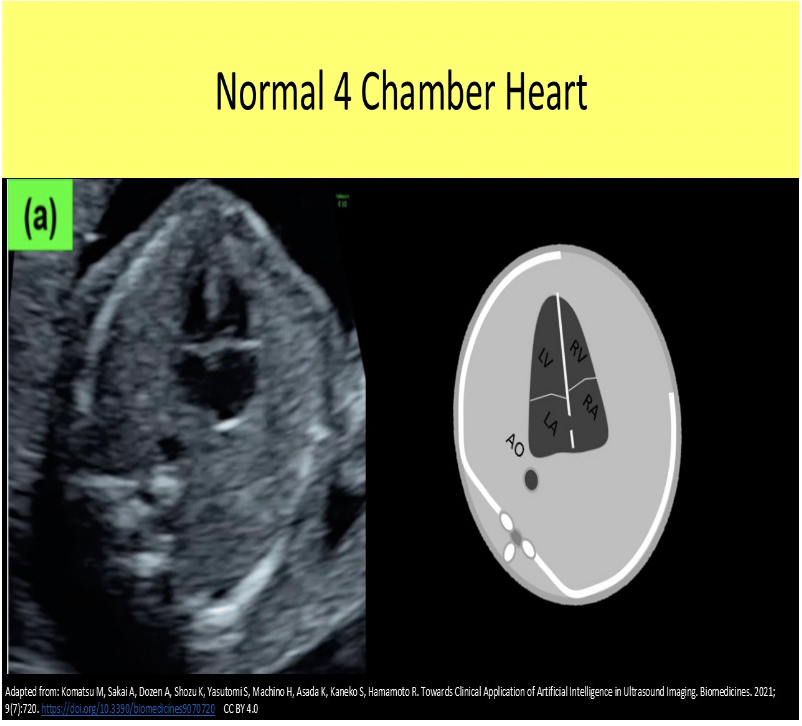
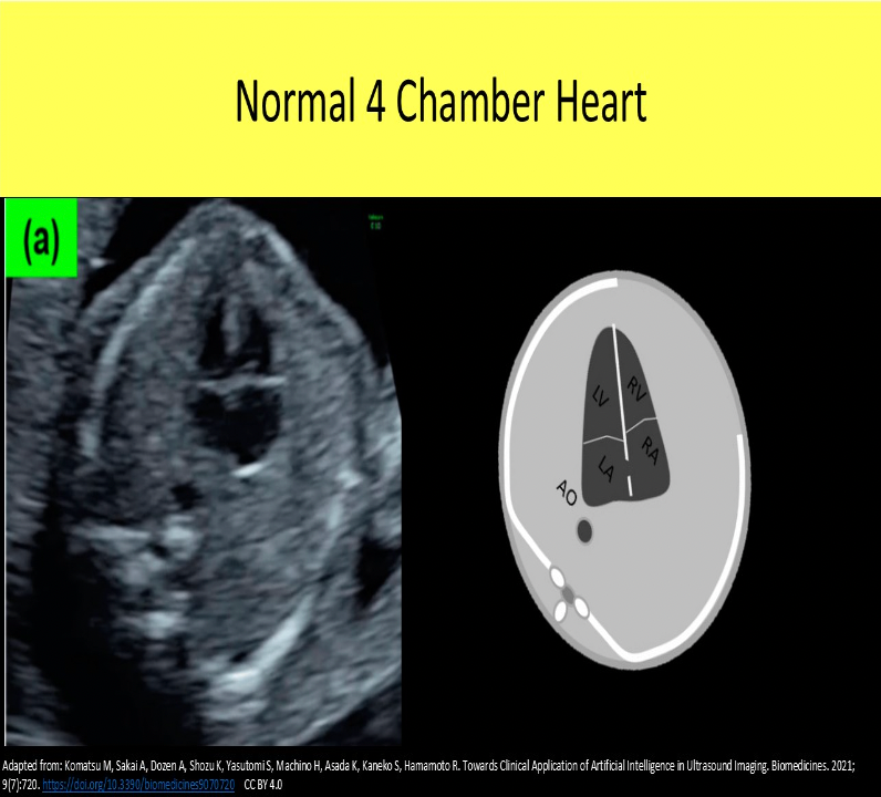
The optimal 4 chamber view of the fetal heart should include:
A. At least one rib in profile of the left side of the chest
B. Cross section of two ribs on both sides of the chest
C. A single rib in profile on both sides the of the chest
D. Cartilage between the ribs, with no bony shadowing present on both sides of the chest
C. A single rib in profile on both sides the of the chest
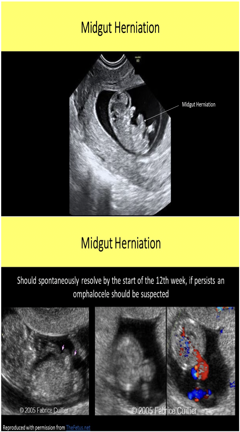
Peristalsis of the fetal bowel should normally be identified
A. in the late 2nd trimester
B. after 35 weeks gestation
C. before the midgut herniation retracts
D. just after midgut herniation retracts
A. in the late 2nd trimester
Where is Wharton jelly found?
A. fossa of Waldeyer
B. cervical mucous plug
C. umbilical cord
D. amniotic fluid
C. umbilical cord
The umbilical cord is composed of 2 arteries and 1 vein surrounded by Wharton jelly and covered by amnion.
Which of the following terms describes the opening in the interatrial septum that allows normal shunting between the atria?
A. patent ductus arteriosus
B. foramen ovale
C. muscular septal defect
D. patent ductus venosus
B. foramen ovale
The foramen ovale normally closes very soon after birth. The ductus arteriosus describes the connecting vessel from the main pulmonary trunk to the descending aorta in fetal circulation. It functions as a shunt to bypass the fetal lungs. The ductus venosus describes the vessel that shunts blood flow around the fetal liver in fetal circulation. Both ductus connections also close shortly after birth.
Herniation of the midgut is an abnormal finding in gestations over _____ in age.
A. 8 wks
B. 10wks
C. 12wks
D. 14wks
C. 12wks
Herniation of the midgut begins at week 8 and retraction occurs by week 12 in a normal gestation.
If the fetus is in breech position, where is the left lobe of the thalamus located. If the structure is normally evaluated in a different imaging plane, select the purple box
The fetus is in breech position and facing the maternal left side. The fetal left side is posterior and the fetal right side is anterior on the image. The thalamus has two lobes and appears as a butterfly shaped structure in the central cerebral tissue. The falx cerebri separates the two lobes.


What fetal structure is indicated by #2?
A. cisterna magna
B. nuchal skin fold
C. caudate nucleus
D. cerebellum
A. cisterna magna
cerebellum
cisterna magna
thalamus
On a fetal ultrasound, the LVOT refers to the refers to the _____ and the RVOT ______
A. aortic valve, pulmonic valve
B. descending aorta, peripheral pulmonary arteries
C. ascending aorta, pulmonary artery
D. aortic arch, pulmonary vein
C. ascending aorta, pulmonary artery
On a fetal ultrasound the LVOT refers to the ascending aorta and the RVOT refers to the pulmonary artery. In pediatric and adult echocardiography, the LVOT refers to the portion of the left ventricle just proximal to the aortic valve and the RVOT refers to the portion of the right ventricle just proximal to the pulmonary valve.
Which type of breech position is defined as fetus with one or both feet in the lower uterine segment?
A. Footling
B. Frank
C. Complete
D. Partial
A. Footling
Frank breech = butt down legs straight up in front of abdomen
Complete breech = butt down legs folded, knees bent, ankles crossed
Footling breech = feet down, baby "standing" on cervix
Advanced maternal age refers to a pregnant woman who is greater than _____ of age.
A. 45yrs
B. 40yrs
C. 35yrs
D. 33yrs
C. 35yrs
Advanced maternal age refers to a pregnant woman who is greater than 35yrs of age. The risk of chromosomal abnormalities and other fetal defects increases significantly when the mother is over 35yrs.

If the fetus is in breech position, where is the left lobe of the cerebellum located? If the structure is normally evaluated in a different imaging plane, select the purple box.
The fetus is in breech position and facing the maternal left side. The fetal right side is anterior and the fetal left side is posterior on the image. The cerebellum has two lobes and appears as a dumbbell shaped structure in the posterior cranium.
The normal anterior-posterior renal pelvic diameter (APRPD) in a second trimester fetus is _____ and in a 3rd trimester fetus ______ is considered normal.
A. less than 2mm, less than 4mm
B. less than 8mm, less than 16mm
C. less than 4mm, less than 7mm
D. less than 10mm, less than 20mm
C. less than 4mm, less than 7mm
The normal anterior-posterior renal pelvic diameter (APRPD) in a second trimester fetus is less than 4mm and in a 3rd trimester fetus less than 7mm is considered normal pyelectasis. Severe hydronephrosis is indicated in the 2nd trimester with an APRPD of more than 10mm. Severe hydronephrosis is indicated in the 3rd trimester with an APRPD of more than 15mm. An increase in the APRPD between the second and third trimesters indicates higher possibility of postnatal abnormalities.
Which of the following terms describes the connecting vessel from the main pulmonary trunk to the descending aorta found in fetal circulation?
A. foramen ovale
B. left atrial appendage
C. ductus venosus
D. ductus arteriosus
D. ductus arteriosus
The foramen ovale describes the opening in the interatrial septum that allows normal shunting between the atria. It functions to as a shunt for the fetal flow to bypass the fetal lungs. The foramen ovale normally closes very soon after birth. The ductus arteriosus describes the connecting vessel from the main pulmonary trunk to the descending aorta in fetal circulation. It functions as a shunt to bypass the fetal lungs. The ductus venosus describes the vessel that shunts blood flow around the fetal liver in fetal circulation. Both ductus connections also close shortly after birth.
Locate the foramen ovale. The structure may or many not be visible in the image

Locate the ascending aorta
The level of amniotic fluid continues to rise throughout pregnancy until approximately weeks in gestational age where it will plateau then decline until birth.
A. 16
B. 20
C. 28
D. 33
D. 33
The level of amniotic fluid continues to rise throughout pregnancy until approximately 33 weeks in gestational age. First it will plateau, then a mild decline occurs a few weeks before birth.
Locate the right pulmonary artery
The fetal heart normally sits at a _____ degree angle in the chest with the apex pointed toward the left anterior chest wall.
A. 30
B. 45
C. 60
D. 90
B. 45
At what gestational age does the fetus completely take over the amniotic fluid production?
A. 24 wks
B. 12 wks
C. 20 wks
D. 16 wks
D. 16 wks
Up until 16wks gestation, the chorion, amnion and placenta produce the amniotic fluid. After 16 weeks, fetus takes over production of fluid.
In a small amount of adults, Meckel's diverticulum can be identified as a remnant of fetal embryology. What causes this anatomic variation?
A. the yolk sac persists as a diverticulum in the ileum portion of the bowel
B. the placenta persists as a diverticulum in the ileum portion of the bowel
C. the gestational sac persists as a diverticulum in the ileum portion of the bowel
D. the placental umbilical cord attachment persists as a diverticulum in the ileum portion of the bowel
A. the yolk sac persists as a diverticulum in the ileum portion of the bowel
Meckel's diverticulum refers to when the yolk sac persists into adult life as a diverticulum in the ileum portion of the bowel.
Locate the ascending aorta. The structure may or may not be visible in the image
It is not demonstrated in this image. The ascending aorta is demonstrated in the left ventricular outflow tract view and the aortic arch view. The descending aorta is seen here posterior to the left atrium.
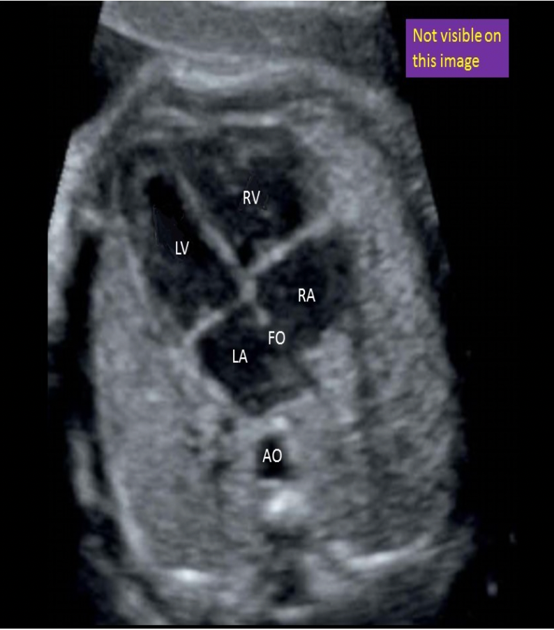
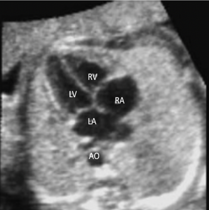
In a normal 4 chamber heart view, what cardiac chamber is closest to the spine?
A. left ventricle
B. left atrium
C. right ventricle
D. right atrium
B. left atrium
The left atrium is the most posterior cardiac chamber and the right ventricle is the most anterior cardiac chamber in the normal heart.
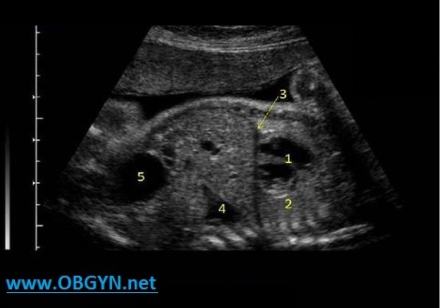
What fetal structure is indicated by #1?
A. gallbladder
B. rib
C. stomach
D. liver
C. stomach
stomach
thoracic spine
umbilical vein
liver
rib
aorta
IVC
The first cardiac chamber to receive oxygenated blood from the placenta is:
A. left ventricle
B. right atrium
C. right ventricle
D, left atrium
B. right atrium
Oxygenated blood from the placenta enters the umbilical vein, then the left portal vein or ductus venosus, then the IVC and finally into the right atrium. The highest concentration of oxygen in the heart is in the right atrium.
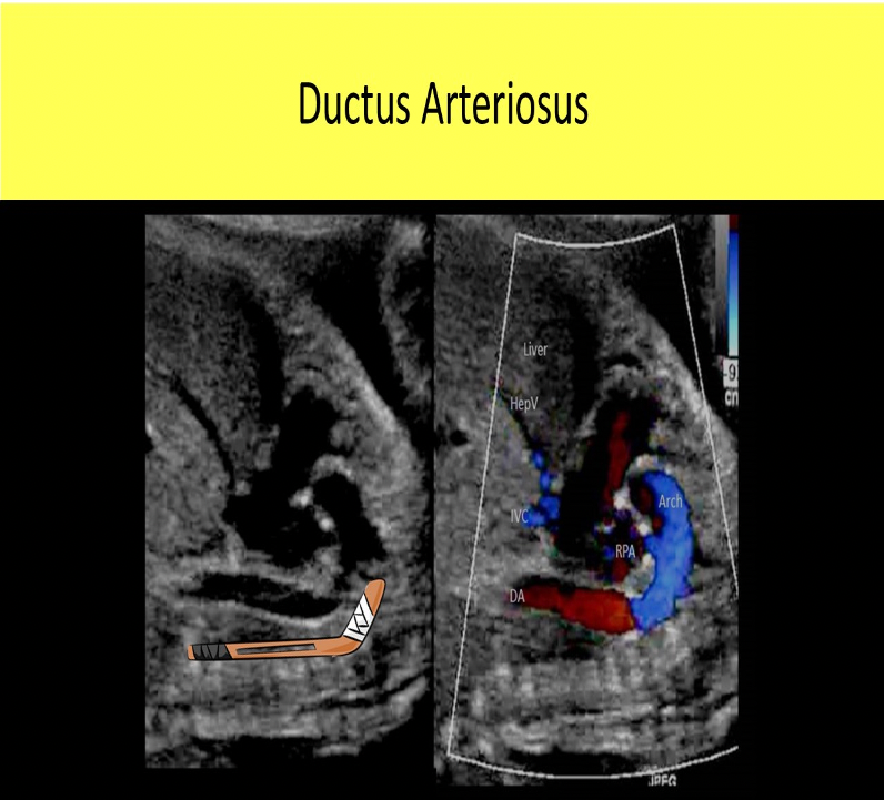
What cardiac structure is described as having a hockey stick appearance on sagittal views of the chest?
A. ductus venosus
B. main pulmonary artery
C. ductus arteriosus
D. aortic arch
C. ductus arteriosus
The ductus arteriosus is described as having a hockey stick appearance on sagittal views of the chest. The aortic arch is described as having a candy cane appearance in sagittal views. The ductus venosus in located in the abdomen.
The left atrium receives blood from what vessel(s)?
A. pulmonary veins
B. pulmonary arteries
C. IVC
D. ductus arteriosus
A. pulmonary veins
The right ventricle sends deoxygenated blood to the lungs through the pulmonary artery. The oxygenated blood leaves the lungs through the right and left pulmonary veins to enter the left atrium. There are usually two sets of pulmonary veins on each side: right upper, right lower, left upper, left lower.

What fetal structure is indicated by #5?
A. left lung
B. diaphragm
C. rib
D. right lung
C. rib
stomach
thoracic spine
umbilical vein
liver
rib
aorta
VC
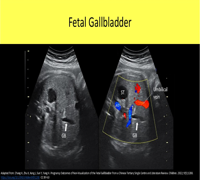
Which of the following correctly describes how to differentiate the umbilical vein and the gallbladder in a fetus?
A. The gallbladder is normally parallel to the abdominal wall while the umbilical vein is perpendicular to the abdominal wall.
B. The umbilical vein is normally parallel to the abdominal wall while the gallbladder is perpendicular to the abdominal wall.
C. Color Doppler will demonstrate flow filling the lumen of the portal vein but flow will only be demonstrated within the walls of the gallbladder.
D. The umbilical vein is located centrally in the abdomen while the gallbladder is located to the right between the right and left lobes of the liver.
D. The umbilical vein is located centrally in the abdomen while the gallbladder is located to the right between the right and left lobes of the liver.
The umbilical vein and the gallbladder are usually oriented perpendicular to the abdominal wall. Color Doppler can be used to differentiate the umbilical and liver vasculature from the gallbladder. No color flow will fill the lumen (or the walls) of the gallbladder. The umbilical vein is located centrally in the abdomen while the gallbladder is located to the right between the right and left lobes of the liver.
The post-partum period usually lasts ____
A. 2-4wks
B. 4-6wks
C. 6-8wks
D. up to 12 wks
C. 6-8wks
Letter B indicates which cardiac structure?
A. right ventricle
B. right atrium
C. left ventricle
D. LVOT
E. RVOT
A. right ventricle
right atrium
right ventricle
left ventricle
LVOT
left atrium
aorta
If two fetuses are different genders, this indicates:
A. monoamniotic twins
B. dizygous twins
C. monozygous twins
D. identical twins
B. dizygous twins
Fetuses of the same gender can be dizygous or monozygous.
Fetuses of different gender must always be dizygous.