A&P I Bones (no skull) Lectures
1/22
There's no tags or description
Looks like no tags are added yet.
Name | Mastery | Learn | Test | Matching | Spaced | Call with Kai |
|---|
No analytics yet
Send a link to your students to track their progress
23 Terms
fetal skull
plates haven’t fully sealed together, sutures haven’t joined the main bones. The gaps where multiple sutures intersect are called fontanelles. There is an anterior fontanelle that is diamond-shaped, situated at the junction of the sagittal, coronal, and frontal sutures. The posterior fontanelle is triangular, located where the sagittal and lambdoid sutures meet.
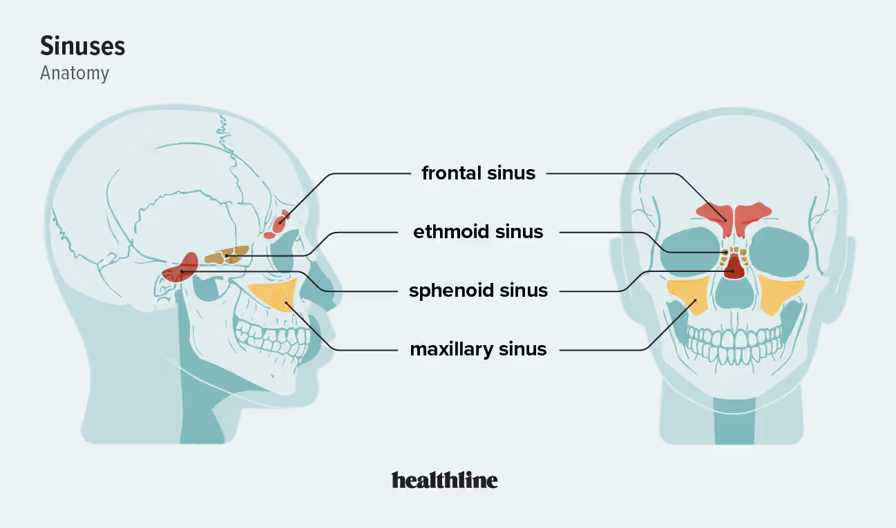
4 paranasal sinuses
maxillary, frontal, sphenoid, ethmoid
number of vertebrae
7 cervical vertebrae (c1-c7) , 12 thoracic vertebrae (t1-t12) , and 5 lumbar vertebrae (l1-l5).
pathology to vertebrae
natural curves to the cervical and lumbar. if exaggerated, condition called hyperlordosis. if there is a lateral curve, condition called scoliosis.
if there is a misalignment of vertebrae that pushes on the nerves on each side, will have muscle pain.
intervertebral discs netweek vertebrae. a herniated disc occurs when swelling on one side pushes on the nerves. a slipped disc occurs when a disc moved out of alignment. can try to use traction to put it back, or need surgery.
cartilage thins with age. stenosis occurs when vertebrae start to fuse together. this often occurs in the neck or low back.
single vertebrae anatomy
spinous process goes to the back. transverse processes to the sides.
if the vertebrae is cervical:
-there is a transverse foramen in the transverse process.
-C1/atlas has no spinous process.
-C2/axis has a thumblike projection called the dens.
thoracic:
-all look like a giraffe. the spinous process lays down.
lumbar:
-spinous process sticks out. looks like a moose.
C1-T12 (roughly) all have a vertebral foramen for the spinal cord to fit into. not so much lumbar.
lumbar puncture
puncture into the epidural space of the meninges of the spinal cord to remove cerebrospinal fluid to test for meningitis or encephalitis. this is invasive. if they flinch you could hit the spinal cord, so it is less common now with other methods.
sacrum
a large, triangular bone at the base of the spine that forms the solid posterior wall of the pelvis. acts as a keystone, supporting the upper body's weight and evenly transferring it through the pelvis to the legs. Formed by the fusion of five individual sacral vertebrae. It connects to the spine via the L5 vertebra, the tailbone via the coccyx, and the hips. Features four sets of holes (foramina) that allow nerves and blood vessels to pass through to the pelvic organs. It is curved/concave on its anterior surface to create room for the pelvic cavity.
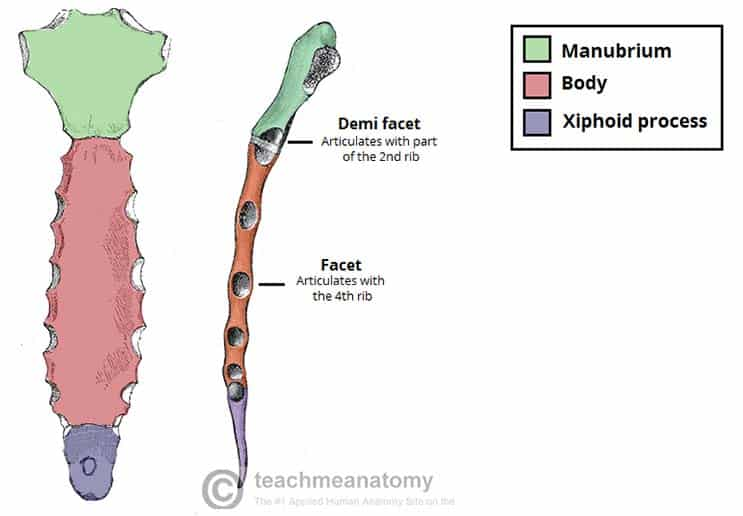
sternum
(breastbone). easily fractures in CPR (cardio-pulmonary resuscitation). a flat, T-shaped bone in the center of the chest that protects your heart and anchors your ribs.
three main parts: the manubrium, the body, and the xiphoid process.
1. Manubrium
- The wide, handle-like superior portion of the sternum.
- Features the jugular notch at the top center. where you do a tracheotomy. It contains facets that connect to your collarbones (clavicles) and the first and second sets of ribs.
2. Body
- The long, flat central section, also called the "blade" of the sternum.
-The sternal angle is the ridge where the manubrium and body meet. The body's sides attach to your third through seventh sets of ribs.
3. Xiphoid Process
-The small, pointed inferior (bottom) tip of the sternum.
-acts as an anchor point for major abdominal muscles
vertebrosternal ribs
attached to the sternum and manubrium. tight bumps on the back. inferior area of bones are pointy, superior is smooth!!!
commonly known as true ribs. the first seven pairs of ribs in the human body. They are named for their direct connections to both the vertebrae and the sternum via their own individual strips of hyaline cartilage, known as costal cartilage.
all connect to thoracic vertebrae posteriorly. connect to sides of sternum anteriorly.
vertebrochondral ribs
ribs 8, 9, and 10. the false ribs located just above the floating ribs. They lack a direct attachment to the sternum. Instead, the costal cartilage of each of these ribs attaches to the cartilage of the rib immediately superior to it.
ribs 11 and 12
floaters. do not attach to anything. small and rounded on the ends. they are only lumbar. sit right above the top of kidneys to protect
craniosynostosis
fontanelles close/are replaced with bone too quickly in fetal skull. causes microcephaly, where the brain has no room to grow.
note that hydrocephaly would occur if the brain were too big due to water fluid around it.
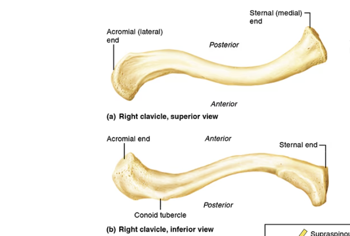
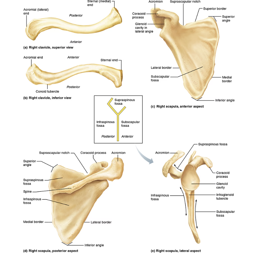
clavicle
a slender, slightly curved long bone located at the base of the neck. connects arm to ribcage, acting as a strut to keep the shoulder joint in place. runs horizontally from breastbone to the top of shoulder.
distinct "S" shape. The half closest to the center of your chest curves forward (convex), while the half closest to your arm curves backward (concave)
Medial (Sternal) End: rounded, thicker end connects to the center of your chest at the top of your breastbone (sternum) to form the sternoclavicular joint.
Lateral (Acromial) End: This flatter, thinner end connects to the outer tip of your shoulder blade (acromion) to form the acromioclavicular (AC) joint.
scapula
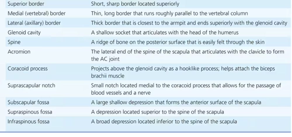
lateral border is thick, medial border is thin
glenoid cavity goes laterally
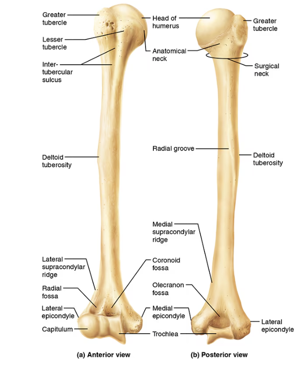
humorus
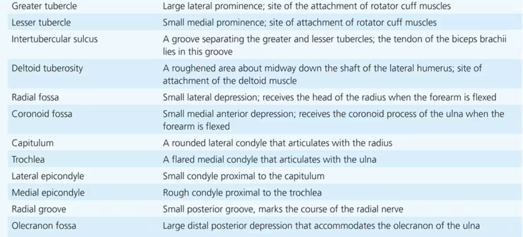
greater tubercle is lateral, lesser tubercle is medial. greater actually looks smaller to me but it says large.
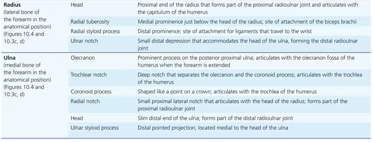
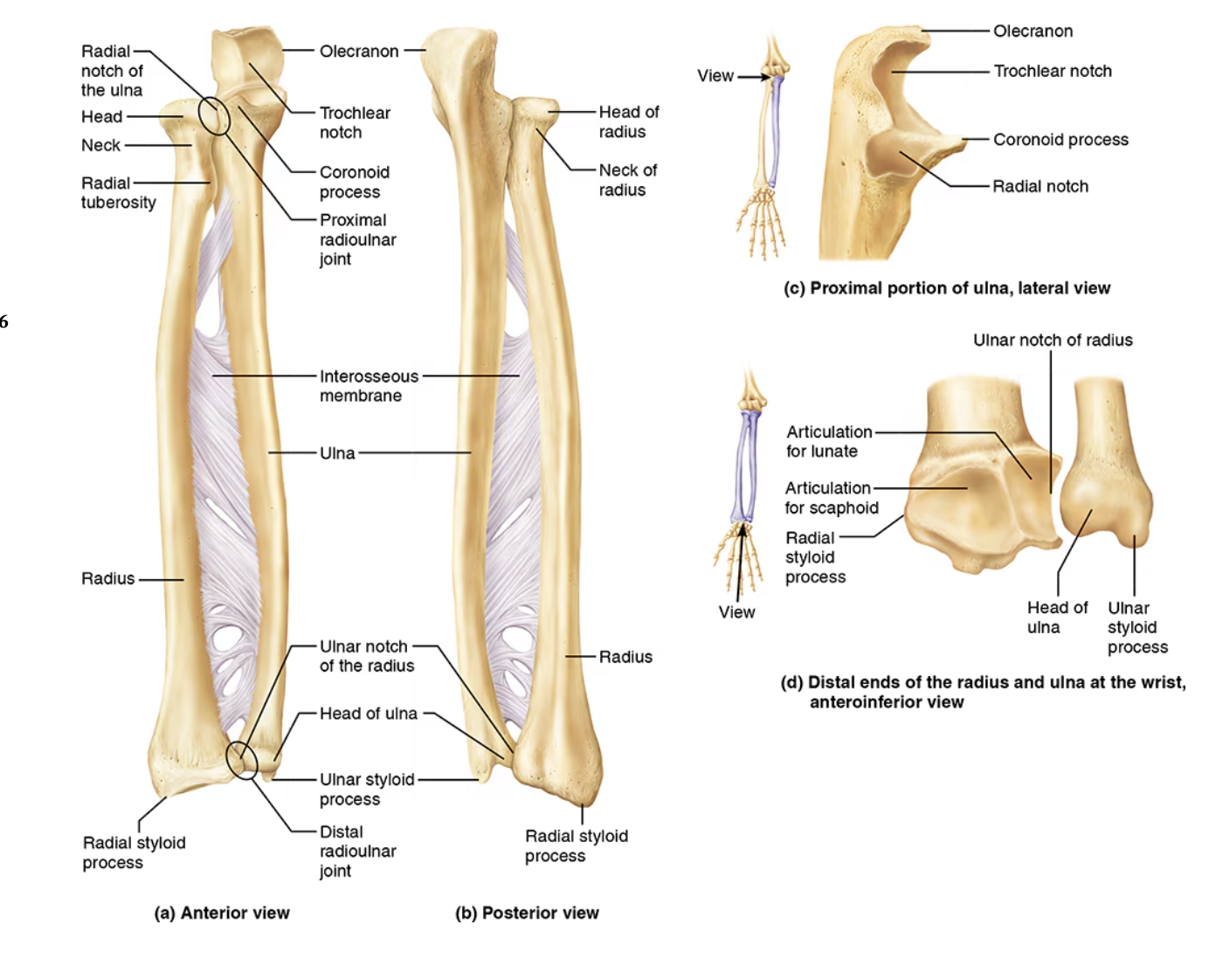
radius and ulna
hand
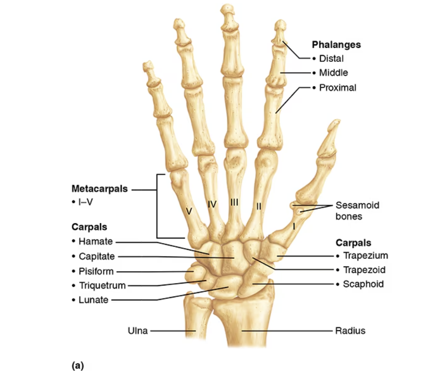
wrist/carpus: the proximal portion of the hand. the eight bones composing it are the carpals. The carpals are arranged in two irregular rows of four bones each. In the proximal row (lateral to medial) are the scaphoid, lunate, triquetrum, and pisiform bones; the scaphoid and lunate articulate with the distal end of the radius. In the distal row (lateral to medial) are the trapezium, trapezoid, capitate, and hamate. (here comes the thumb going towards the thumb)
The metacarpals, numbered I to V from the thumb side of the hand toward the little finger, radiate out from the wrist like spokes to form the palm of the hand. The bases of the metacarpals articulate with the carpals; their more bulbous heads articulate with the phalanges distally. When the fist is clenched, the heads of the metacarpals become prominent as the knuckles.
The fingers are numbered from 1 to 5, beginning from the thumb (pollex) side of the hand. The 14 bones of the fingers, or digits, are miniature long bones, called phalanges (singular: phalanx). Each finger contains three phalanges (proximal, middle, and distal) except the thumb, which has only two (proximal and distal).