BIO 315 Lab Exam 3: Digestive System
1/90
There's no tags or description
Looks like no tags are added yet.
Name | Mastery | Learn | Test | Matching | Spaced | Call with Kai |
|---|
No analytics yet
Send a link to your students to track their progress
91 Terms
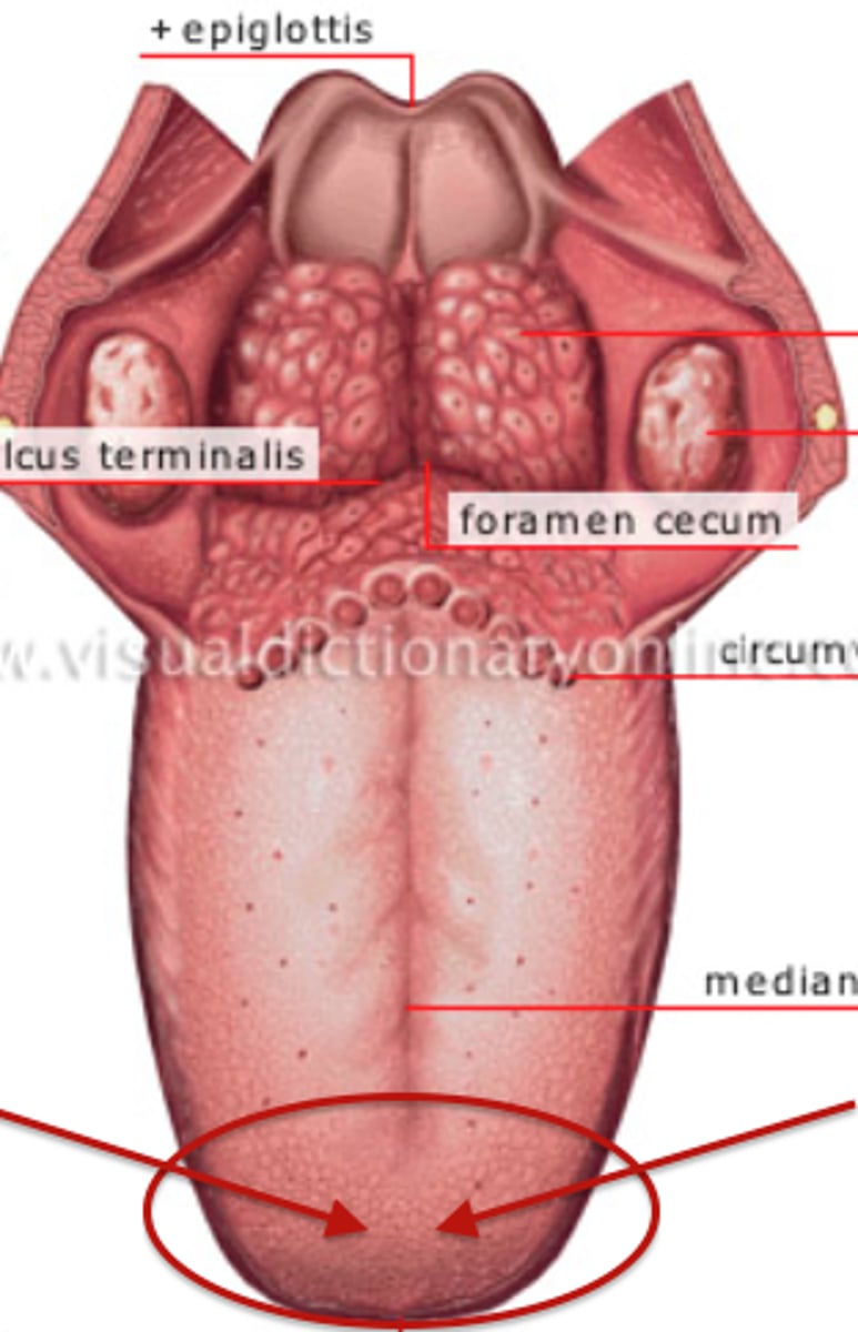
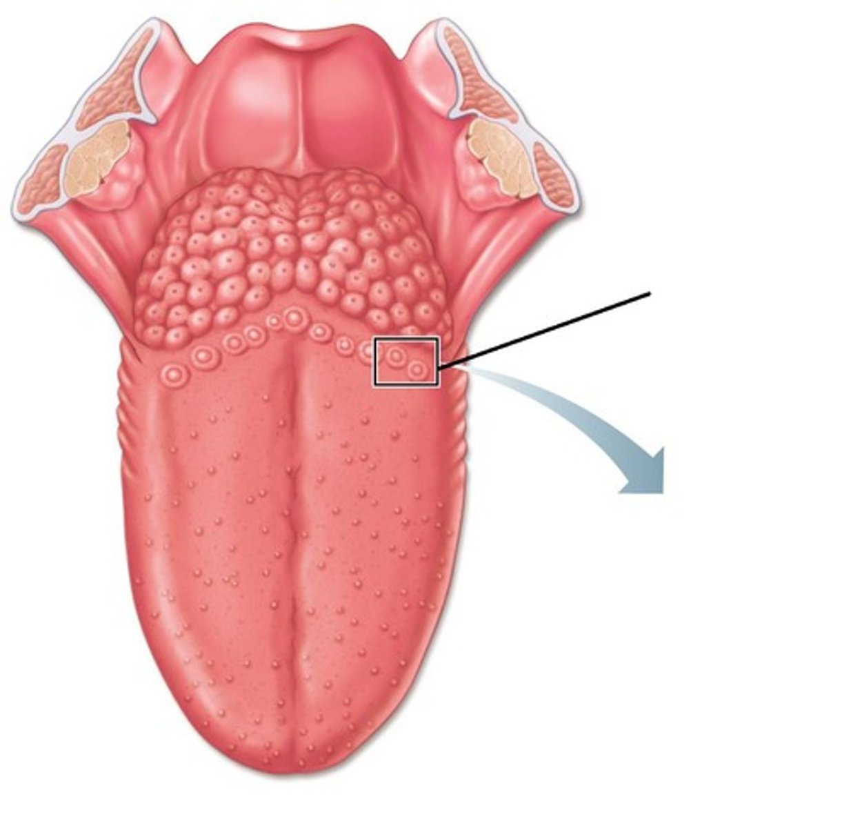
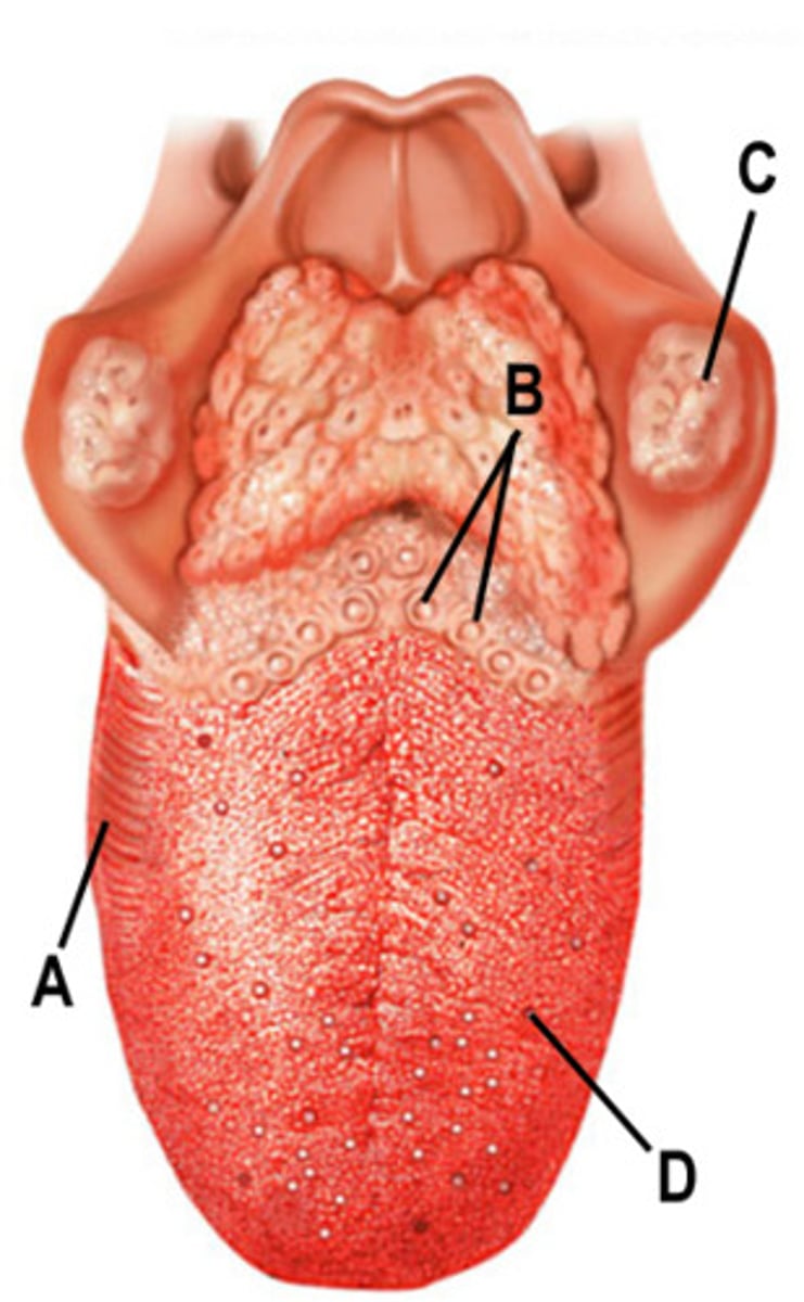
Root of the tongue
Feature
- Gray line under the tongue
Tip of the tongue
Feature
- Very front of the tongue
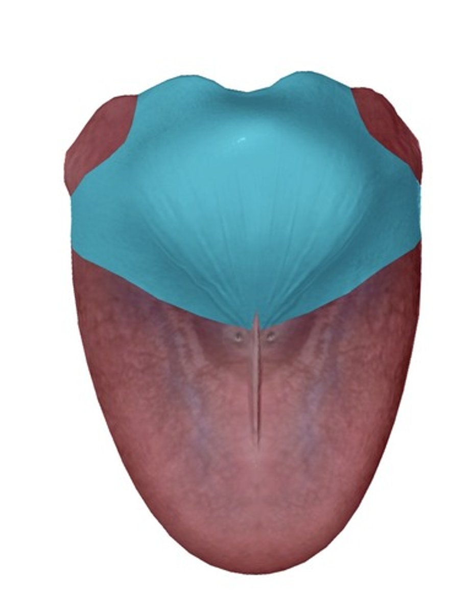
Body of the tongue
Feature
- All the pink above the black
Dorsum of tongue
Surface
- Whole top part of tongue
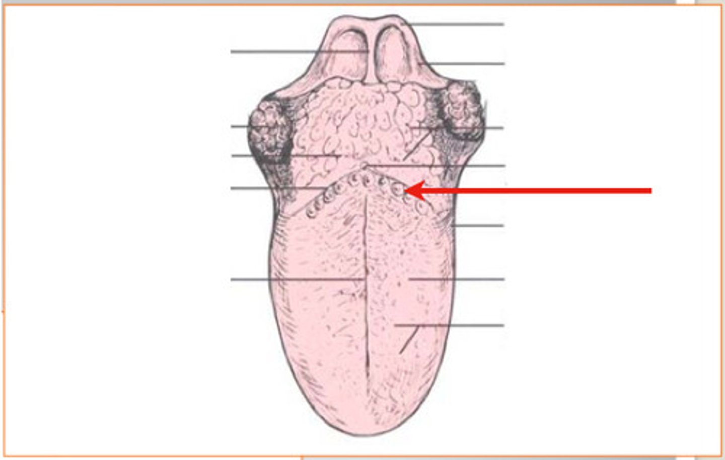
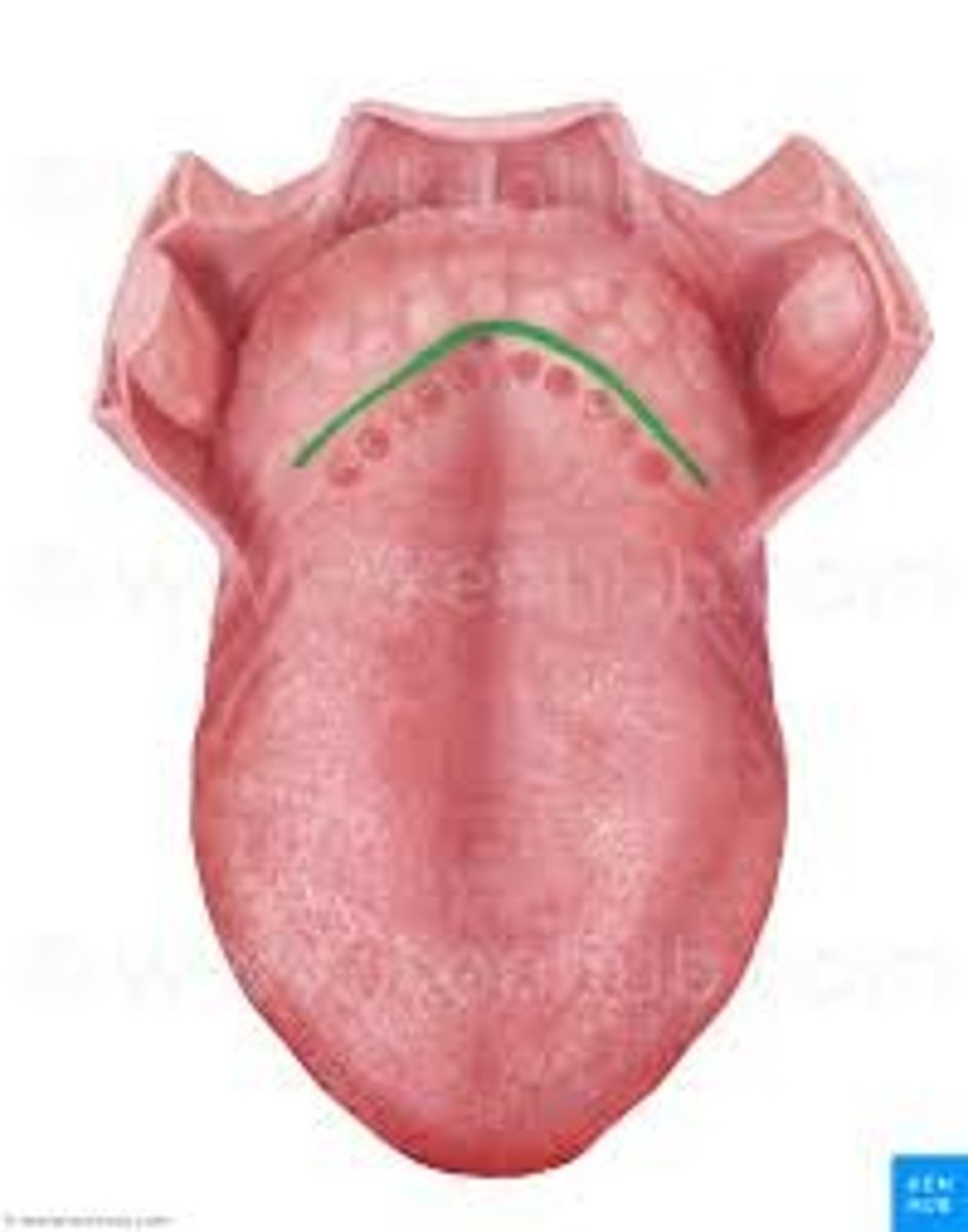
Terminal sulcus
Depression
- Valley in tongue
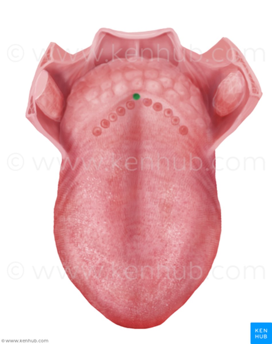
Foramen cecum
Hole
- In center back of tongue
Lingual tonsil
Strucutre
- Black bumps on the back of tongue
Filliform papillae
Collective strucutre
- First third of bumps on the tip of the tongue

Fungiform papillae
Collective structure
- Middle taste buds
- Look like mushrooms
Vallate papillae
Collective structure
- Tastebuds on the back of the tongue
- Biggest ones up on ridge
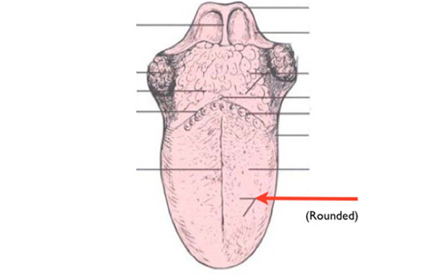
Inferior surface of tongue
Surface
- Underside of tongue
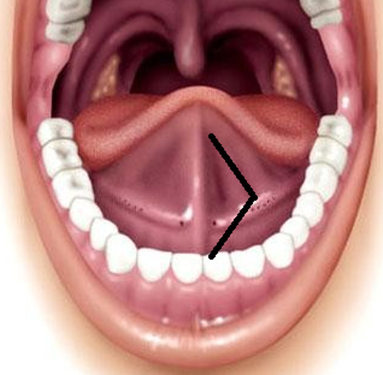
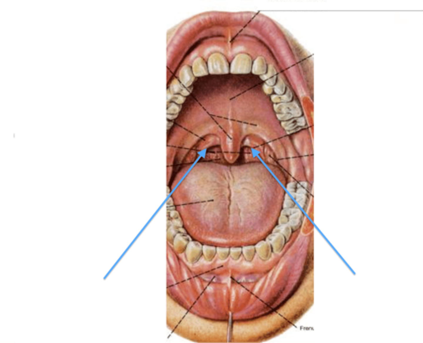
Frenulum of tongue
Structure
- Webbing under tongue
Genioglossus muscle
Structure
- Square shaped muscle right under the tongue
Hyoglossus muscle
Structure
- Connects the hyoid bone to the side of the tongue
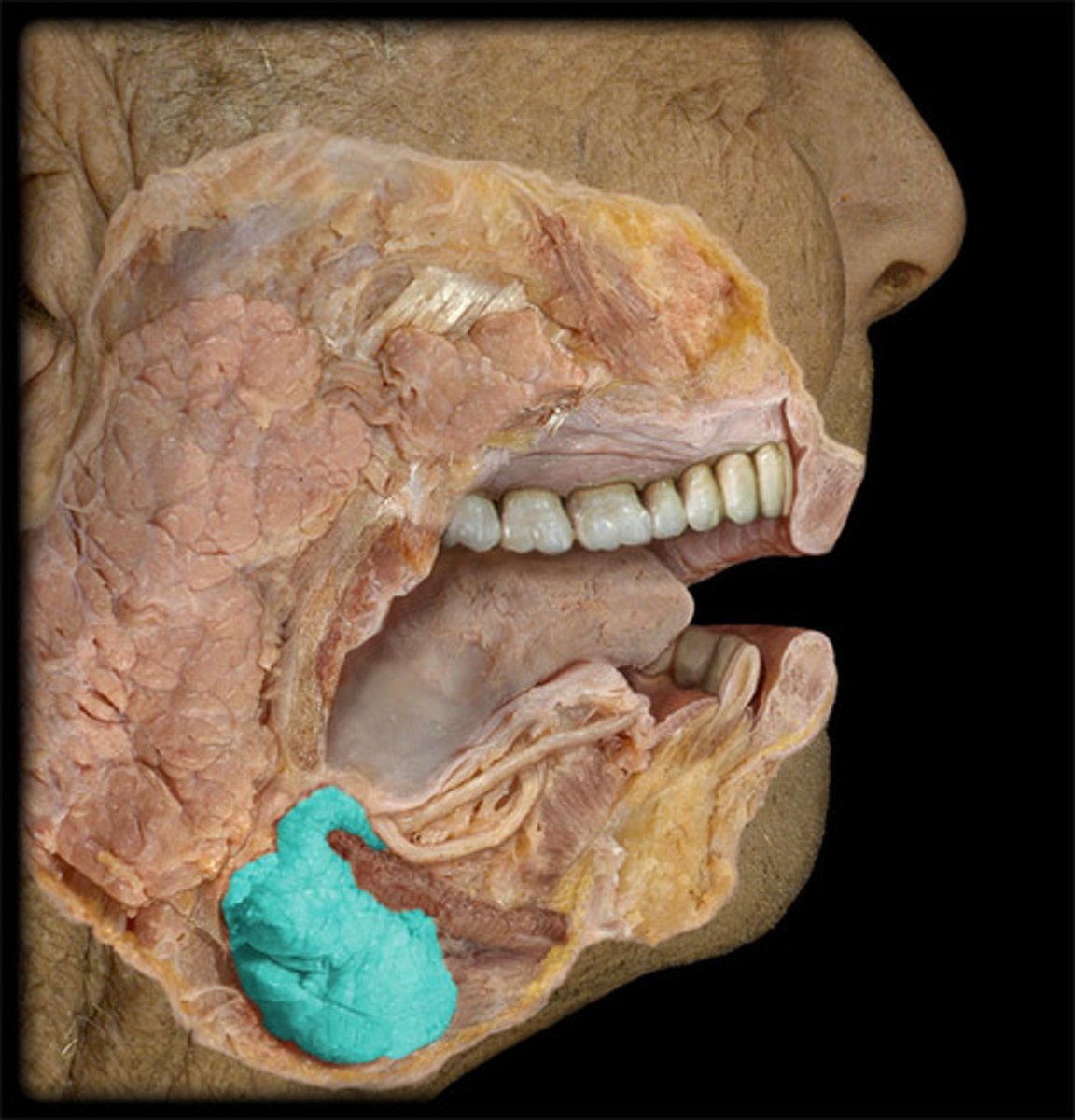
Submandibular gland
Structure
- On side of the neck
- Bulb
- Oval blob under the jaw
- TA will stick probe in it to show
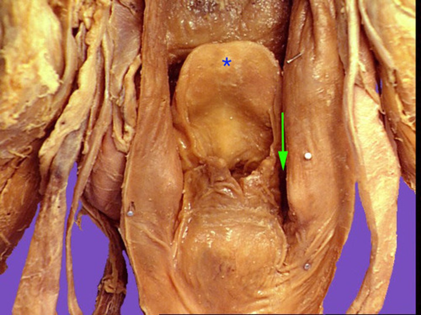
Palatoglossal arch
Strucutre
- arch formed by 2 thick muscles connecting
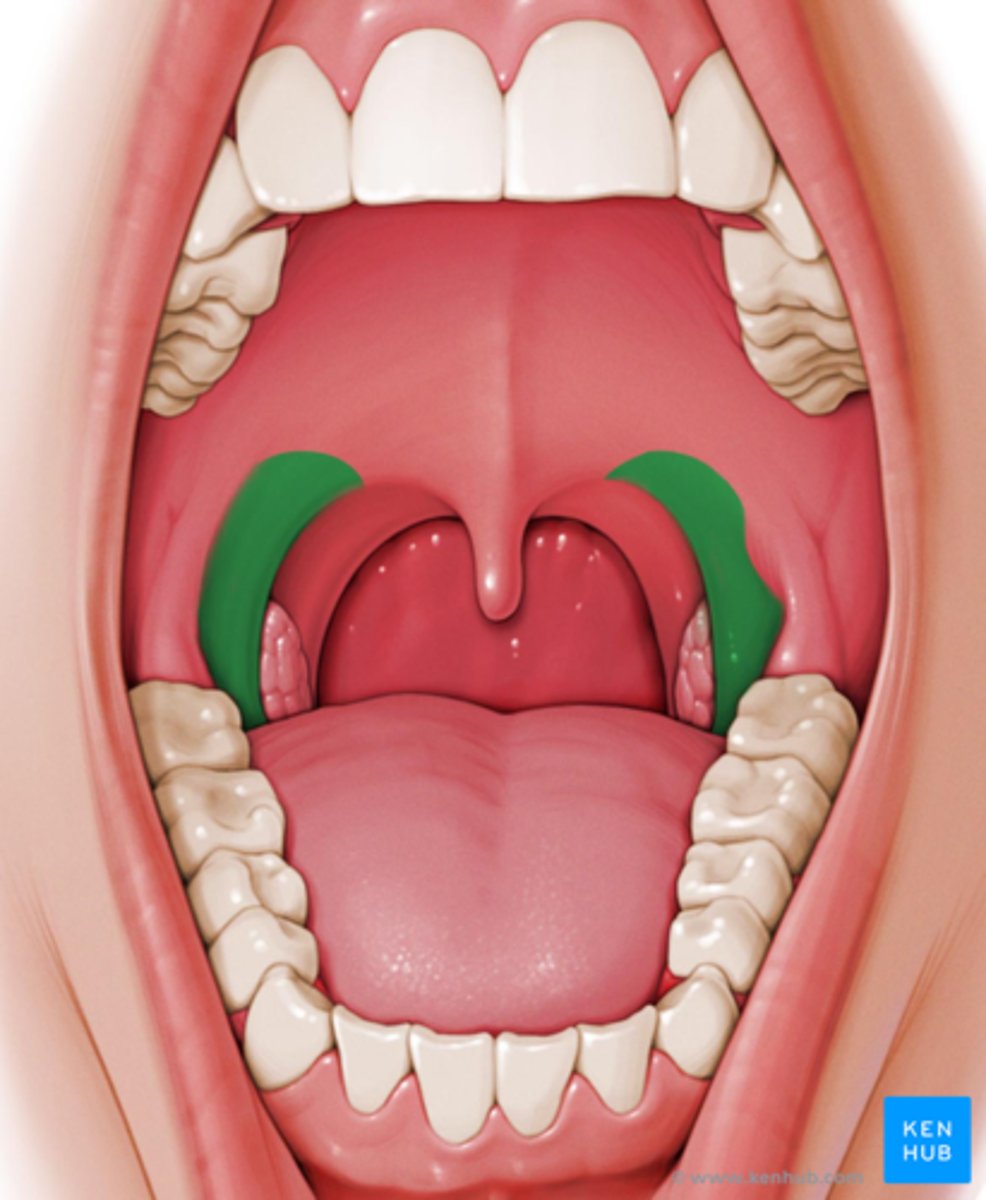
Palatopharyngeal arch
Structure
- arch formed by 2 thin muscles connecting
Tonsillar fossa
Depression
- In between arches on side of mouth
- Under the epiglottis
Palatine tonsil
Entire structure
- pink bumps lateral to black bumps
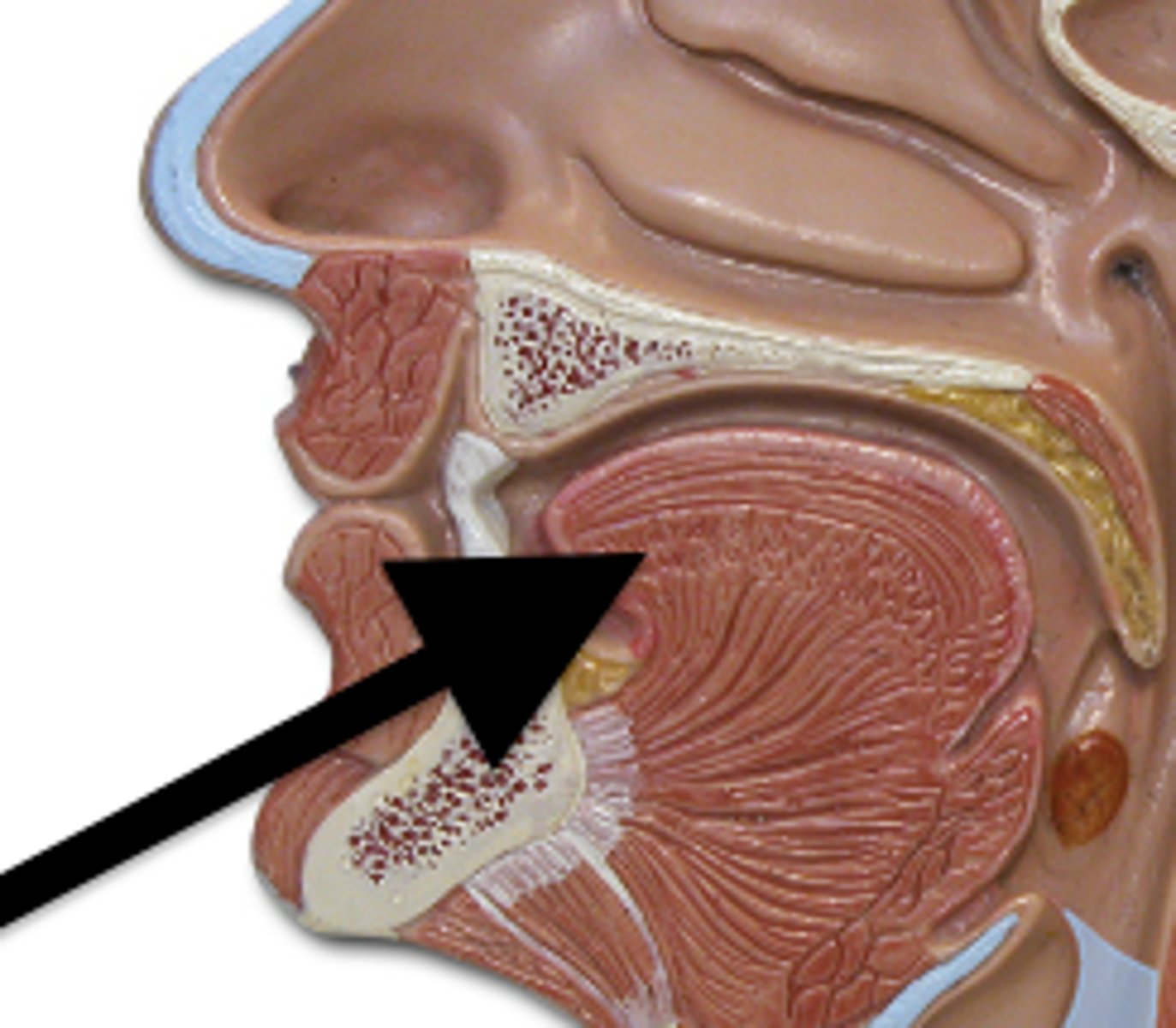
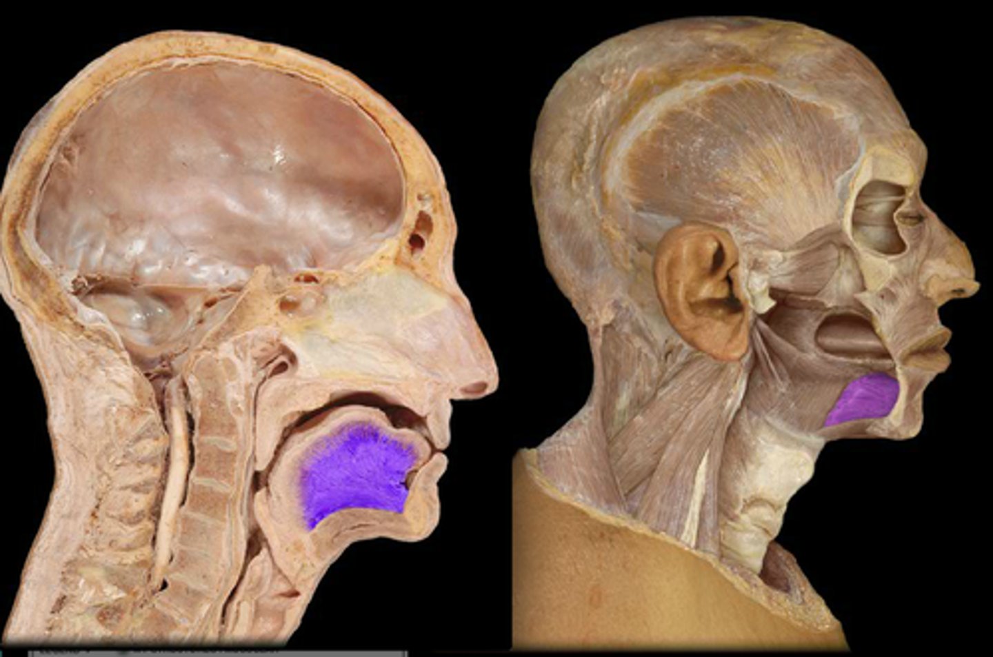
Nasopharynx
Boundary
- On top portion of head
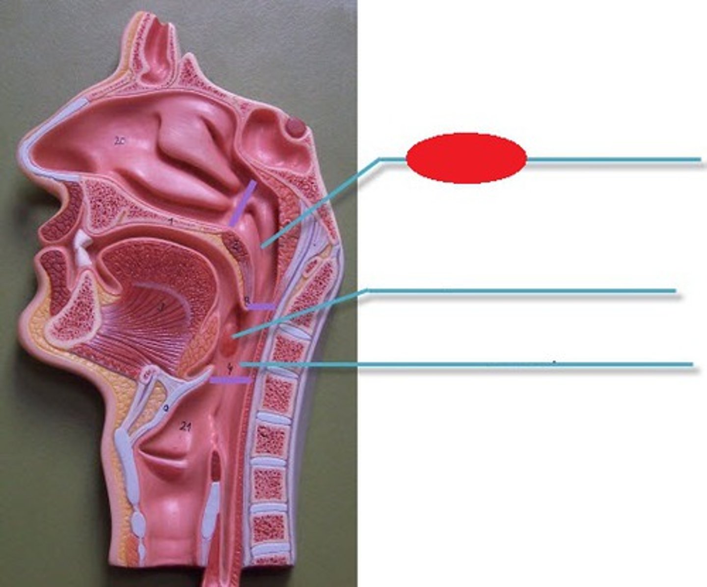
Oropharynx
Boundary
Middle part where mouth is
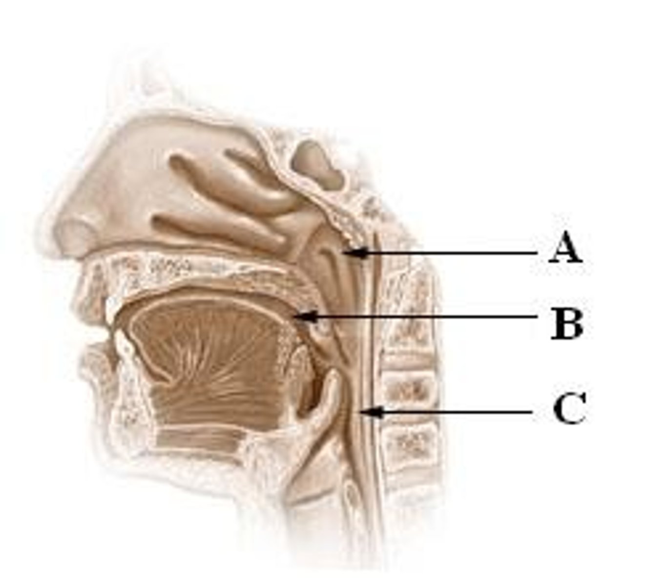
Laryngopharynx
Boundary
- On neck

Piriform recesses
Depression
- Above the epiglottis on the side
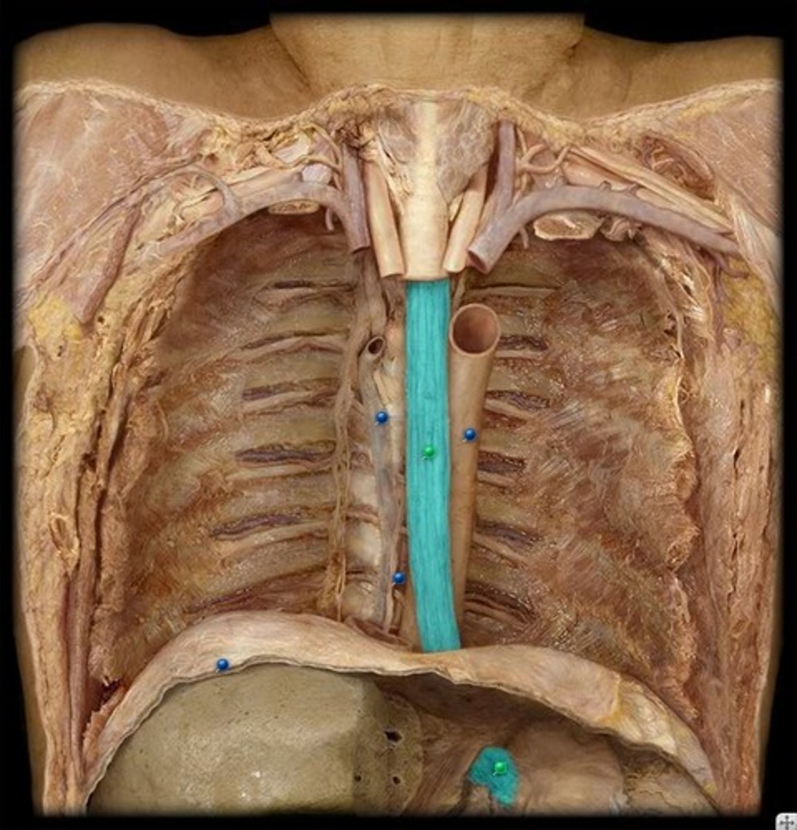
Thoracic esophagus
Structure
- In the middle of the body below the aorta
- In the chest cavity
Greater curvature of stomach
Border
- Convex lateral surface of the stomach
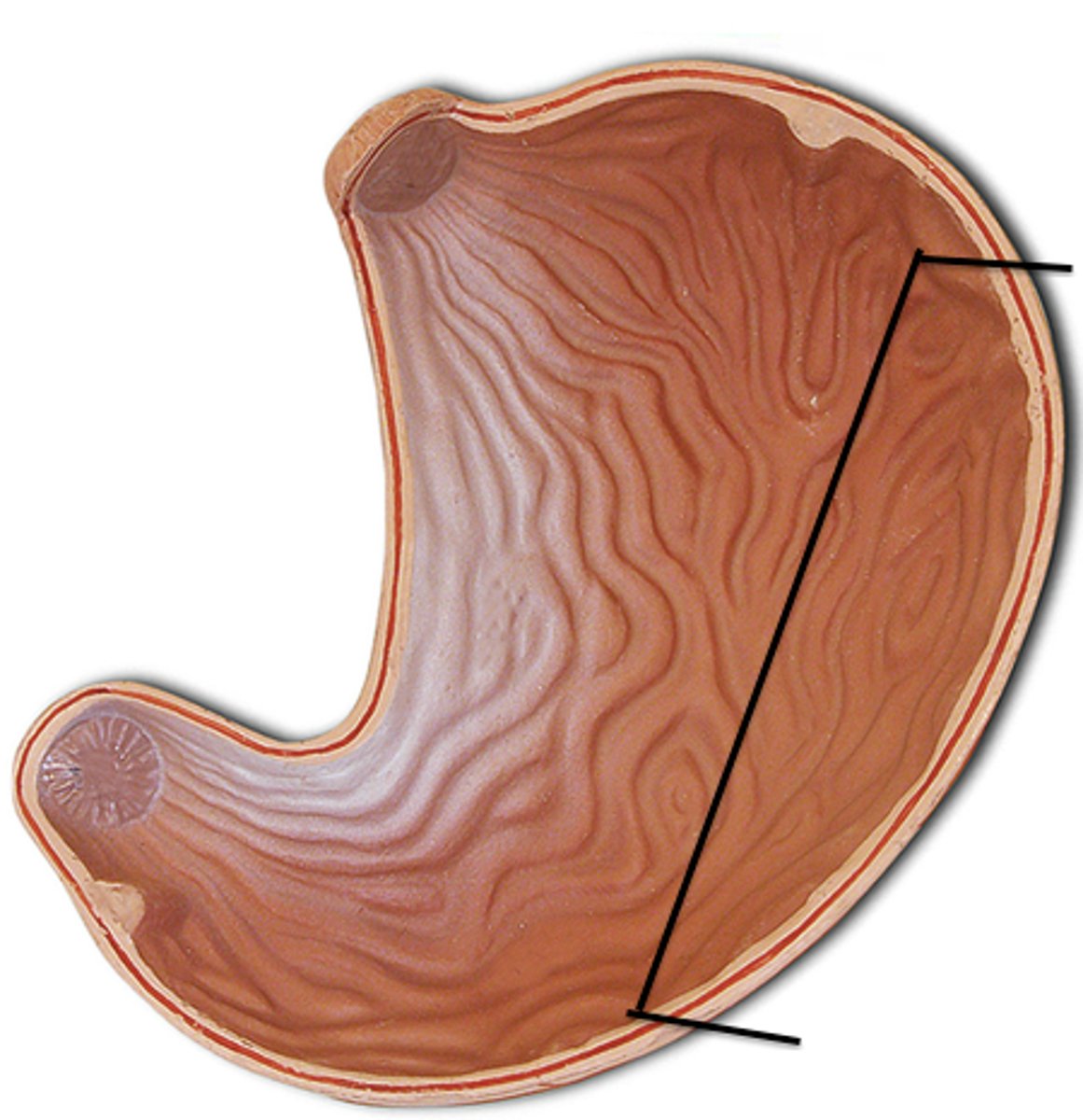
Lesser curvature of stomach
Border
- Concave medial surface of the stomach
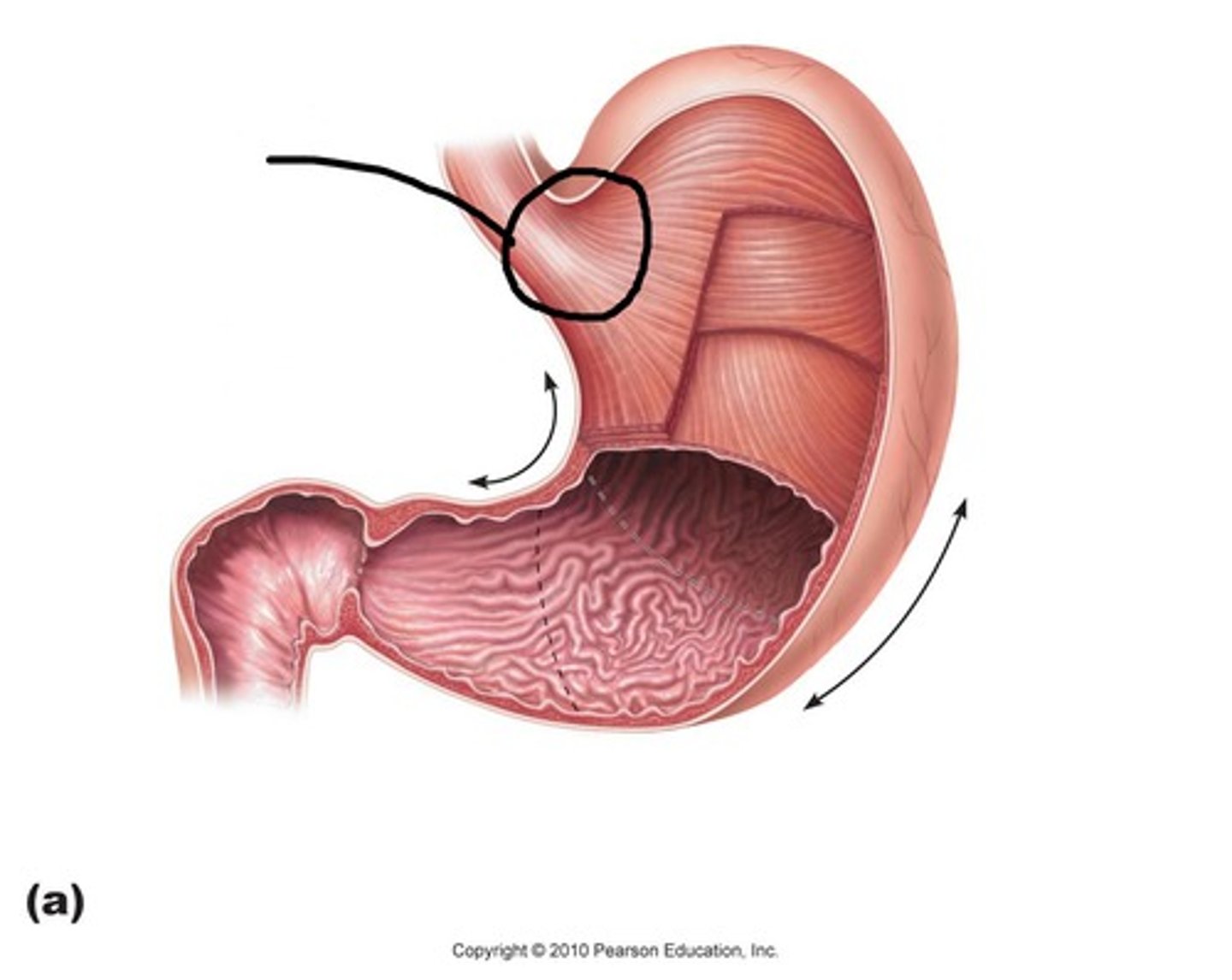
Cardia of the stomach
Feature
- Top portion where esophagus enters
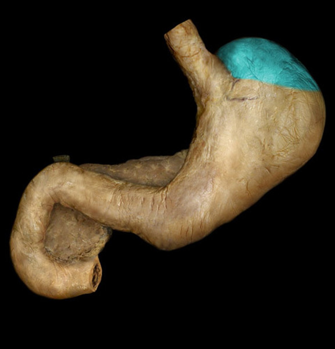
Fundus of the stomach
Balloon
- Top bag-like portion, lateral to the cardia
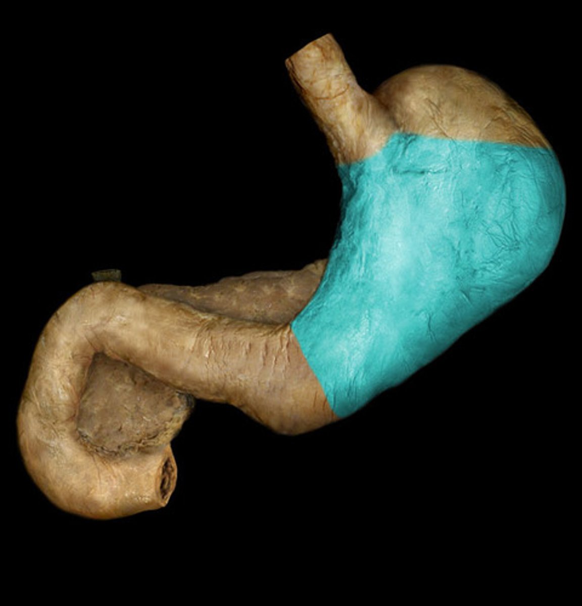
Body of the stomach
Region
- Middle part
- The rest of the stomach
Pyloric antrum
Feature
- Divet before the ball like structure
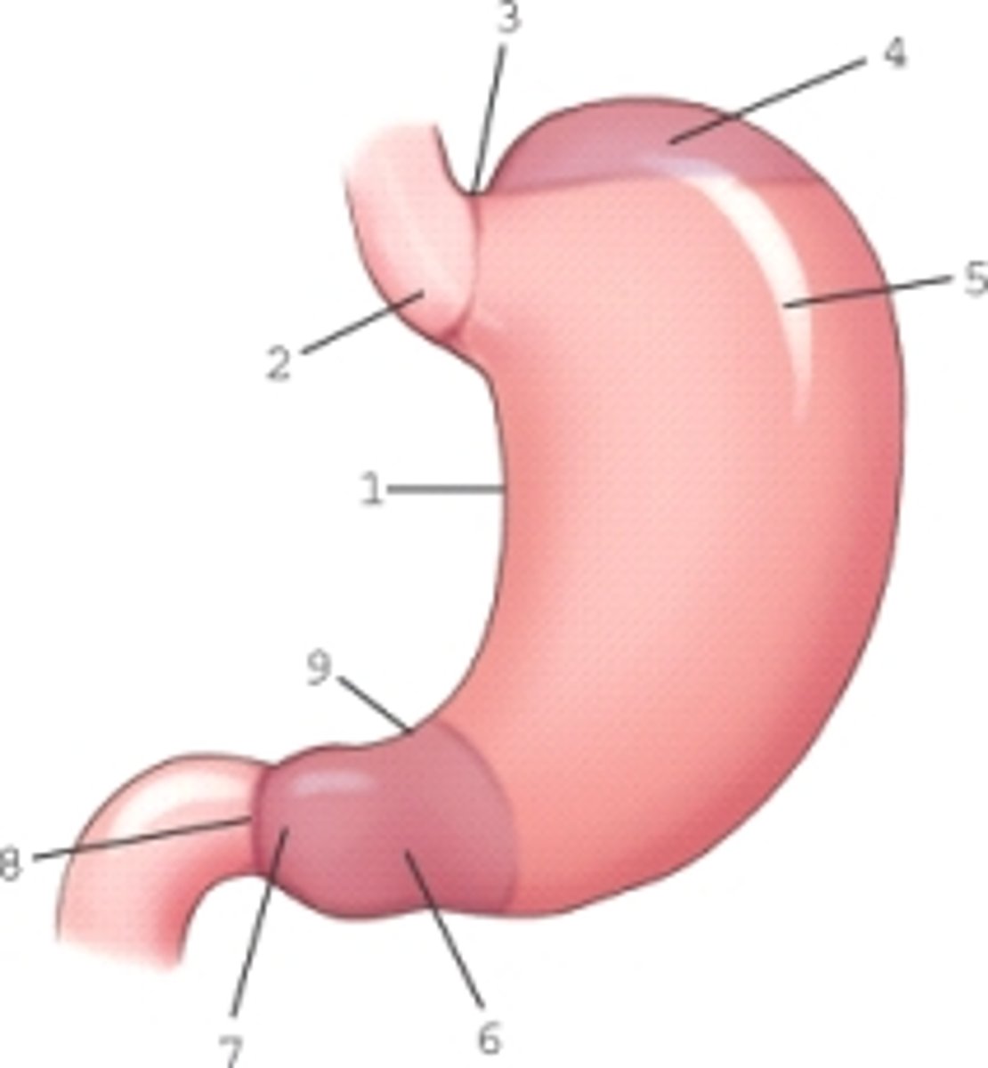
Pylorus
Strucure
- After the pyloric antrum
- Narrow exit part of the stomach
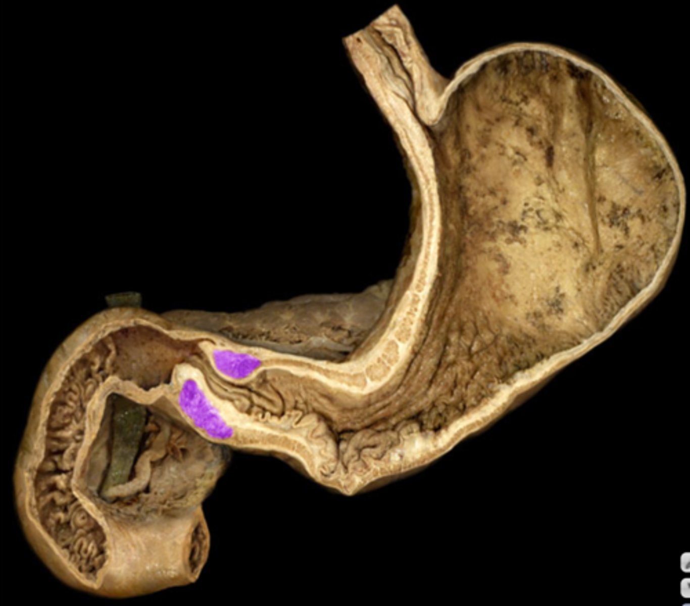
Pyloric sphincter muscle
Internal structure
- ridges
- TA will open the pylorus, and lay probe inside to show
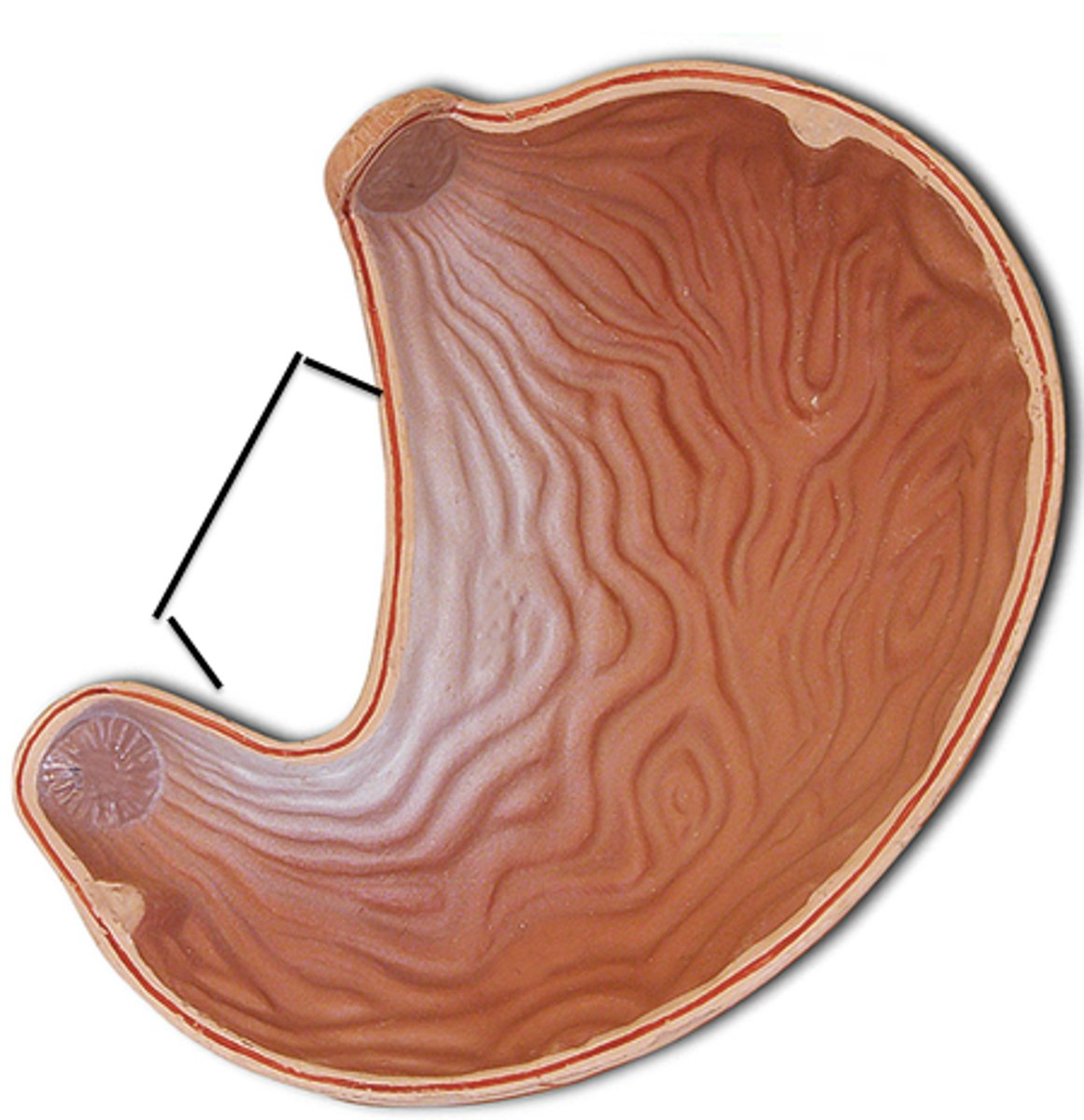
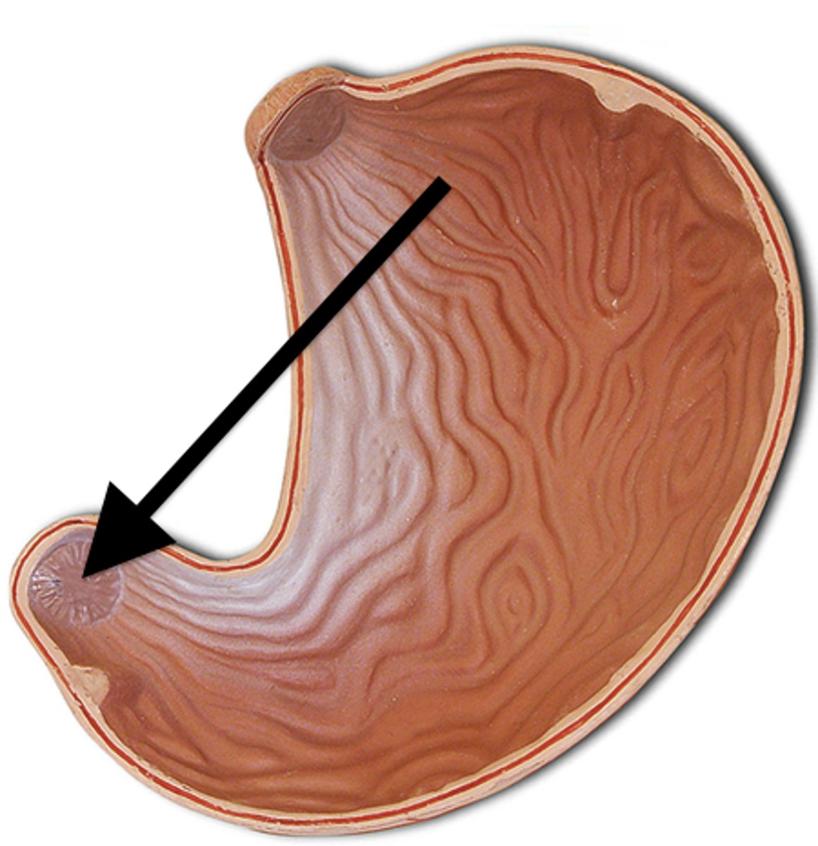
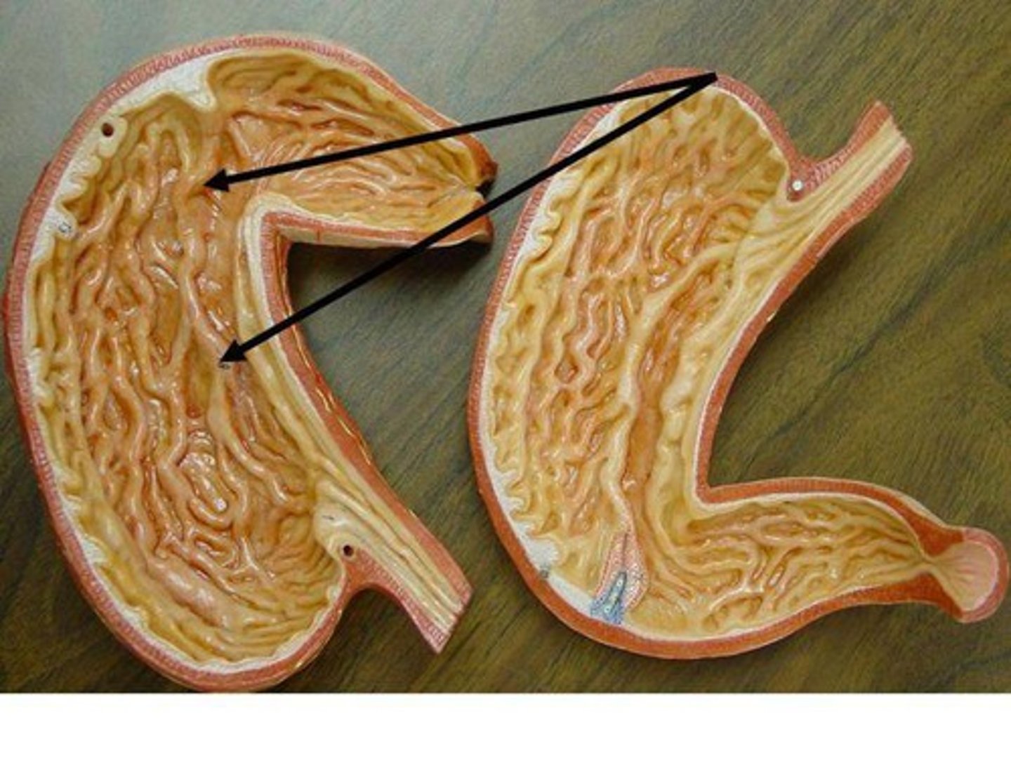
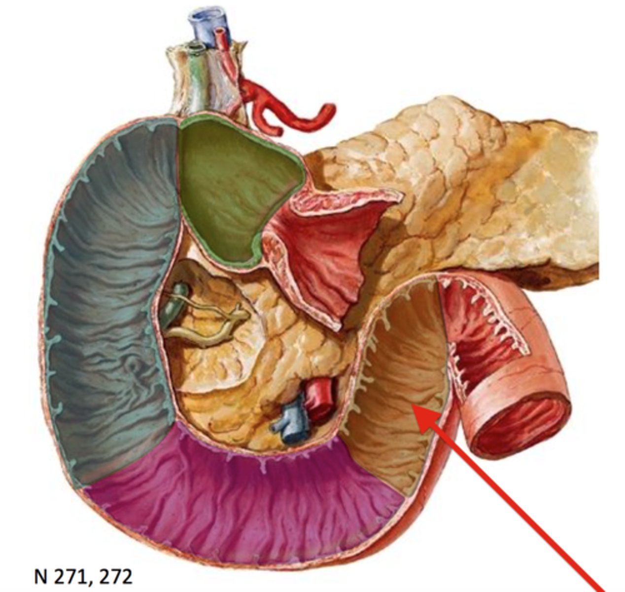
Gastric folds
Internal structure
- Folds on inside of stomach
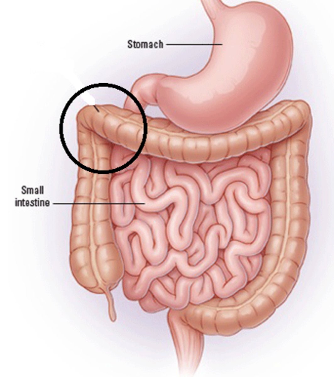
Superior part of the duodenum
Portion
- connects to pylorus of stomach
- Top part
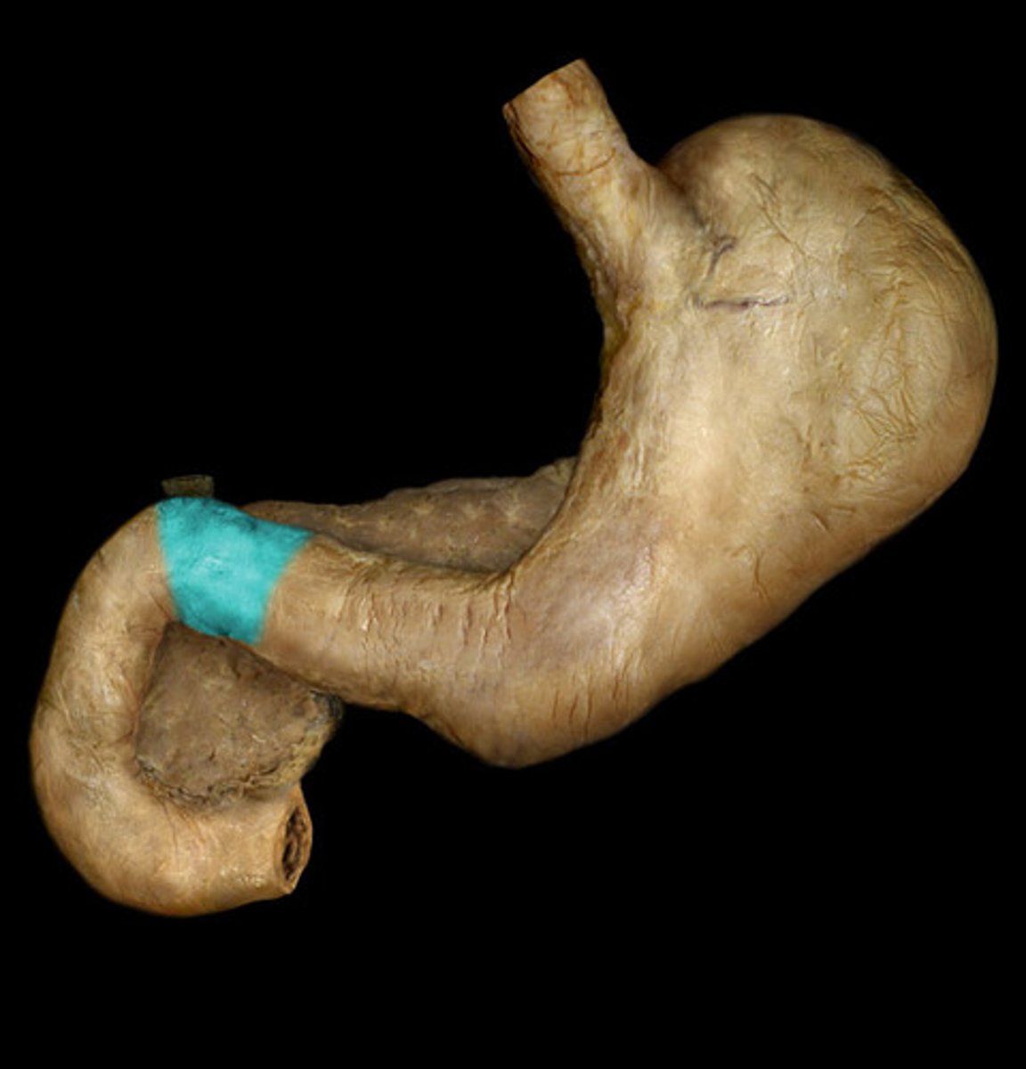
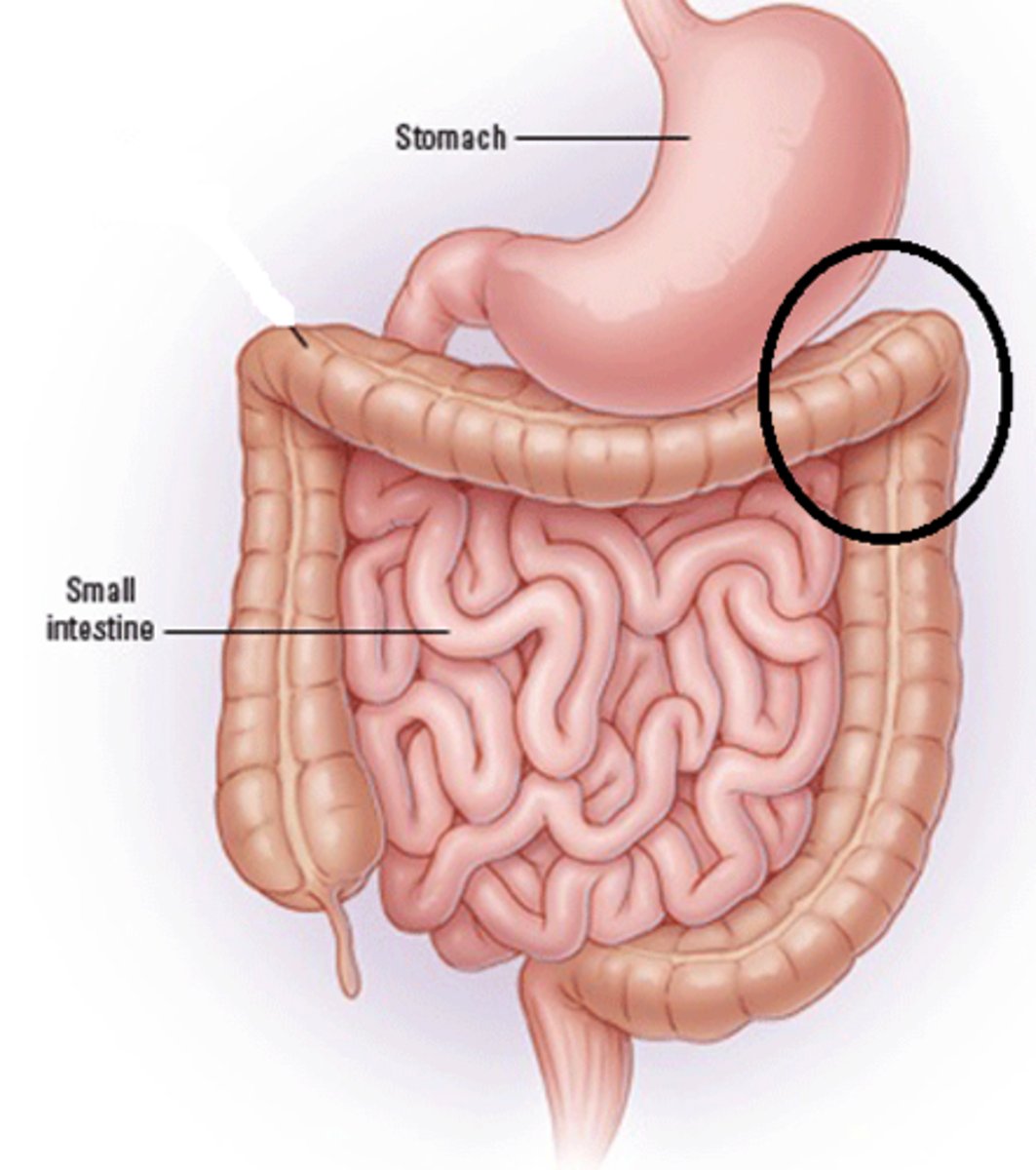
Descending part of duodenum
Portion
- Goes downward
- Wraps around the pancreas
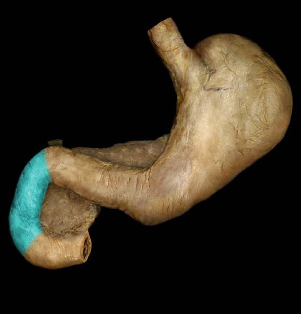
Horizontal part of the duodenum
Portion
- Below the head of pancreas
- Bends 90 degrees
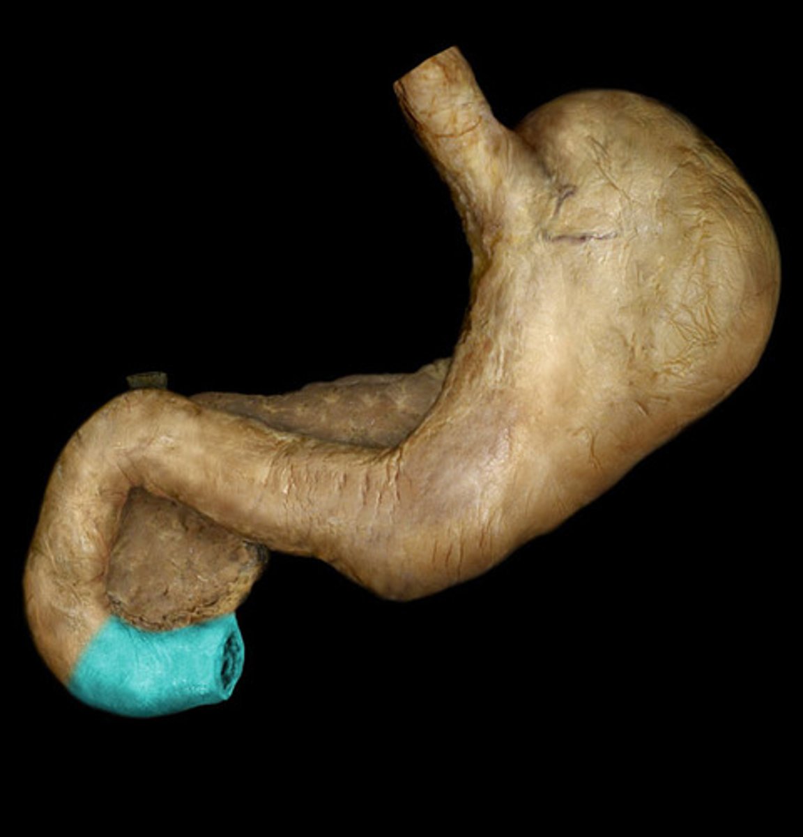
Ascending part of the duodenum
Portion
- Goes up
- Very last part of horseshoe shape
- After the 90 degree section
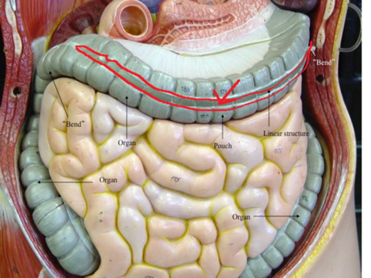
Cecum
Structure
- Ball like structure
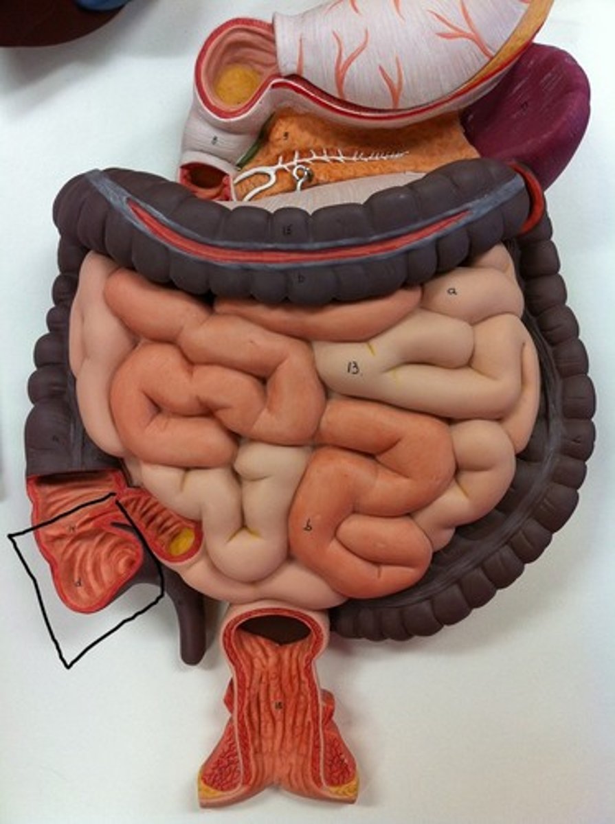
Ileal orifice
Junction
- Between ileum and cecum
- TA will lay probe above the cecum to show
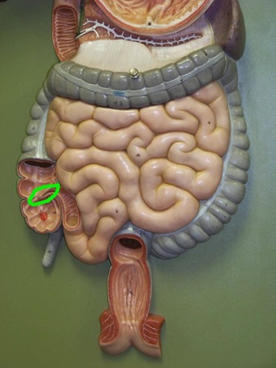
Appendix
Worm
- Between ileum and cecum
- Yellow
- Attached to the cecum
Ascending colon
Structure
- Goes up from the cecum
- Upward portion
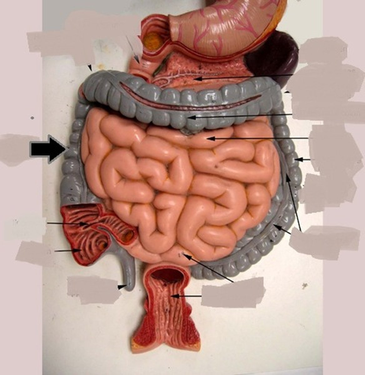
Right colic flexure
Corner
- Upper right corner around liver
Transverse colon
Strucutre
- The horizontal part on the top
- Top of the square
Left colic flexure
Corner
- Upper left corner around spleen
Sigmoid colon
Structure
- Most inferior part
- Bottom of the square
Taeniae coli muscle
Structure
- Shiny bit of smooth muscle in middle of small intestine
- TA will show on the transverse colon
- TA FAVE
Haustra of colon
Chunks
- Curves of the intestine
- The chunky segments
- TA FAVE
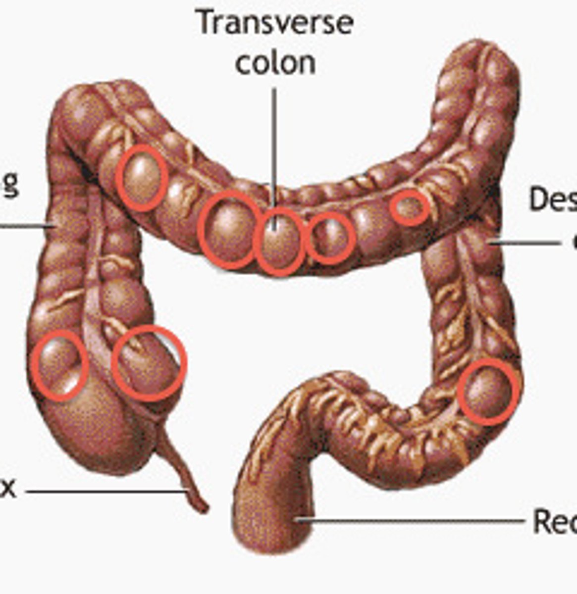
Omental appendices
Feature
- Fat parts hanging off the colon
- Shown on the right lateral side
Transverse folds of the rectum
Internal structure
- All the peaks and points inside the rectum
Anal canal
Opening
- Opening by rectum
- TA will place probe in hole to show
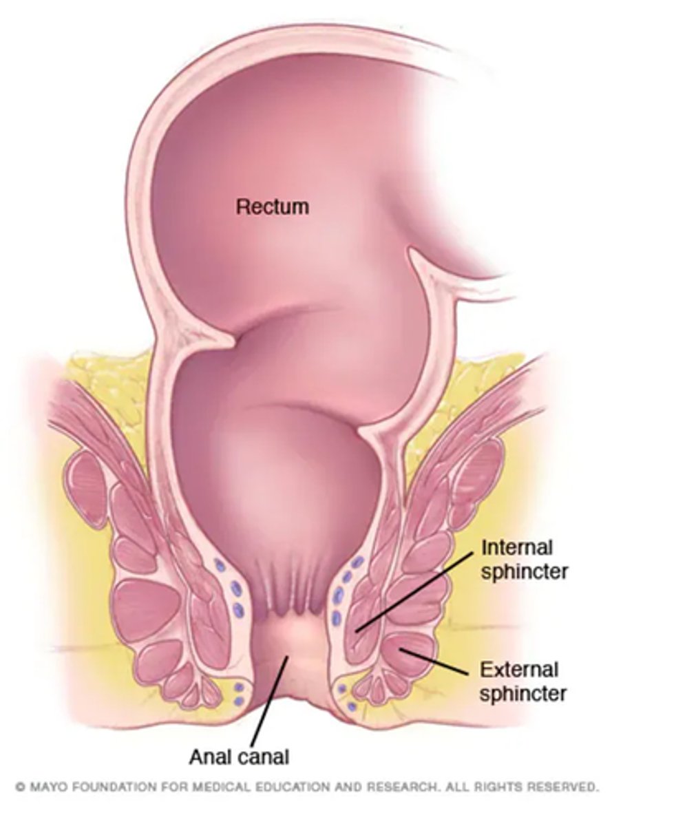
Internal anal sphincter muscle
Strucutre
- Divits on side of anal canal
- Muscle at the end
- #61
- TA will have model open to show

External anal sphincter muscle
Structure
- The circle surrounding anal canal
- TA will close the model and trace outer circle to show

Anal columns
Ridges
- Lighter ridges in anal canal
- Above the internal anal sphincter
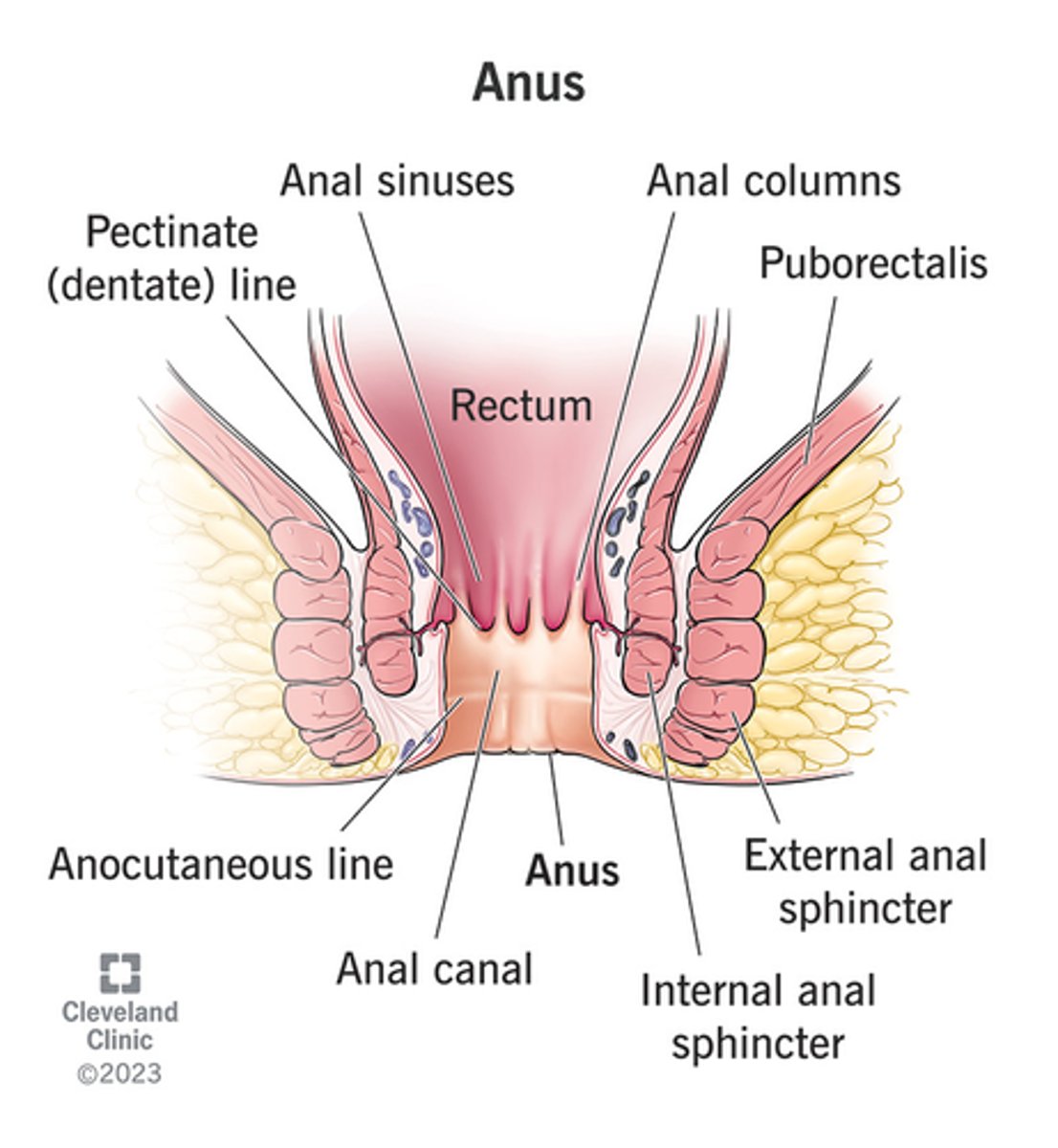
Anal sinuses
Depressions
- Darker areas between the anal columns

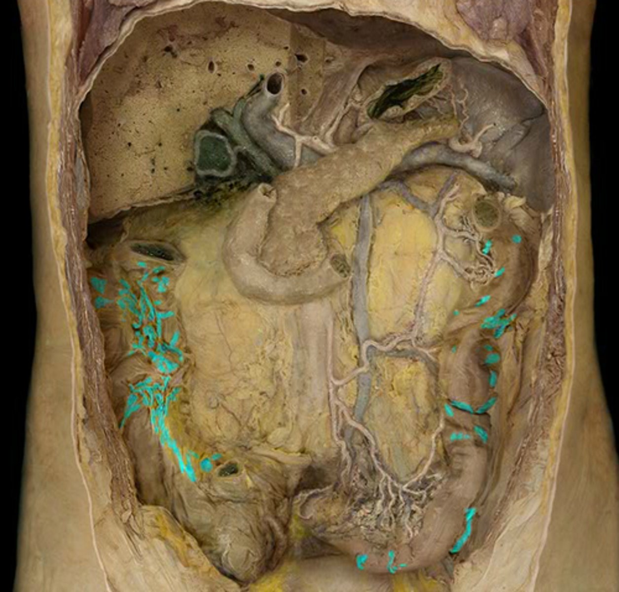
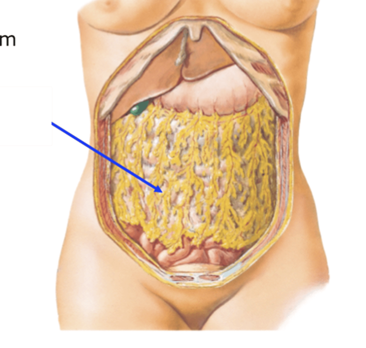
Greater omentum
Covering
- Fat drape over front of body
The mesentery of the small intestine
Connecting structure
- The yellow fat connected to the small intestine
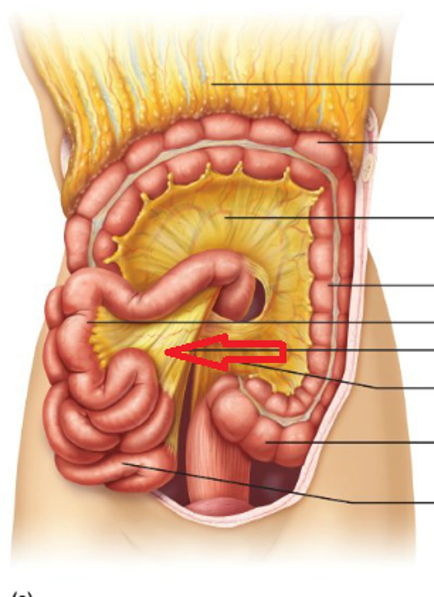
Sigmoid mesocolon
Covering
- trampoline looking thing
-connects sigmoid colon to posterior wall
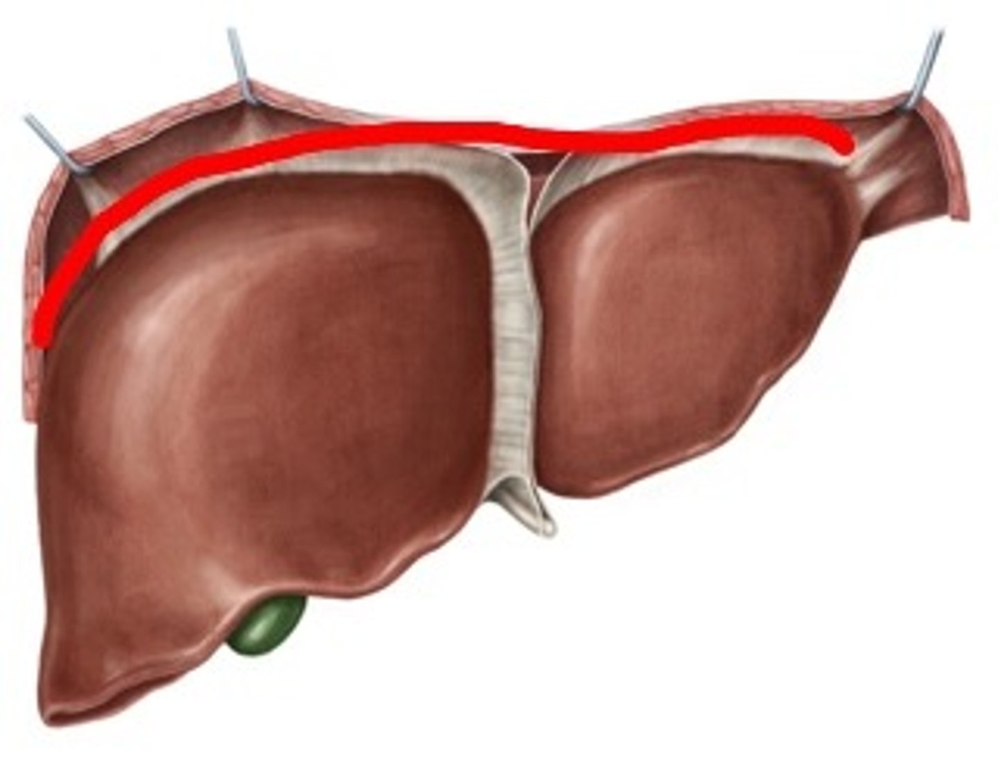
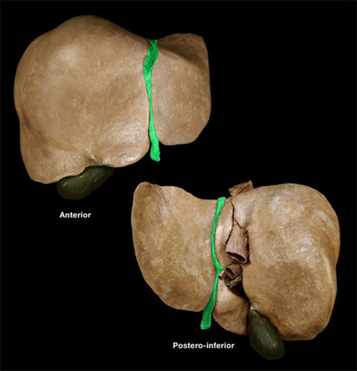
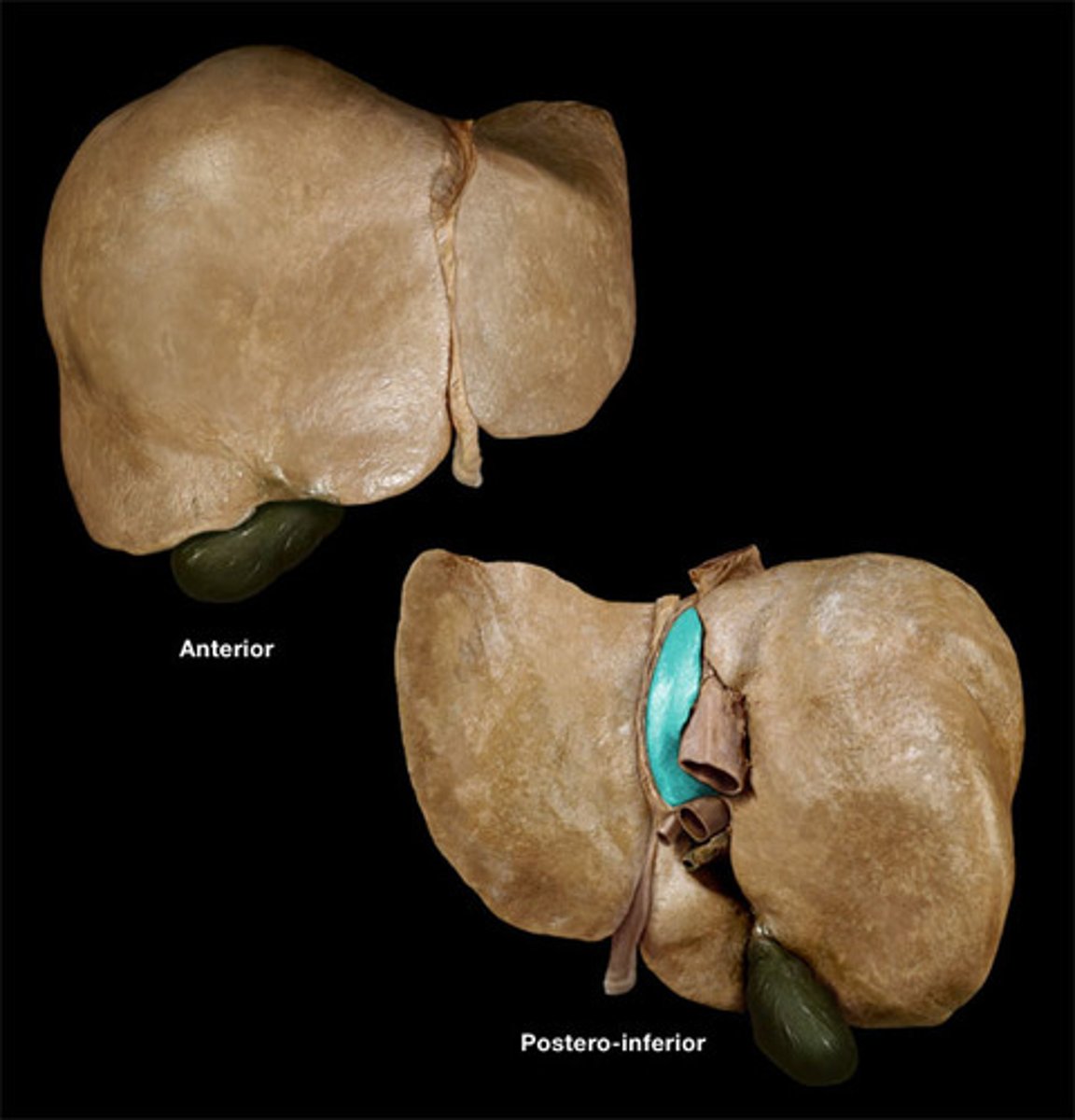
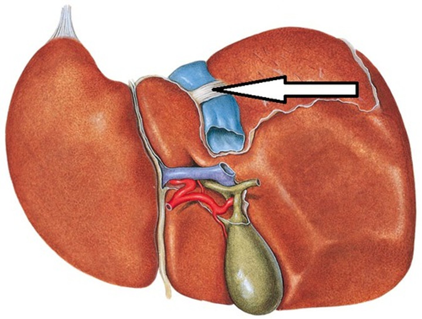
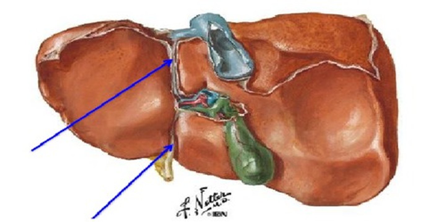
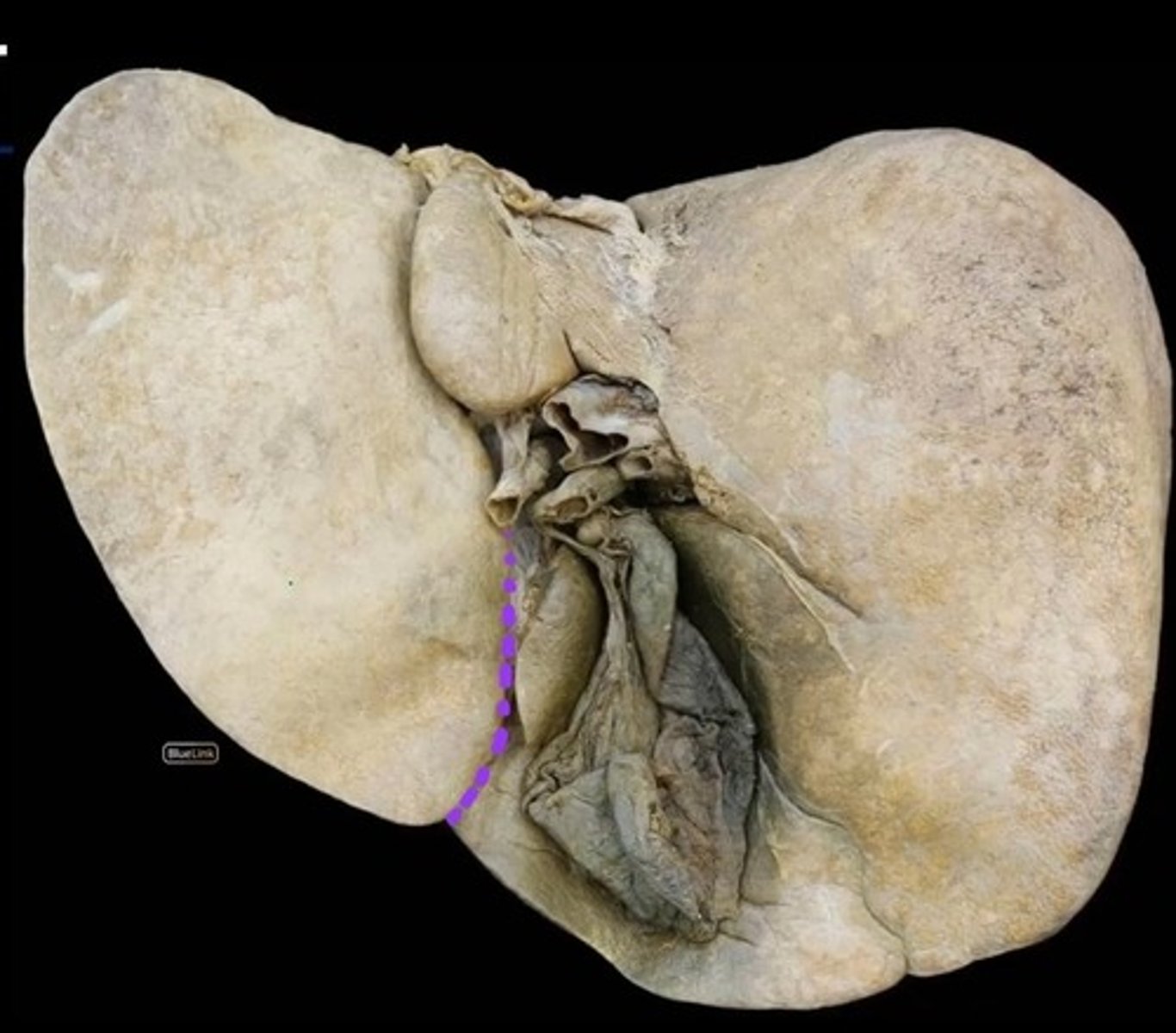
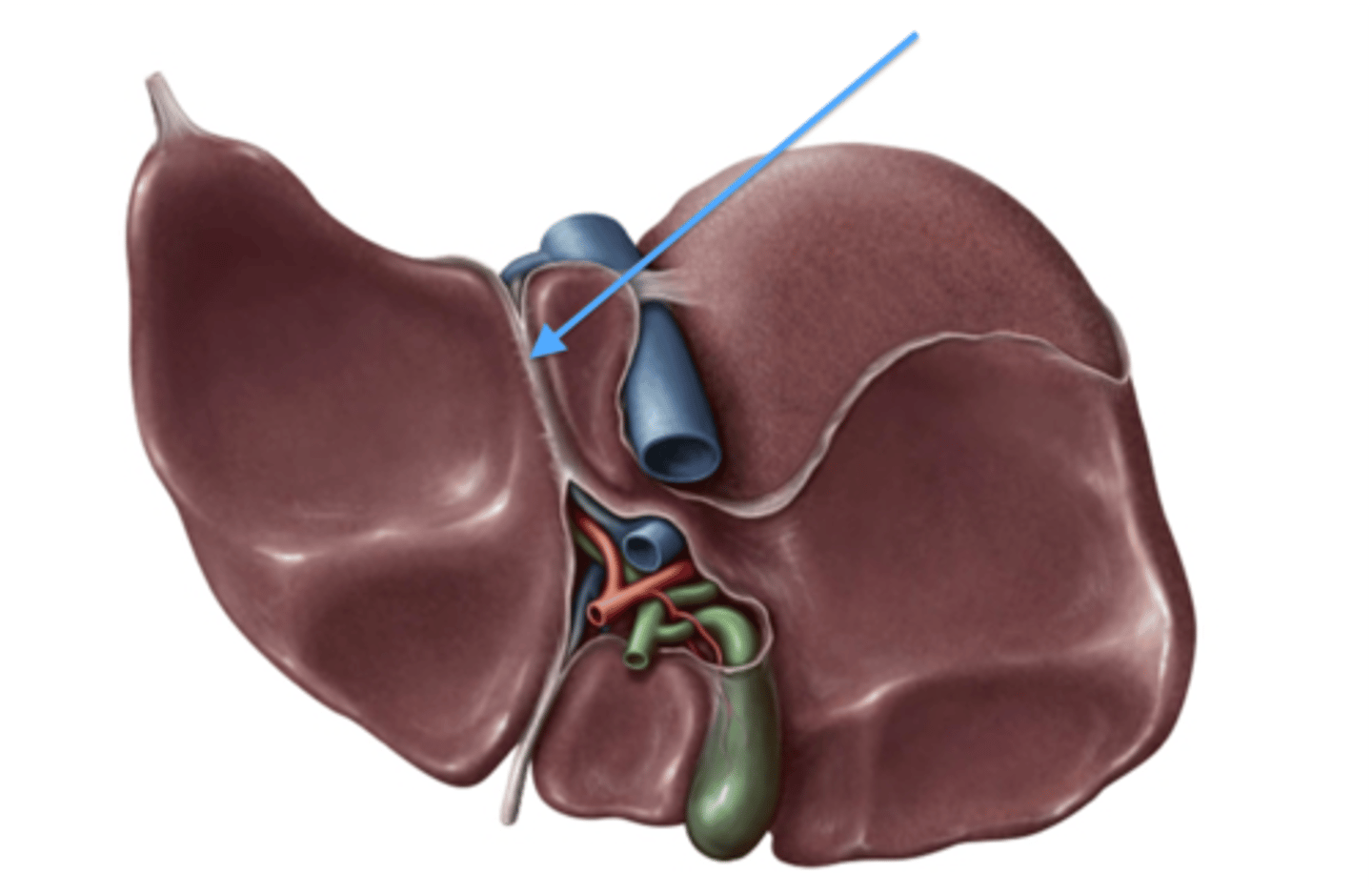
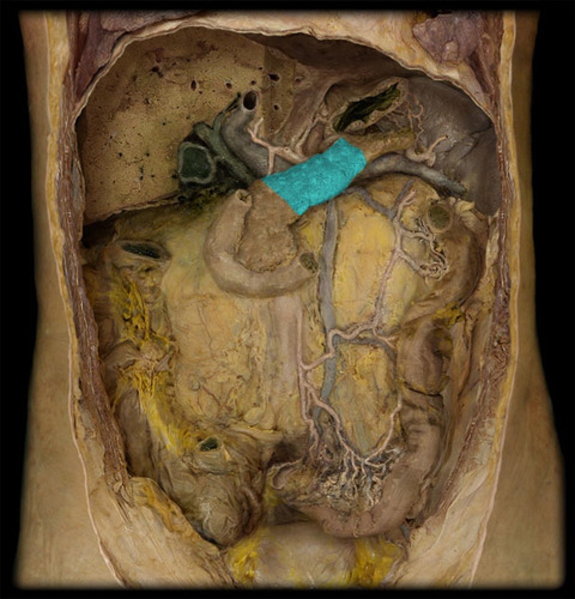
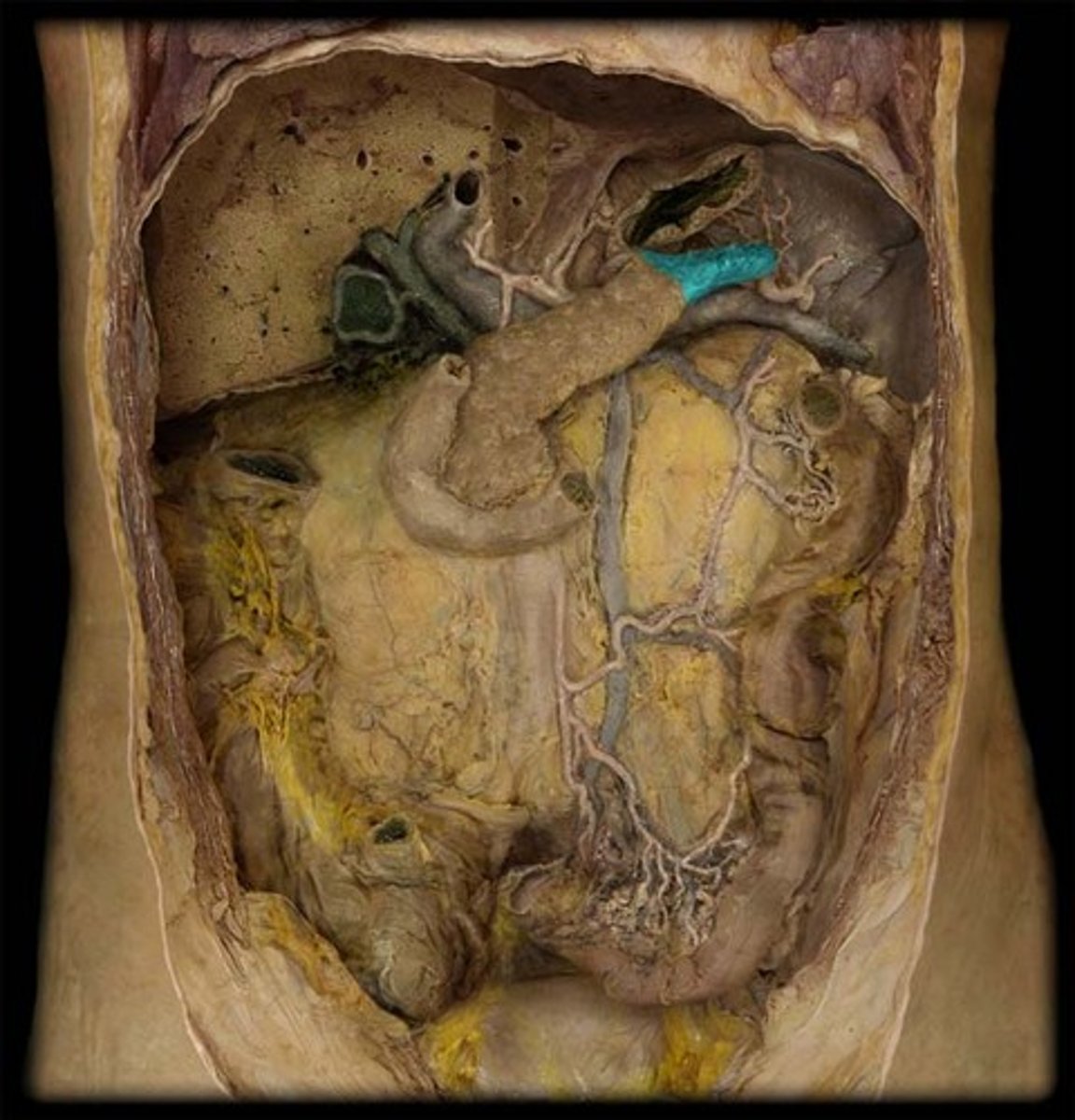
Diaphragmatic surface of the liver
Region
- On top of liver
Visceral surface of the liver
Region
- Bottom and backside of liver
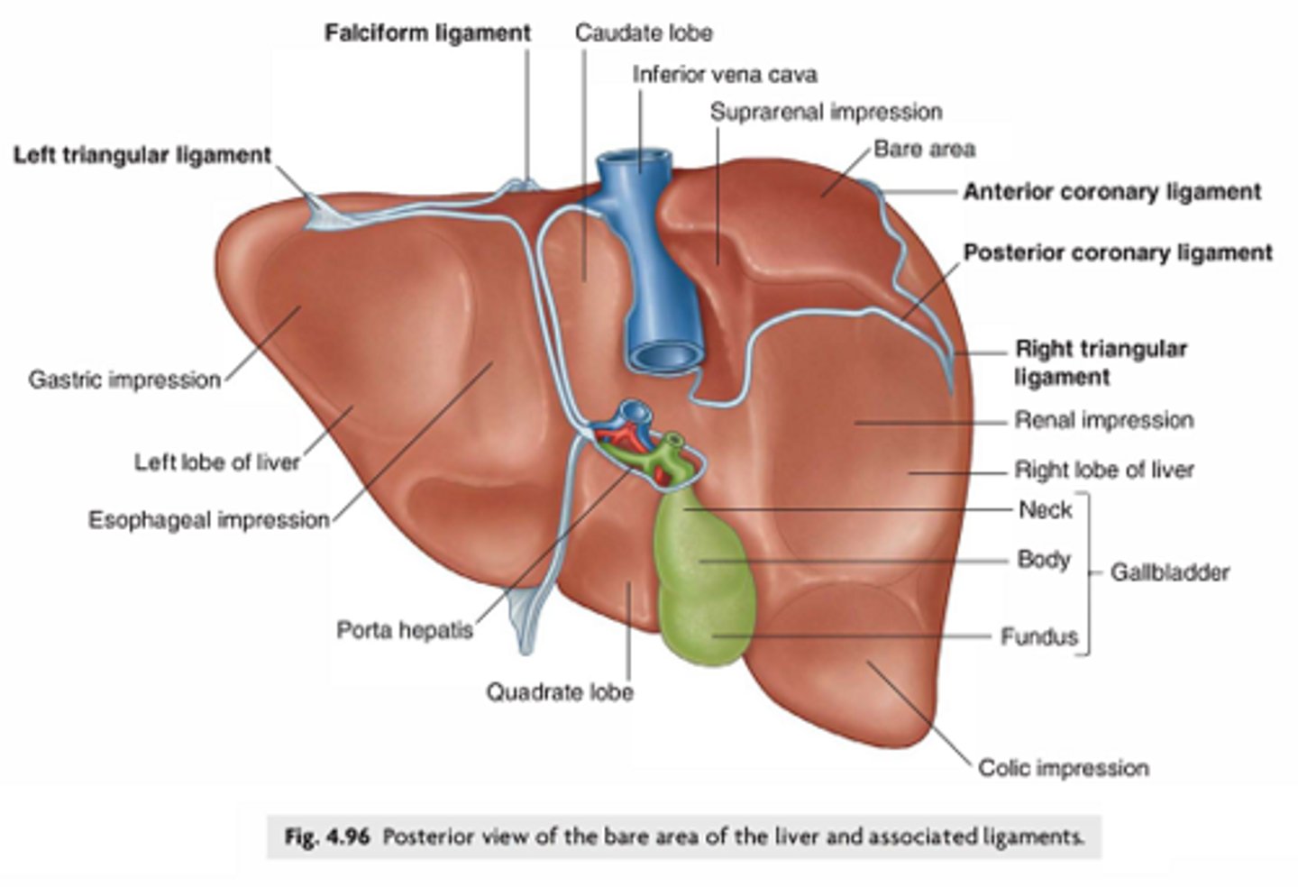
Inferior border of the liver
Boundary
- Bottom of liver, sharper edge
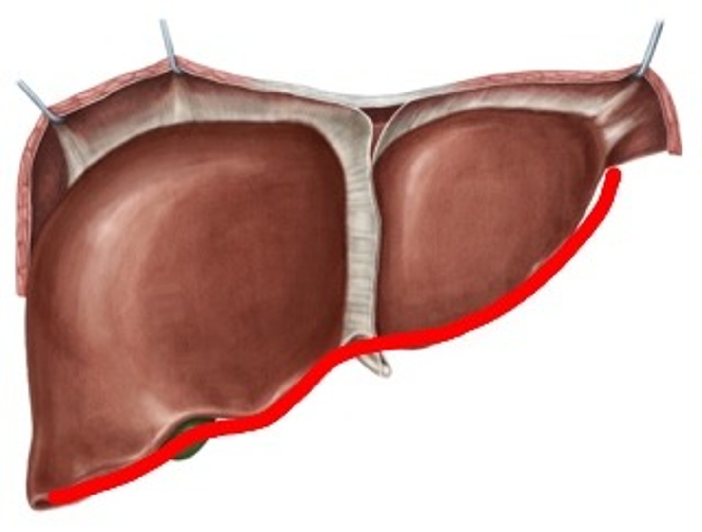
Falciform ligament
Structure
- Structure in between 2 lobes like a web
- Middle flap on the front
Anterior layer of the coronary ligament
Layer
- Connects the diaphragm and liver on the FRONT side
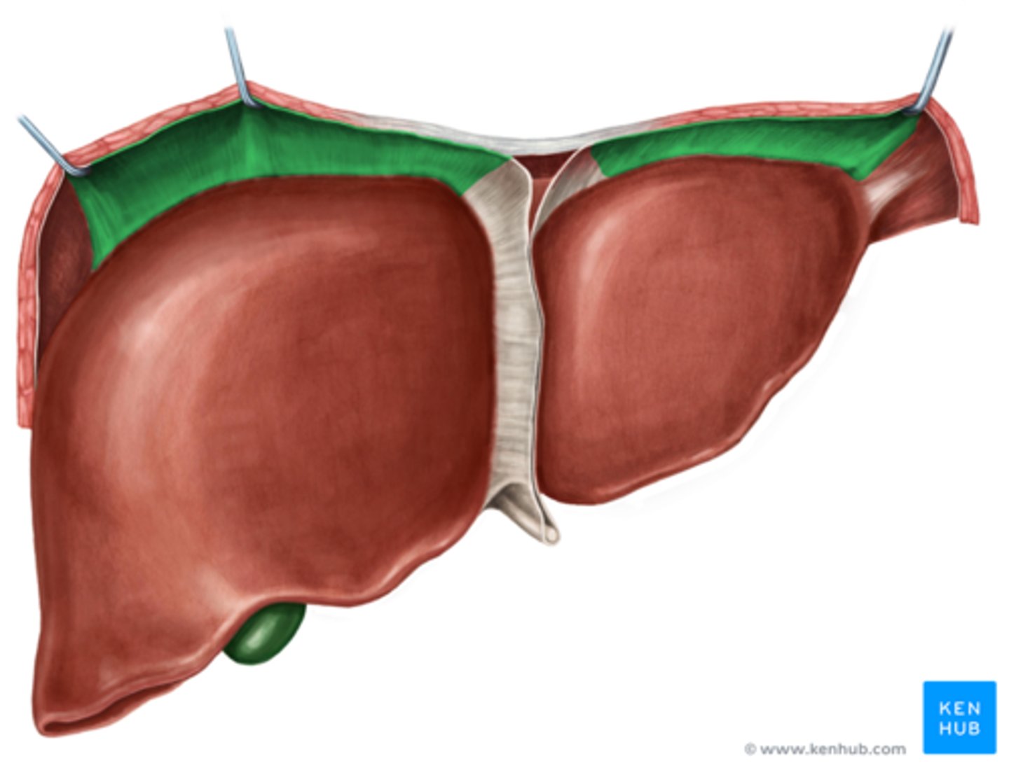
Posterior layer of the coronary ligament
Layer
- Connects diaphragm and liver on the BACK side

Bare area of the liver
Area
- Area that is split so you can see through the diaphragm
- TA will place probe in the slit in the coronary ligament to show
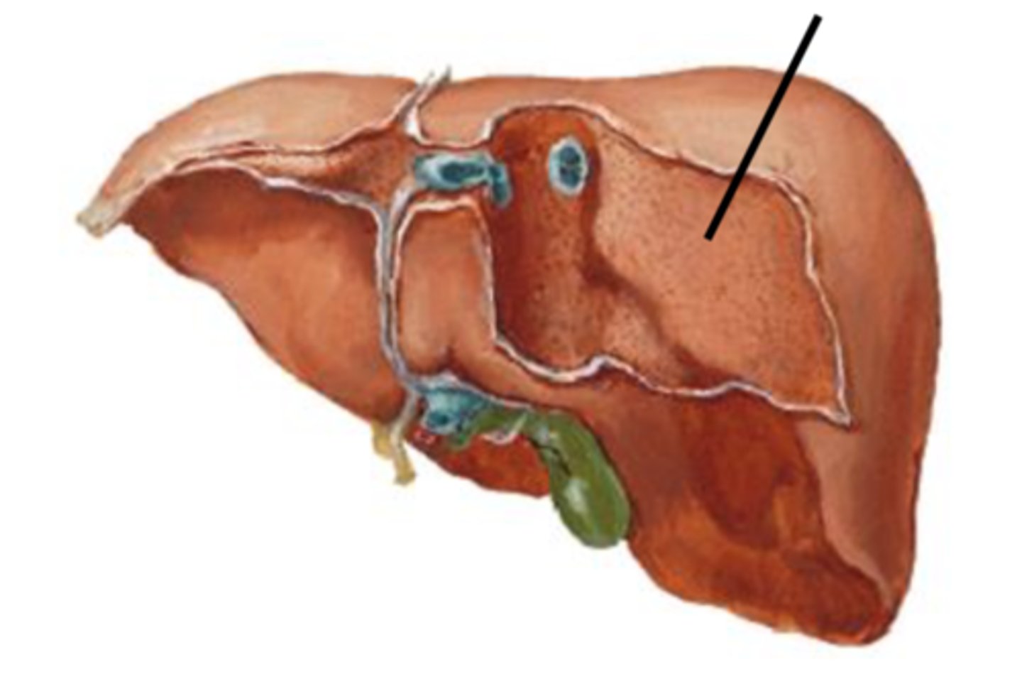
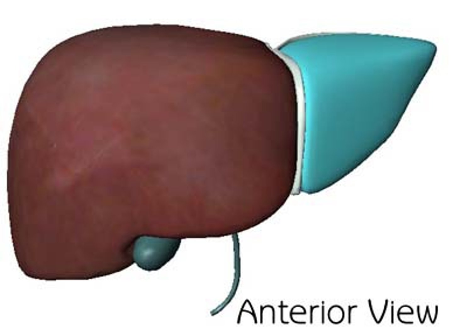
Right lobe of the liver
Structure
- Larger side
- On the right from posterior view
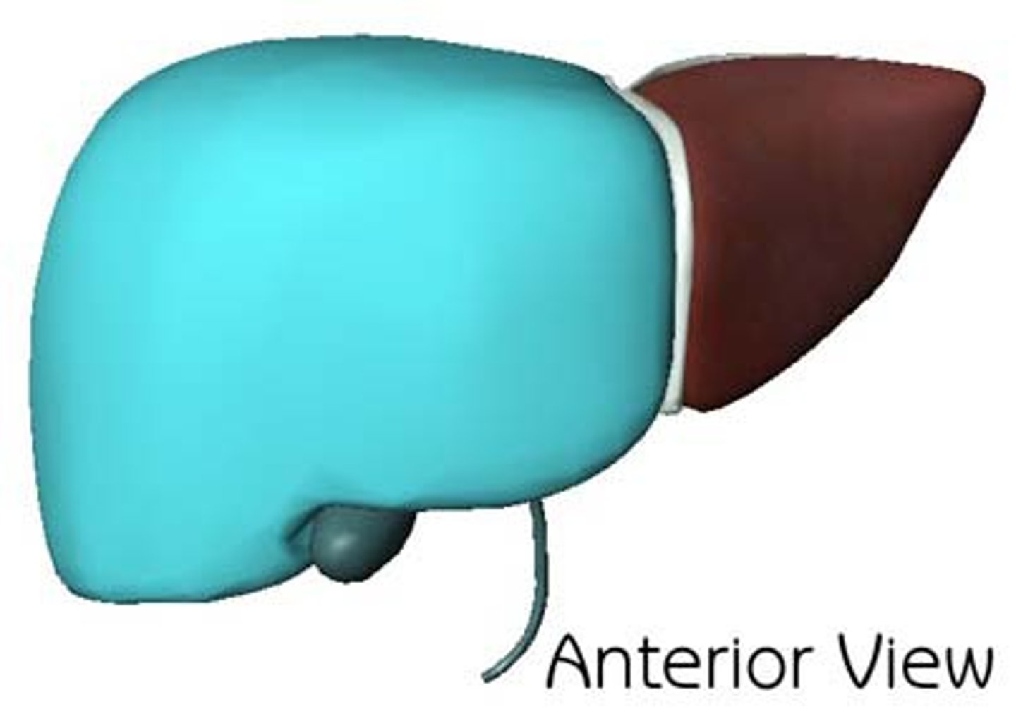
Left lobe of the liver
Structure
- Smaller side
- On left side from posterior view
Caudate lobe
Structure
- Top lobe in the middle
Quadrate lobe
Structure
- Bottom lobe in middle

Right sagittal fissure
Division
- right side of the H

Sulcus for the inferior vena cava
Depression
- Top half of the right side of H
Fossa for the gallbladder
Depression
- Bottom right side of H
- Where the gallbladder would sit
Left sagittal fissure
Divison
- Left side of the H
Fissure for round ligament
Depression
- Bottom left side of H
Round ligament of liver
Structure
- Bottom left side of H
- TA will tap it to show

Fissure for ligamentum venosum
Depression
- Top left of H

Ligamentum venosum
Structure
- That fills that depression
- top left of H
Porta hepatis
Division
- Middle of the H
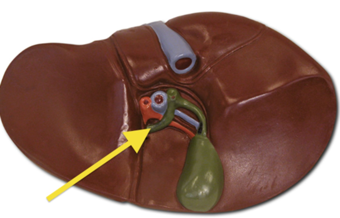
The hepatic "H"
Collective division
- The entire H
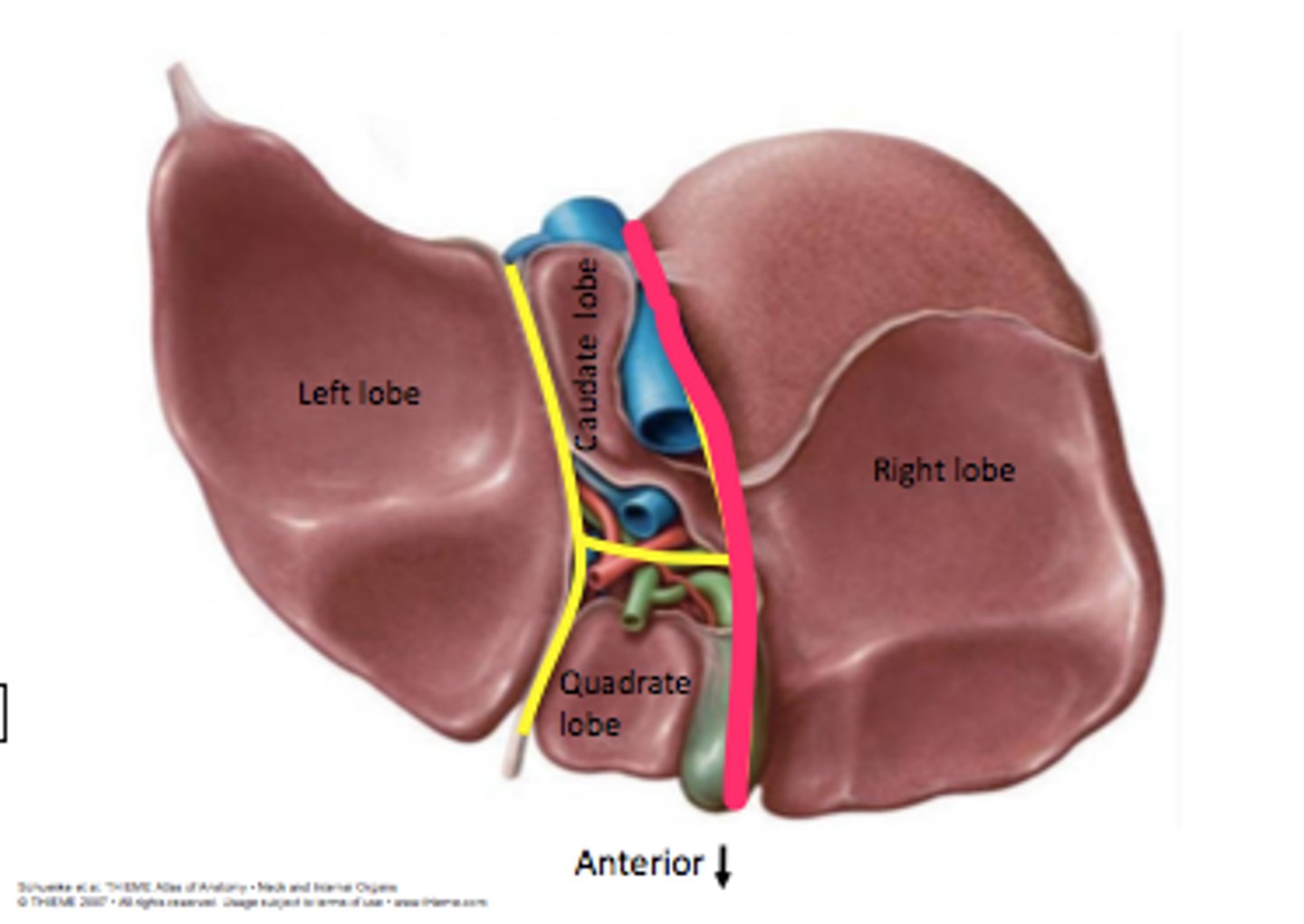
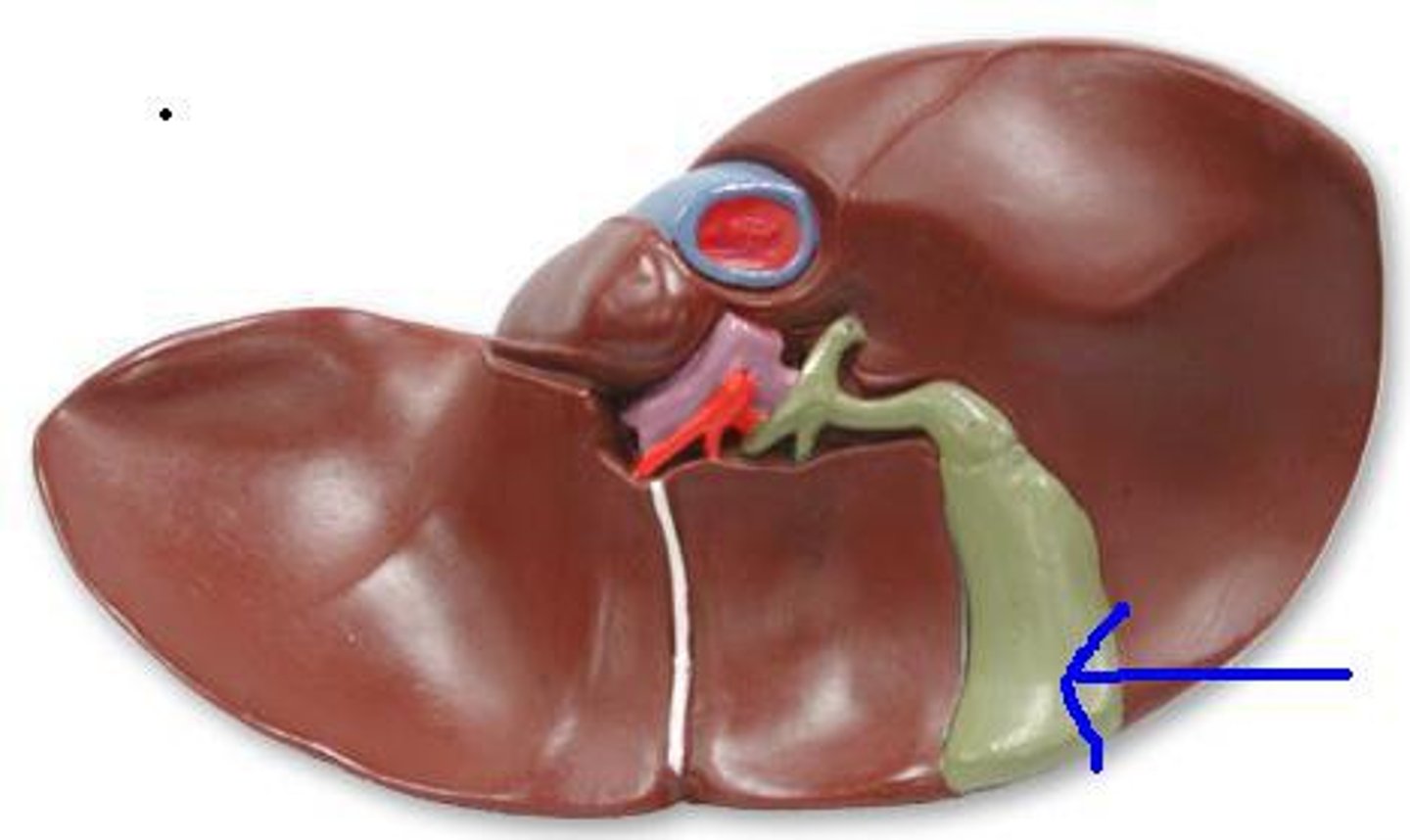
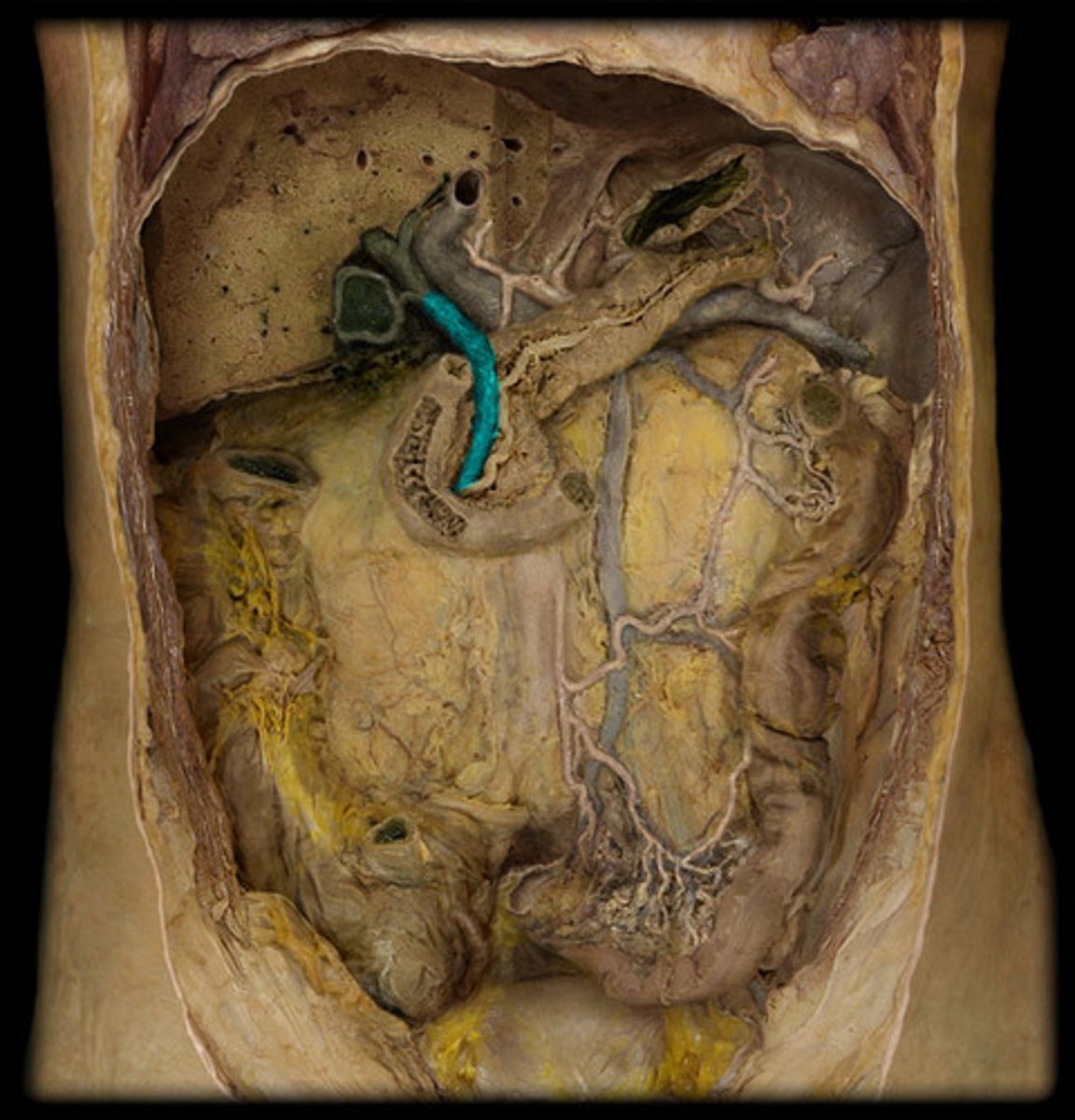
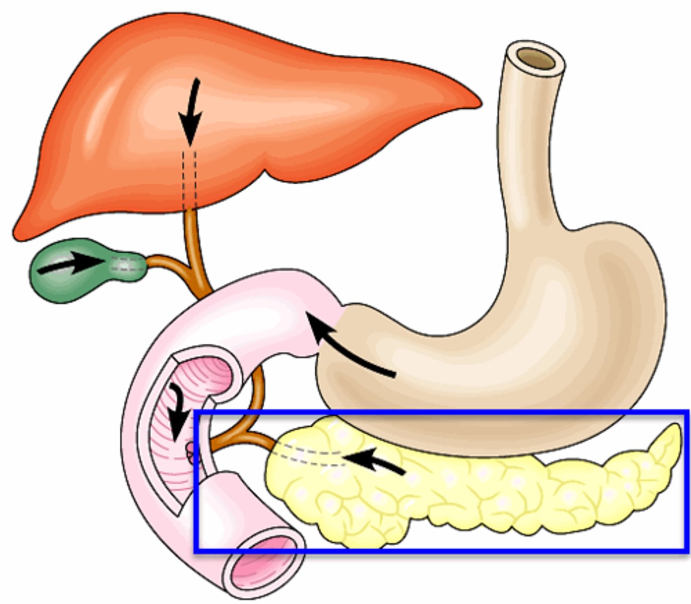
Gallbladder
Structure
- Dark sac
- Deflated balloon
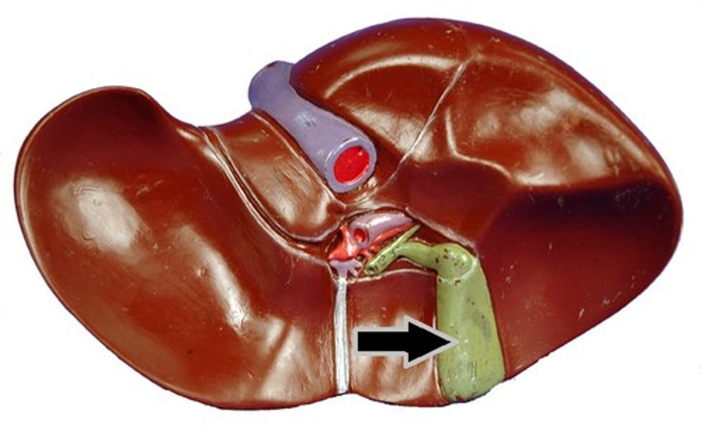
Fundus of the gallbladder
Feature
- Bottom curved portion of the gallbladder
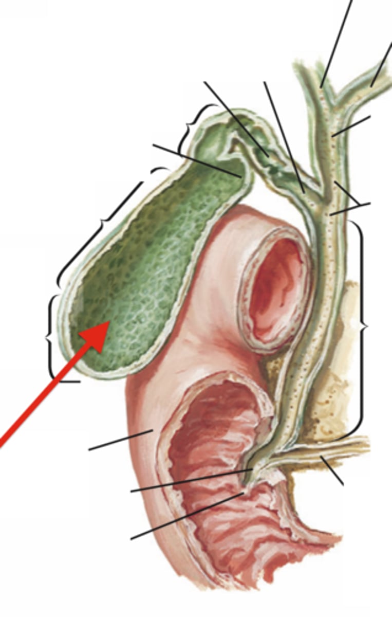
Cystic duct
Structure
- Comes straight off of the gallbladder going up
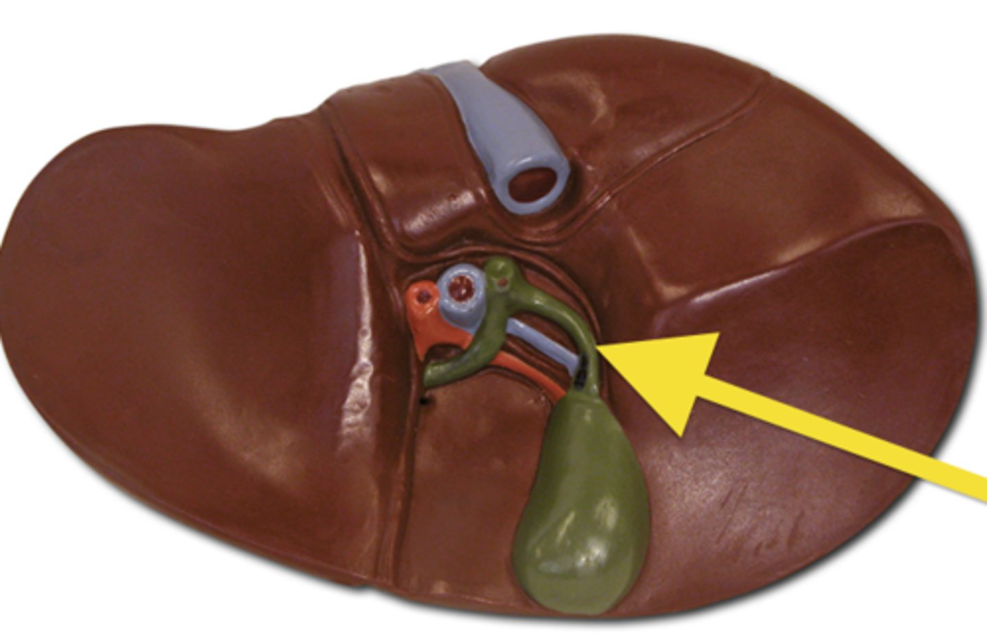
Common hepatic duct
Structure
- Part of the cystic duct that continues straigt up
- Structure comes up from the cystic duct
- TA FAVE
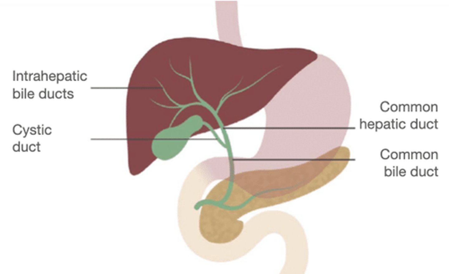
Bile duct
Structure
- Comes off of cystic duct going down
Hepatopancreatic ampulla
Structure
- Bulb at the end of the bile duct and start of the duodenum intestine
Major duodenal papilla
Opening
- On the inside of pancreas
- TA will stick the probe inside the duodenum to show
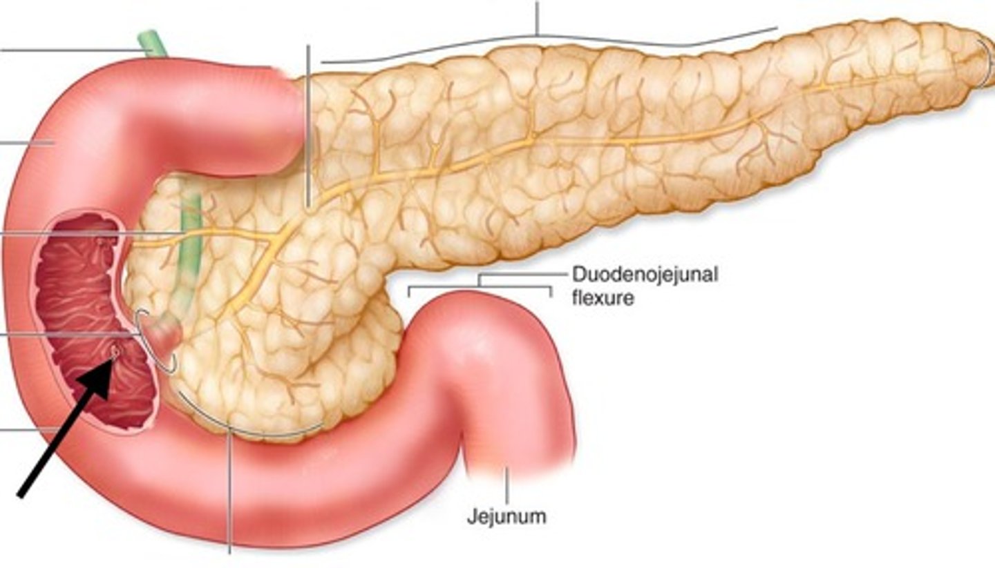
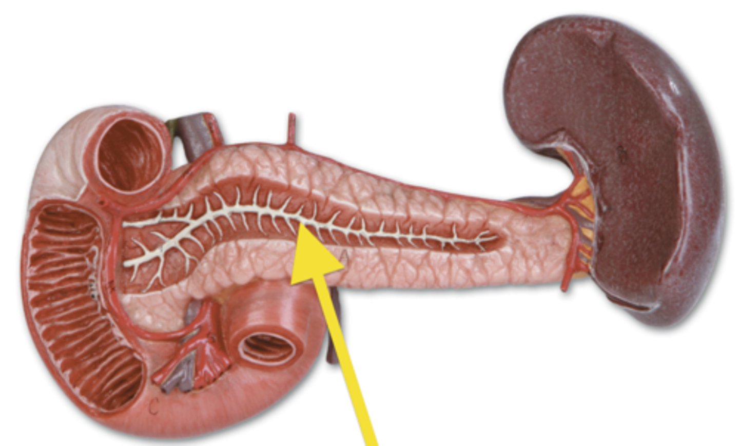
Pancreas
Structre
- Mangled looking structure
Head of the pancreas
Feature
- Shrimp tail at the top
- Medial
Uncinate process of the pancreas
Feature
- Curled part on the head of pancreas
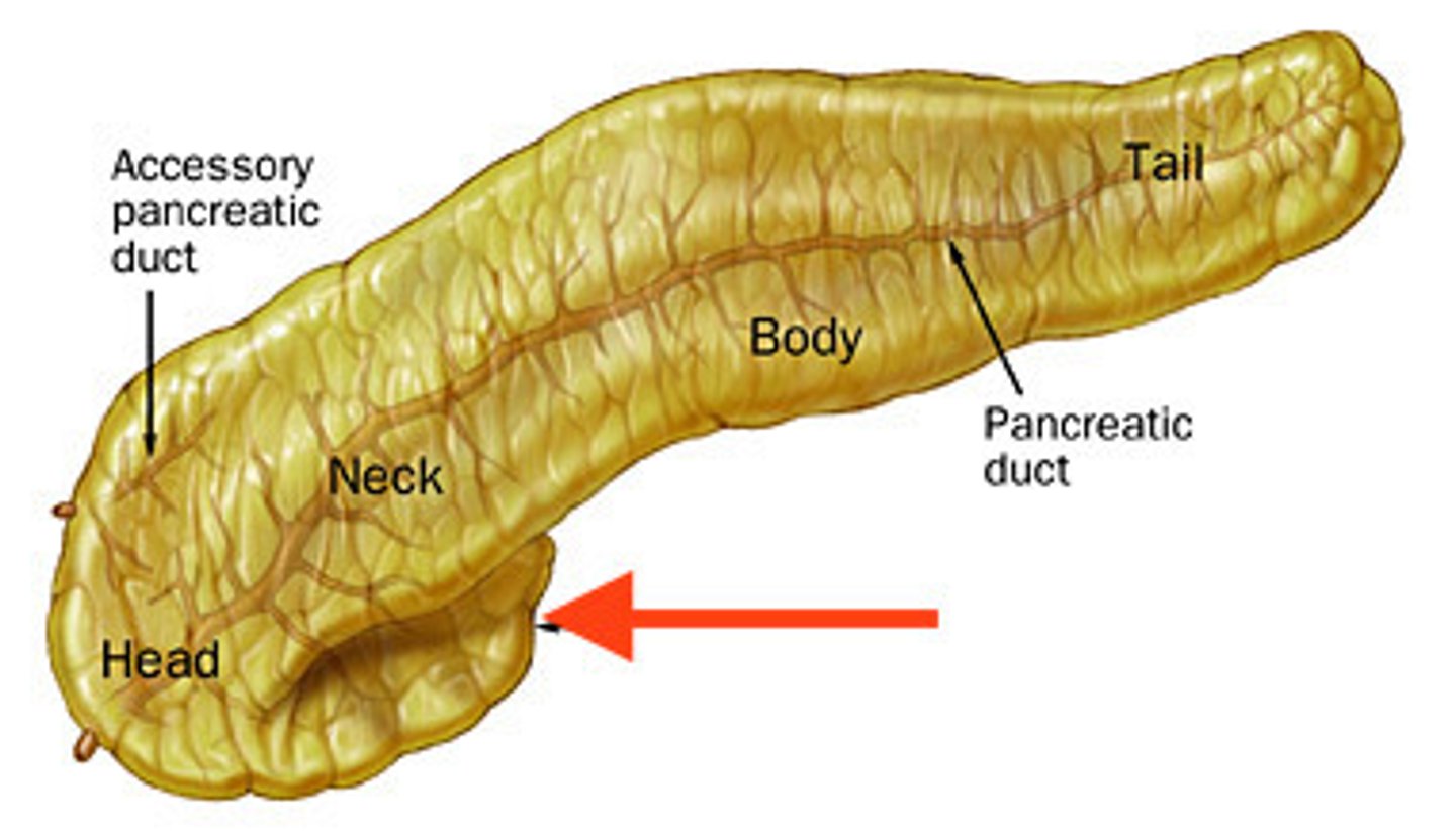
Body of the pancreas
Feature
- Lateral to the head
- Middle portion
Tail of the pancreas
Feature
- Tip of the pancreas
- most lateral
Pancreatic duct
Structure
- TA will flip pancreas over to show
- Middle lighter line