Life Support Lecture 1:
1/62
There's no tags or description
Looks like no tags are added yet.
Name | Mastery | Learn | Test | Matching | Spaced | Call with Kai |
|---|
No analytics yet
Send a link to your students to track their progress
63 Terms
What are the functions of the Respiratory System
Gas Exchange, regulation of pH, Olfaction, Voice production, and protection against microorganisms and pathogens
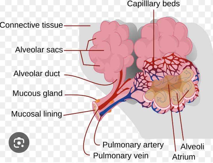
Alveoli
the tiny air sacs in the lungs that have a hige surface area and very thin walls which allow for the diffusion of O2 and CO2. They are surrounded by capilaries
How do the lungs regulate the pH in the body
by controlling the levels of carbon dioxide in the blood. If there is a higher level of CO2 found in the body, it lowers the pH making the body more acidic and vise versa with basic
how does the respiratory system protect the body?
Physical barriers such as nose hairs and mucus, and airflow in the nasal cavity prevent pathogens from coming in
If they do end up entering the body it is removed by mucus trapping particles and cilia (tiny hair-like structures) moves the mucus upwards which leads to the pathogens being swallowed or expelled
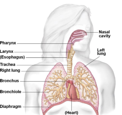
Nasal Cavity:
Prevents pathogens for coming in as it is protected by a mucous membrane that produces mucus. Mucus contains cilia
Together, these components form a protective mechanism, where mucus traps dust, bacteria, and foreign particles, while cilia sweep this mixture toward the throat to be swallowed
What is the Respiratory Tract made up of?
Upper respiratory tract (nose to larynx) and Lower respiratory tract (trachea to lungs)
Why is an infection in the lower respiratory tract worse than one in the upper respiratory tract?
This is because they affect the lungs and airways, directly impairing oxygen exchange and their dysfunction cannot be easily compensated (replaced), making damage life-threatening
Dysfunction of gas exchange leads to low oxygen levels in the blood → Hypoxia → lead to organ failure
Treat with antibioitics
Hypoxia
a dangerous medical condition where body tissues are deprived of adequate oxygen supply, potentially leading to organ damage or death
Nose (Upper Tract)
Wars and moistens air, it has a palatine bone which seperates the nasal cavity from the mouth
Nostrils have stiff guard hairs, or vibrissae that prevent insects and large airborne particles from entering
Nasal Septum
A wall that separates the left and right nostril, a rich blood supply leads to nose bleeds
Paranasal Sinuses
Air filled cavities in the skill that open (drain) mucus into the nasal cavity through small openings called ostia.
There are 4 pairs/cavities in the sinuses → Frontal, Ethmoid, Sphenoid and Maxillary
What are the functions of the sinuses?
lighten the skull by replacing dense, heavy bone with air-filled cavities, acting as structural "hollows" within the facial skeleton
The four paired paranasal sinuses—frontal, sphenoid, ethmoid, and maxillary—drain mucus into the nasal cavity through small openings called ostia (This drainage helps maintain humidification and immune defense)
Sites where air is warmed and its velocity is slowed to permit particles to optimise airflow and allows olfactory sensations to occur
Maxillary Sinuses:
Largest sinuses located under the eyes in the cheek area
Maxillary Bones
The maxillae are the upper jaw bones.They hold the upper teeth and form part of the nose and eye sockets
The maxillary sinuses are inside the maxillary bones
Pharynx/Throat (Upper Respiratory Tract)
Muscles of the pharynx facilitate swallowing and contribute to speech
What are the 3 Components of the Pharynx and their Functions?
Nasopharynx: Functions with air passage and located behind nasal cavity
Oropharynx: Passage of air, fluids and liquids and located behind the mouth
Laryngopharynx: has a shared pathway for food and air which then splits into the Larynx and the Oesophagus
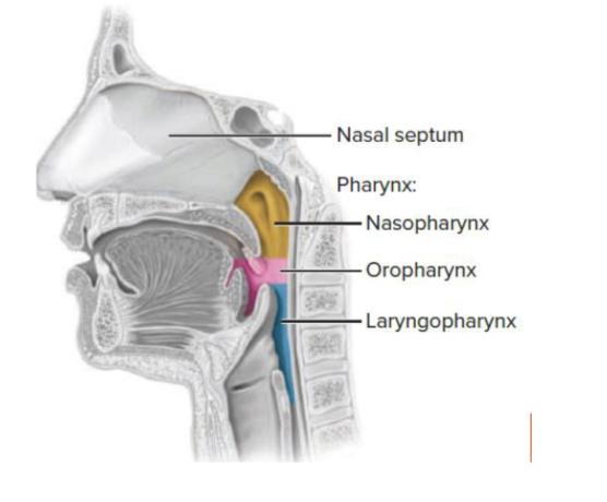
What system are the Oropharynx and the Laryngopharynx apart of?
Since they work with passage of food they are part of the digestive system and they are lined with stratified squamous epithlium (a multi-layered protective tissue)
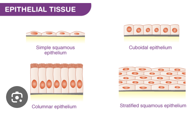
What is an Epithelium?
a fundamental, avascular tissue type composed of densely packed cells that covers external body surfaces (skin), lines internal cavities, and forms glands
What is the Epiglottis?
a flap of cartilage that during swallowing the flap folds down over the larynx (airway and voice box) which prevents food from entering the lungs and food is directed down the oesophagus
If food enters the larynx it triggers a cough reflex to expel it
What is the Larynx?
chamber composed mainly of cartilage (9) and muscle and it acts as a vital pathway for air, produces sound via vocal cord vibration, and protects the lower airways by closing during swallowing
What is the Glottis?
the part of the larynx consisting of the vocal cords and the opening between them. It affects voice modulation through expansion or contraction.
Opening is caused by airflow through the glottis which vibrates the vocal cords and produces sound
Sounds are produced when muscles in the larynx are close/tensed
(Not a cartilage)
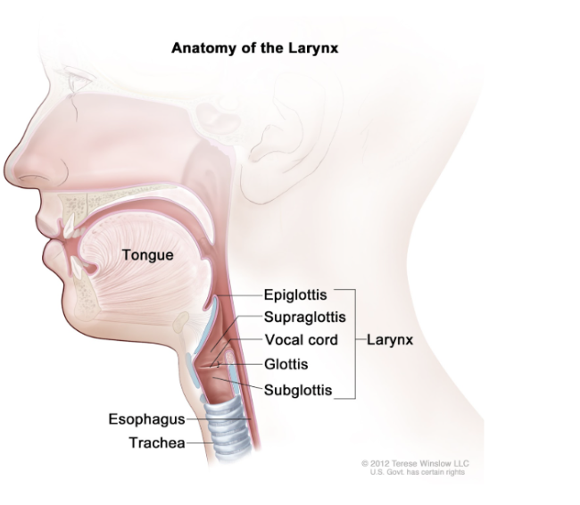
What is the Hyoid Bone
It is where the tongue is located
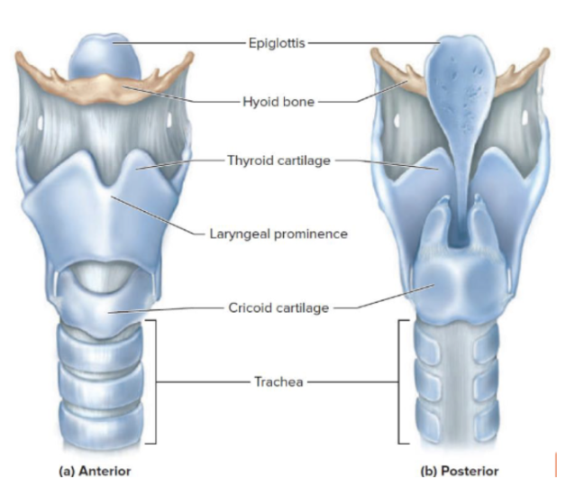
Trachea (Lower Respiratory Tract)
Acts as the main airway, transporting air between the larynx and lungs and it 12 cm long and 2.5cm in diameter
At its inferior (bottom) end, it forks into right and left bronchi
What is the Hyaline Cartilage?
Its what makes up the trachea in 16-20 C-Shaped rings
It is imporant as it keeps the trachea open during inhalation. When we inhale the pressure inside our airway drops and without support the tube could collapse
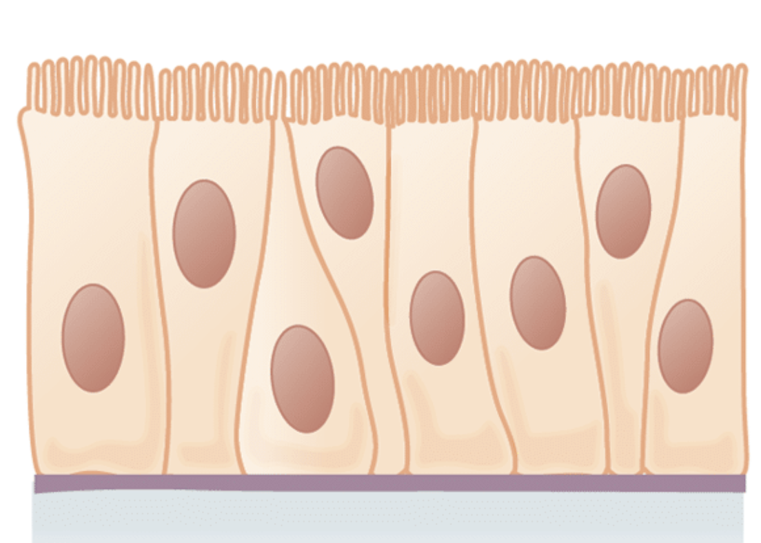
What type of tissue is lined in the trachea
It's lined with a ciliated pseudostratified columnar epithelium rich in mucus-producing goblet cells
Pseudostratified: Looks like multiple layers, but actually all cells touch the basement membrane so it is one single layer
Columnar: Cells are tall and rectangular
Goblet cells: Specialized epithelial cells that in this case produce and secrete mucus
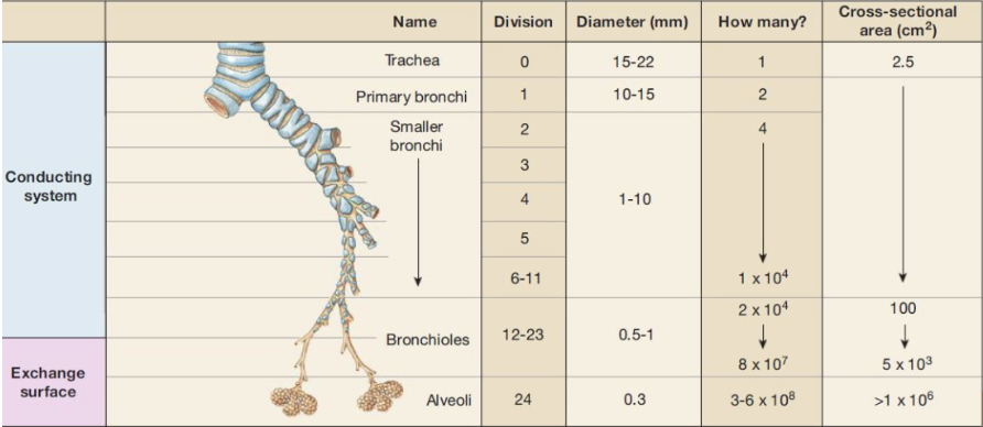
What is the Bronchial Tree?
a series of branching passages that carry air from trachea to gas-exchange surfaces in the lungs (alveoli) (includes trachea, bronchi and bronchioles)
bronchi branch into smaller passageways leading to tubes called bronchioles and terminal bronchioles
Conducting Pathway: Trachea → Primary bronchi (right & left) → Secondary (lobar) bronchi → Tertiary (segmental) bronchi → Bronchioles → Terminal bronchioles
What is the Function of the Lungs?
facilitate gas exchange through alveoli, bringing oxygen into the bloodstream and removing carbon dioxide waste through breathing
It is cone shaped with an apex and base
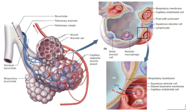
What is the function of Alveoli?
This is done through the alveoli (sacs within the lungs where gas exchange occurs and is surrounded by capillaries which allows for the diffusion of gas
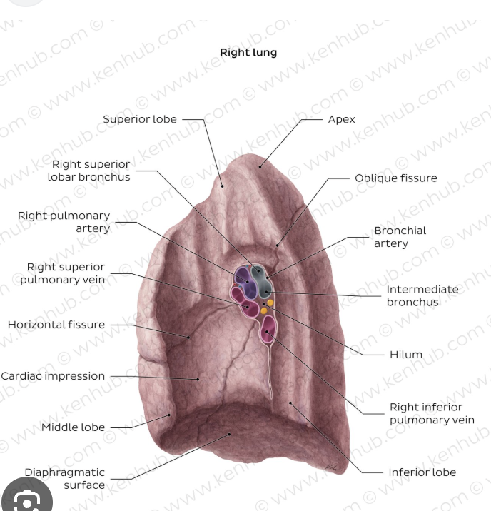
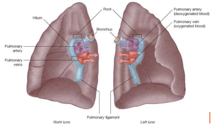
What is the Hilum?
a slit on the lung on the mediastinal surface through which it receives and exits bronchi, blood vessels, nerves and lymphatics
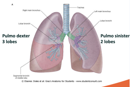
What are the lobes on the lungs that make them asymmetrical?
Right lung (on patient): There are 3 lobes, the superior, middle and inferior
Pulmo dexter refers to the right lung
Left lung (on patient): only two lobes because heart is also occupied here, the superior and inferior
Pulmo sinister refers to the left lung
What is the Pleurae?
Two-layered membrane that surrounds the lungs
Parietal layer connects to and lines the thoracic wall (outer layer)
Puncture of parietal pleurae may result in collapsed lung (pneumothorax)
Visceral layer connects to and covers the lung (inner layer)
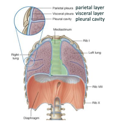
What is the Thoracacic Wall?
musculoskeletal cage (ribcage) that protects the thoracic cavity's vital organs, including the heart, lungs, and great vessels
What is the Pleural Cavity (Located on Photo Above)
Pleural cavity contains thin layer of fluid allowing lungs to move freely against thoracic wall
The fluid functions as lubrication (reduces friction during breathing), adhesion (keeps lungs “stuck” to chest wall) and helps lungs expand with the thoracic cavity
The pleural cavity has negative pressure which keeps the lungs expanded and prevents collapse. Therefore a puncture would replace the fluid/negative pressure and lead to a collapse.
Extra Information on the Alveoli
Each lung contains 150 million alveoli, which provide a large surface area for gas exchange
Each alveolus is covered with a web of blood capillaries
The capillaries create extremely close contact between air and blood
Alveoli produce surfactant through the alveolar epithelial cells which coats the alveoli and reduces surface tension
Provide a very large surface area for gas exchange
How do Oxygen and Carbon Dioxide diffuse in and out of the body?
Oxygen is diffused into alveoli → capillary → red blood cells
Carbon Dioxide moves from blood → alveoli → exhaled
What is the Function of the Red Blood Cells in Oxygen Exchange?
The red blood cells contain hemoglobin which bind to oxygen once it diffuses into capillaries. It then transports the oxygen to the tissues
What is the Function of the White Blood Cells in Oxygen Exchange?
White blood cells are active immune cells that protect the alveoli as they are sensitive
They patrol the alveolar surface so no pathogens reach the alveoli, but if something does the white blood cells can act against it
If alveoli does gets destroyed it causes issues by reducing surface area and can impair gas exchange which leads to hypoxia (low oxygen levels)
The Effect of COVID on Alveoli
SARS-CoV-2 infects alveolar epithelial cells, particularly type II cells, leading to inflammation and fluid accumulation in the alveoli.
alveolar epithelial cells produce surfactant and help repair alveoli
This thickens the respiratory membrane and reduces gas exchange efficiency, causing hypoxia.
Although some repair is possible, severe damage can result in fibrosis and long-term impairment, contributing to respiratory failure.
What is Pulmonary Ventilation?
Breathing it includes inspiration (inhaling) and expiration (exhaling)
One complete inspiration and expiration is called a respiratory cycle
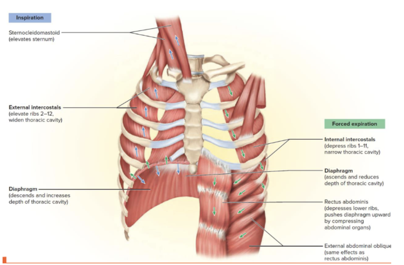
What Muscles are involved in the Respiratory Cycle?
Diaphragm (below the lungs and heart) and Intercostal muscles (External and Internal Intercostals which are located between ribs)
What is Intrapulmonary Pressure?
Pressure difference between the lungs and the atmpsphere which allow for airflow
Airflows from high pressure to low pressure
What happens during Inspiration?
During inspiration the diaphragm contracts (moves downwards and flattens) and the external external intercostal muscles contract (ribs move up and outwards)
Results in thoracic cavity volume increases, pressure inside lungs decreases and air flows into the lungs
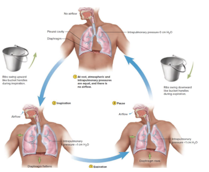
What happens during Expiration (non-forced)?
During expiration the diaphragm relaxes (moves upward into a dome shape) and the ribcage moves downwards and inwards
Results in thoracic cavity volume decreasing, pressure inside lungs rises above atmospheric pressure and air flows out of the lungs
What happens during Expiration (Forced)?
During forced expiration internal intercostal contract (depress the ribs) and abdominal muscles contract (push the diaphragm upwards) may assist
What is the Hilum?
Functions to act as the critical anatomical gateway and central "root," connecting the lungs to the mediastinum
Each lung has its own hilum and mediastinum
Bronchi (airways), Pulmonary arteries, Pulmonary veins, Nerves and Lymphatic vessels all pass through the hilum
What are Arteries
Carry blood away from the heart
What are Veins?
Carry blood towards the heart
What is the Pulmonary Trunk?
Large vessel leaving the right ventricle
Splits into pulmonary arteries
What is the Pulmonary Artery?
Carry deoxygenated blood
From heart to lungs
What are the Pulmonary veins?
Carry oxygenated blood
From lungs to left atrium of heart
What is Stridor?
a high-pitched, wheezing sound caused by disrupted airflow due to a narrowed or obstructed upper airway
What does the anterior part of the laryngopharynx lead to?
Leads to larynx → trachea → lungs (air) (is larger on the diagram)
What does the posterior part of the laryngopharynx lead to?
Leads to oesophagus → stomach (food)
What is the difference between airways of adults and children
Airway of children is narrower and shorter than adults
Disproportionally bigger tongue of children
Epiglottis is more floppy of children
What can cause an obstruction to the airway?
Tongue
Fluids → vomit, mucus and blood
Foreign Materials → food and toys
Swelling of tissue → infection, anaphylaxis (allergy) and burns
External Compression → tumor, thyroid, abscess and trauma
What is Aspiration?
occurs when food, liquid, saliva, or stomach contents enter the airway and lungs instead of the oesophagus, often causing coughing, wheezing, or choking
In unconscious people he epiglottis closes the airway
What can Aspiration Cause?
Leads to aspiration pneumonia which is a chemical lung injury that causes severe inflammation in the alveoli leading to impaired gas exchange
Usually a cough reflex occurs, but in unconscious patients (since they can’t use reflexes) gastric contents can enter the airway and reach the lungs
What are Adjuncts?
devices used to establish and maintain a patent airway during basic and advanced life support, preventing the tongue or soft tissues from obstructing the pharynx
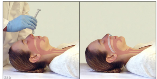
Oropharyngeal Airway:
Administered through the mouth and prevents the tongue from falling back and obstructing the airway
Only administered on unconscious patients as it triggers the gag reflex
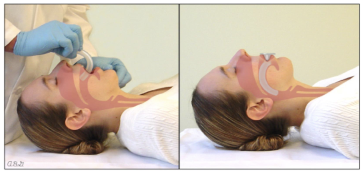
Nasopharygeal Airway:
Is inserted through the nose into the pharynx
Used on patients that are semi-conscious and have an intact gag reflex
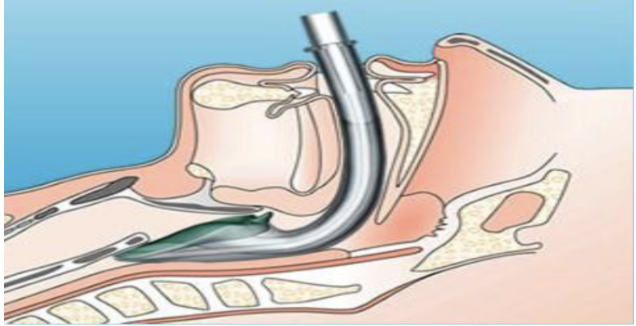
Laryngeal Mask Airway:
Is inserted through the mouth to above the larynx
Used on patients who are unconscious (no active gag reflex)
Forms a seal around the laryngeal inlet
Allows ventilation without intubation
However Does NOT fully protect against aspiration
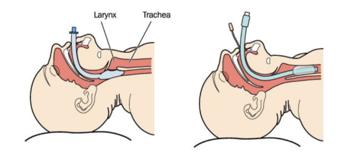
Endotracheal Tube:
Is inserted through the mouth and past vocal cords into trachea
A cuff is inflated inside the trachea and below the vocal chords to seal the trachea
There is a balloon at the end of the tip which is inflated behind the vocal chords so that the tube cannot move
With the balloon in place it pushes down the oesophagus a little bit, and it does not allow gastric contents to flow into the lungs which means aspiration is avoided
What occurs when none of the tubes work?
If we are not able to place one of these airways or can’t get air in or out of the patient, they do a surgical procedure where an incision is made over the throat and it’s membrane and a tube is placed directly into the trachea