Gross 2 Practical 1 All Wet Lab Structures
1/239
There's no tags or description
Looks like no tags are added yet.
Name | Mastery | Learn | Test | Matching | Spaced | Call with Kai |
|---|
No analytics yet
Send a link to your students to track their progress
240 Terms
platysma muscle
identify this muscle
sternocleidomastoid muscle
identify this muscle
external jugular vein
identify this structure
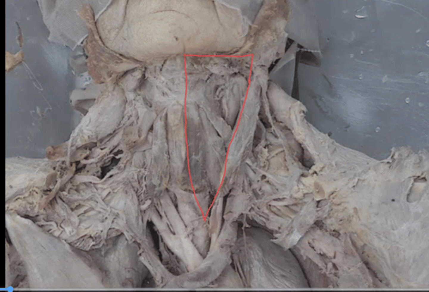
anterior triangle
identify this triangle
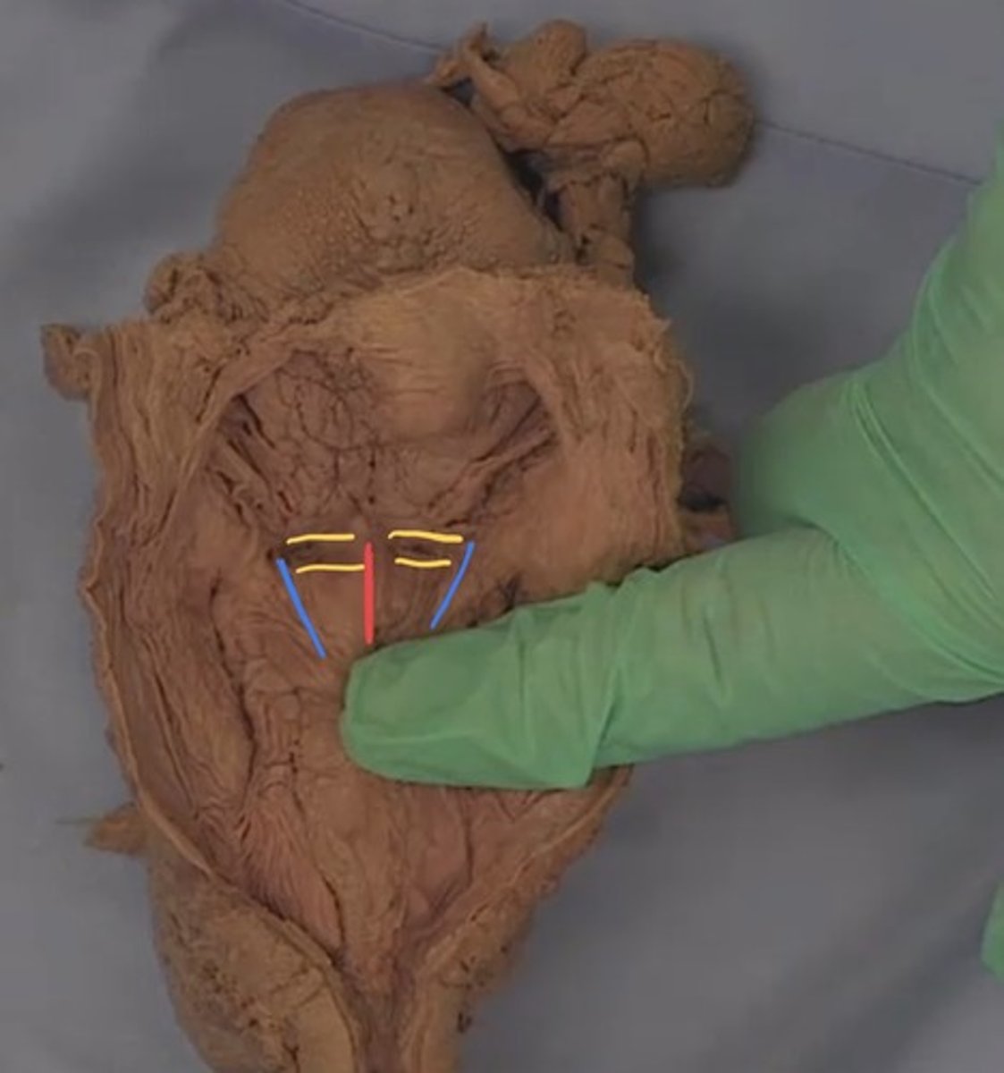
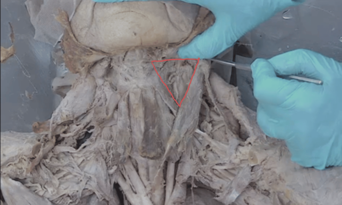
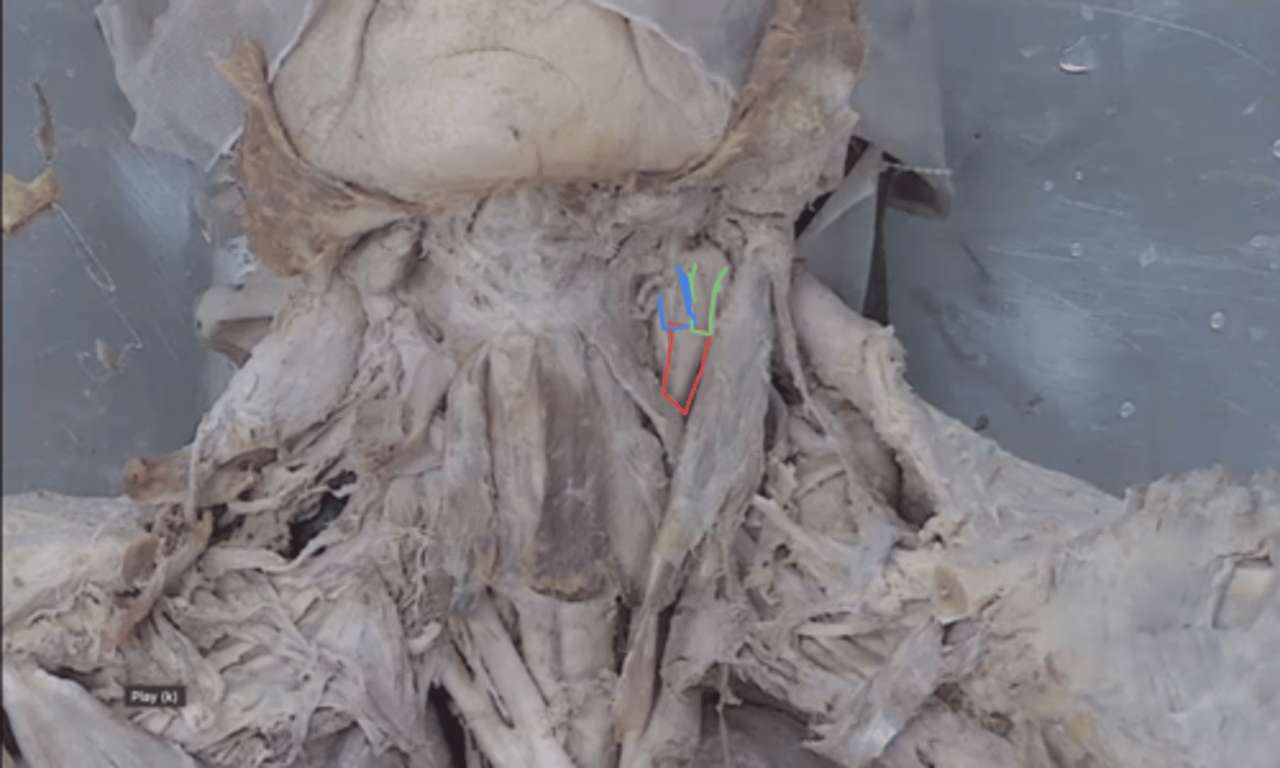
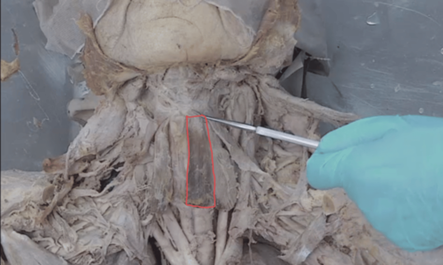
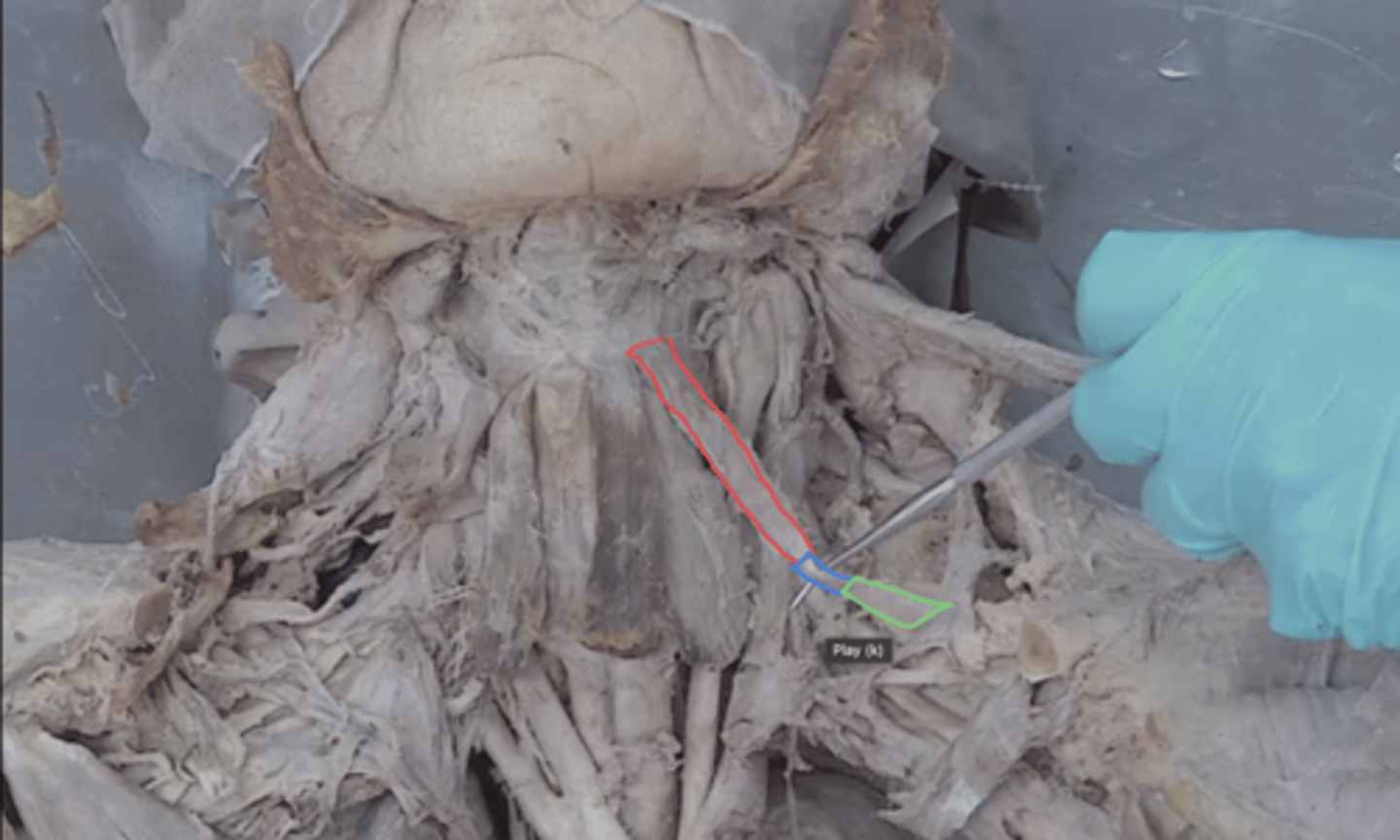
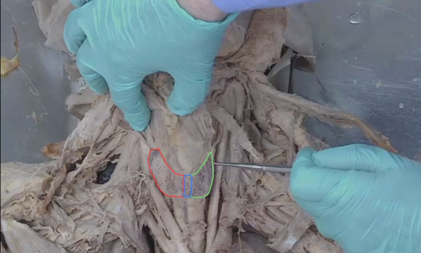
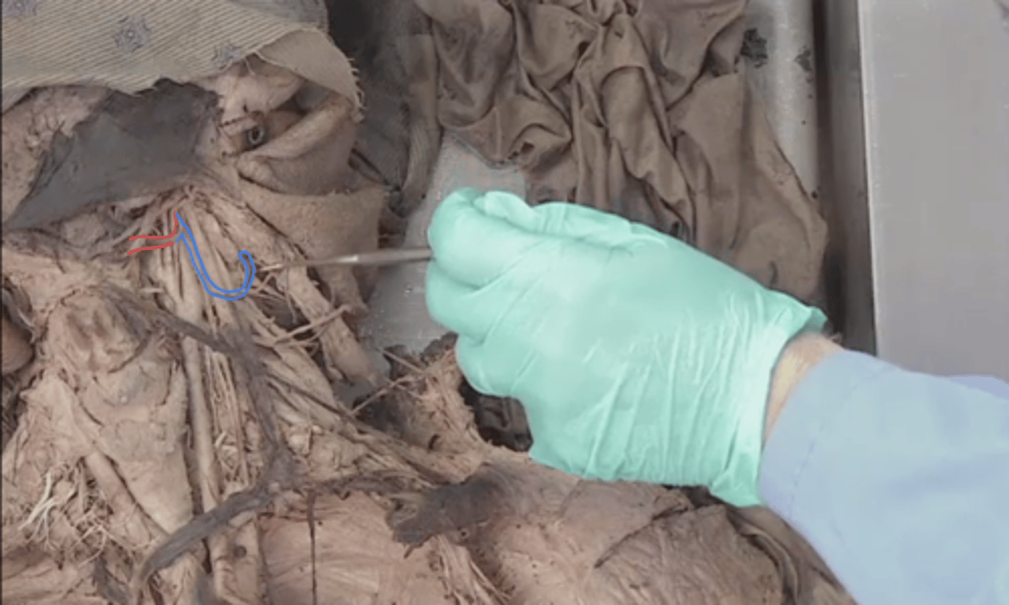
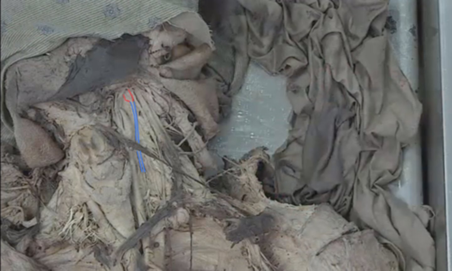
anterior belly of digastric muscle
what muscle is the red outline
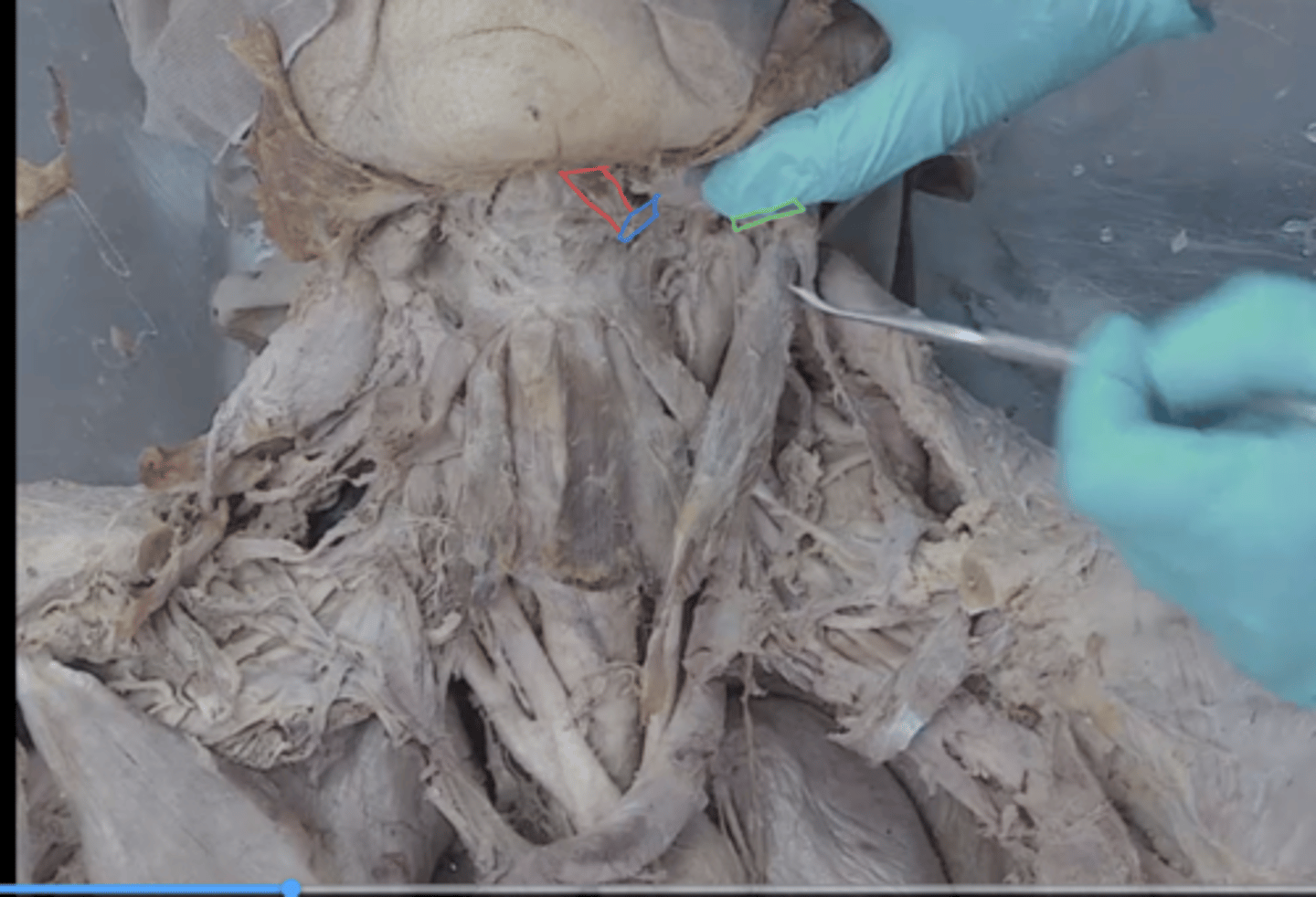
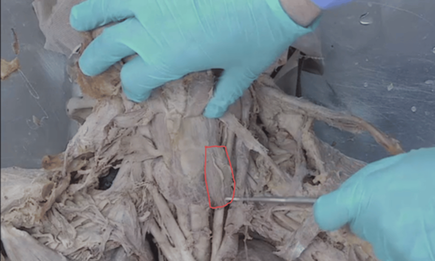
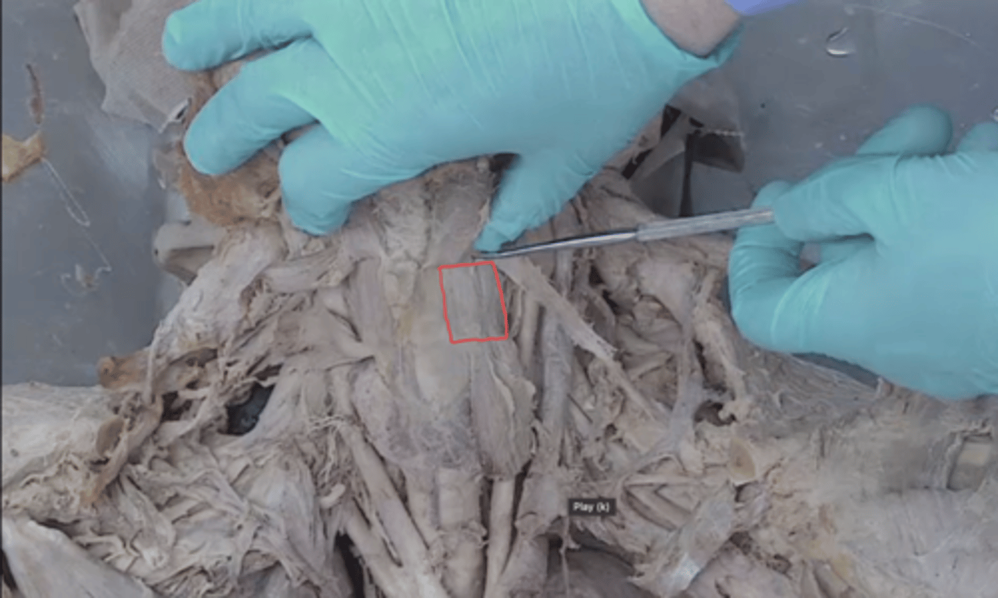
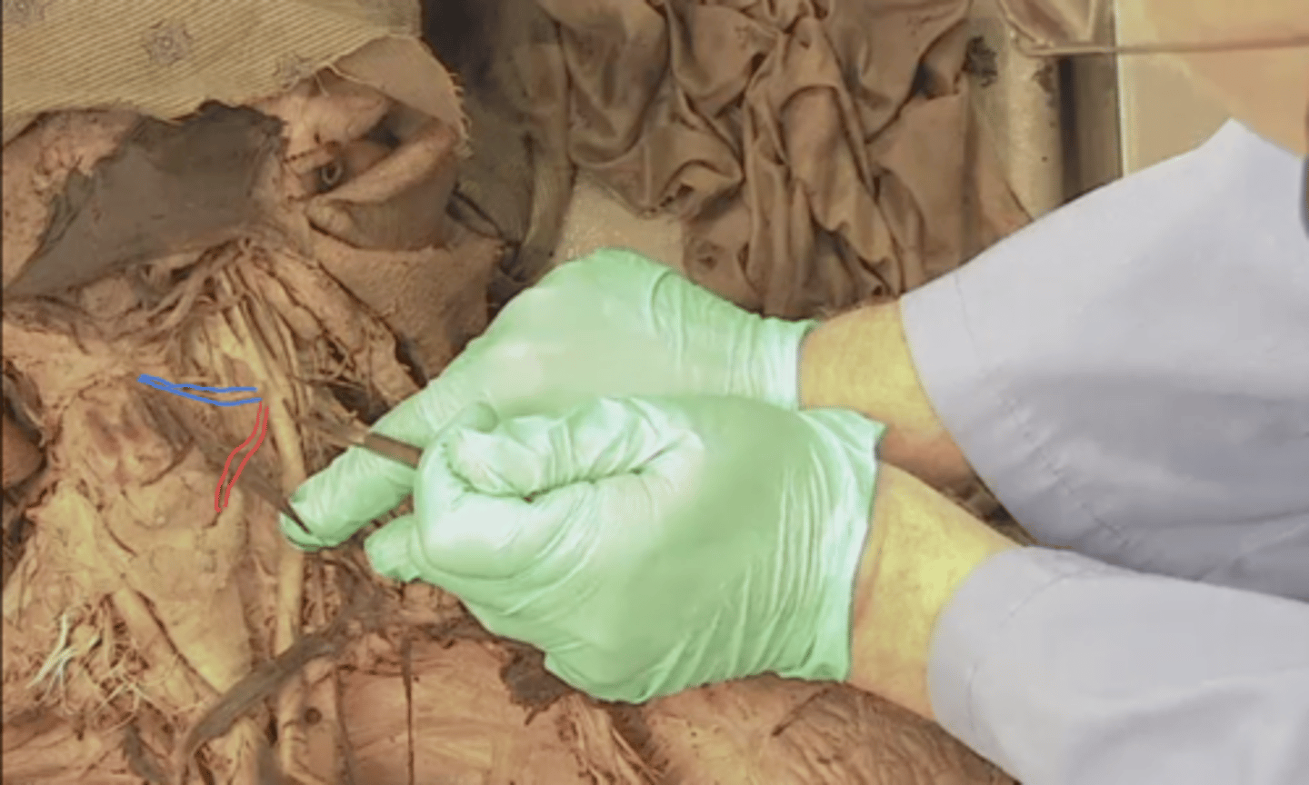
intermediate tendon of digastric muscle
what structure is the blue outline

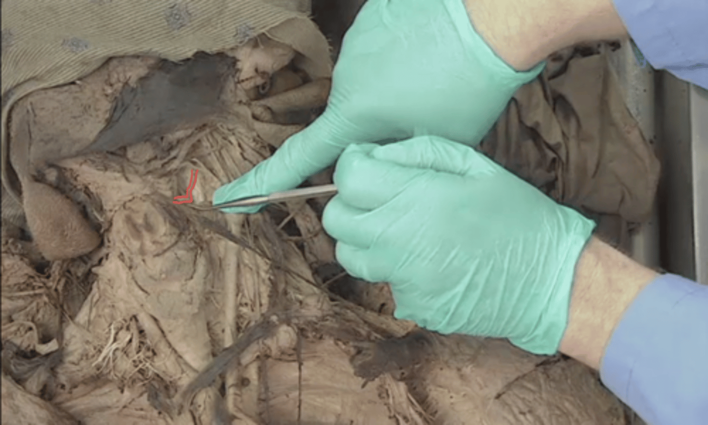
posterior belly of digastric muscle
what muscle will be seen here in green if I dive deep with probe

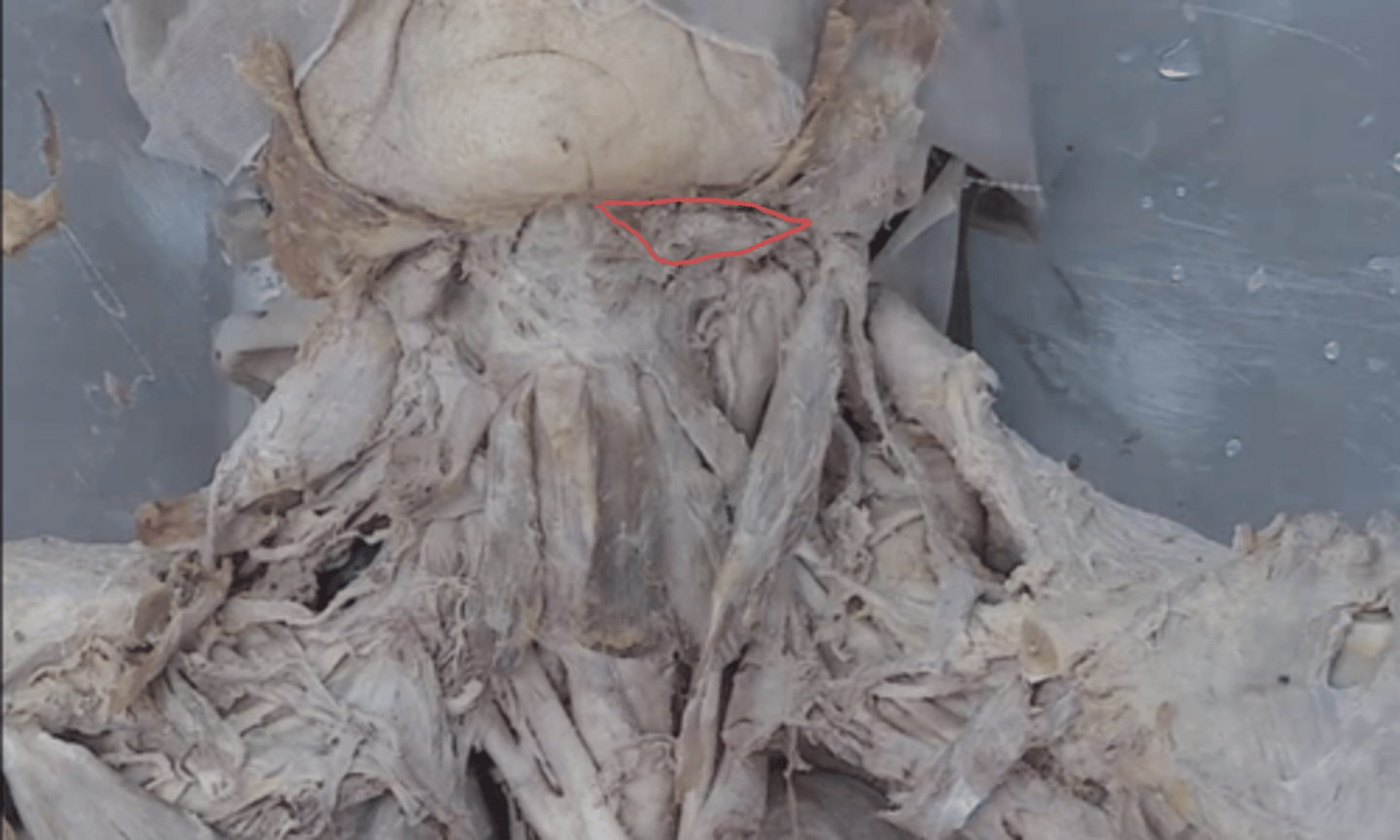
submandibular triangle
identify this triangle
submandibular gland
identify the structure outlined in blue

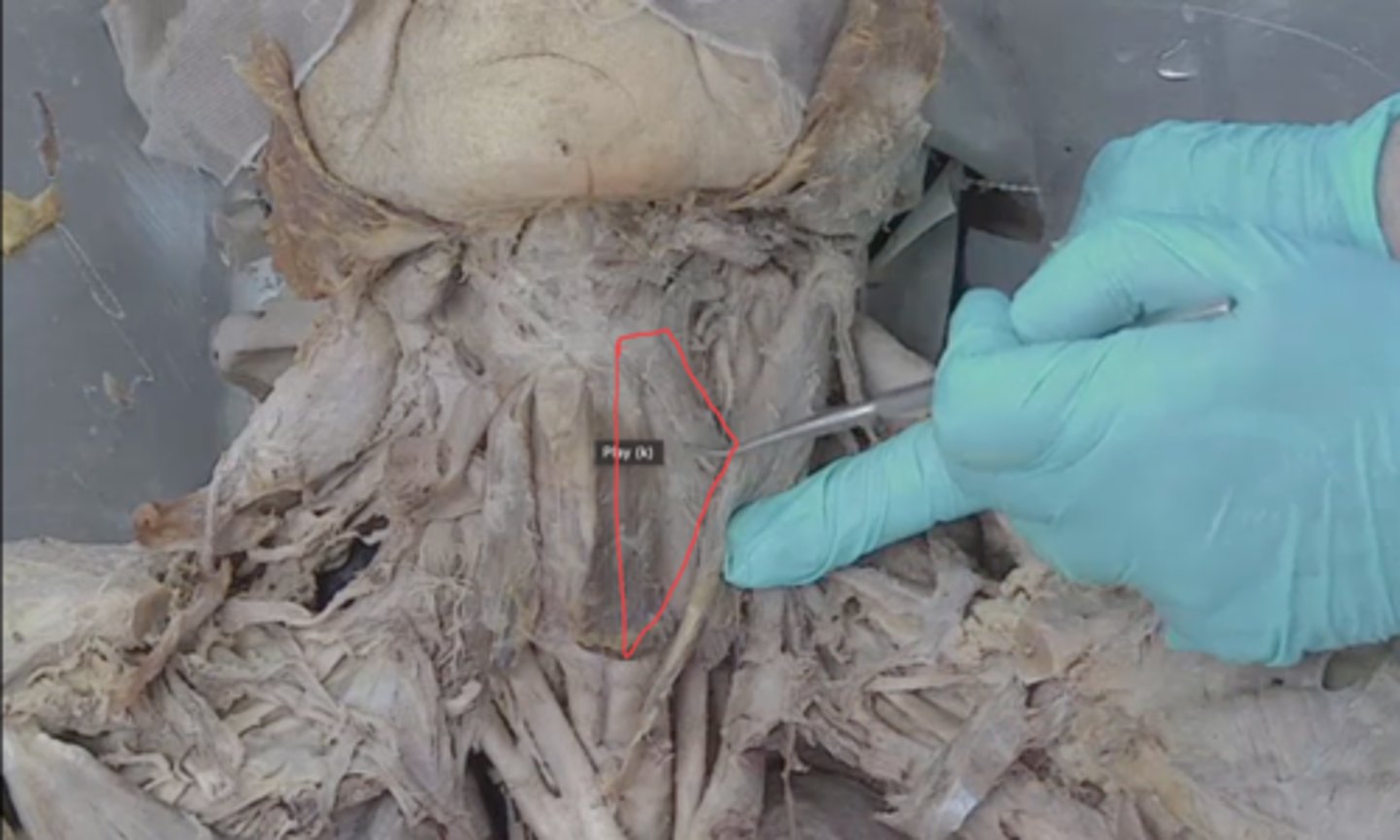
muscular triangle
identify this triangle
carotid triangle
identify this triangle
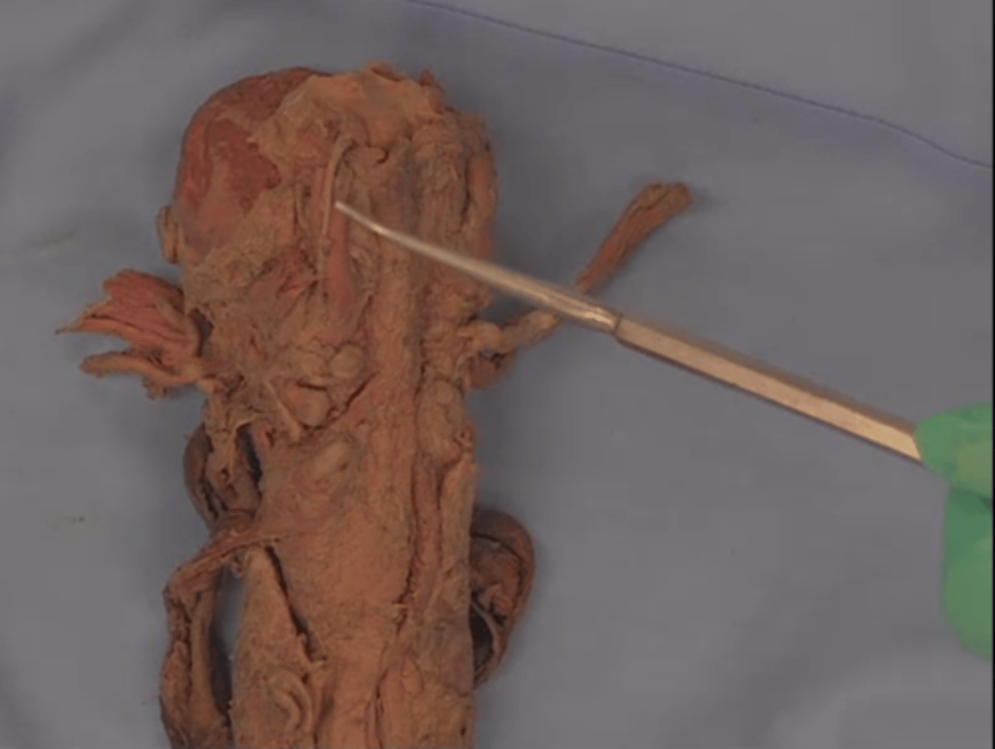
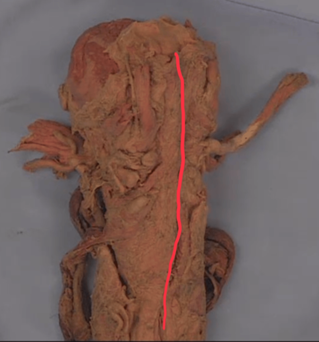
common carotid artery
identify structure in red
external carotid artery
identify structure in blue

internal carotid artery
identify structure in green

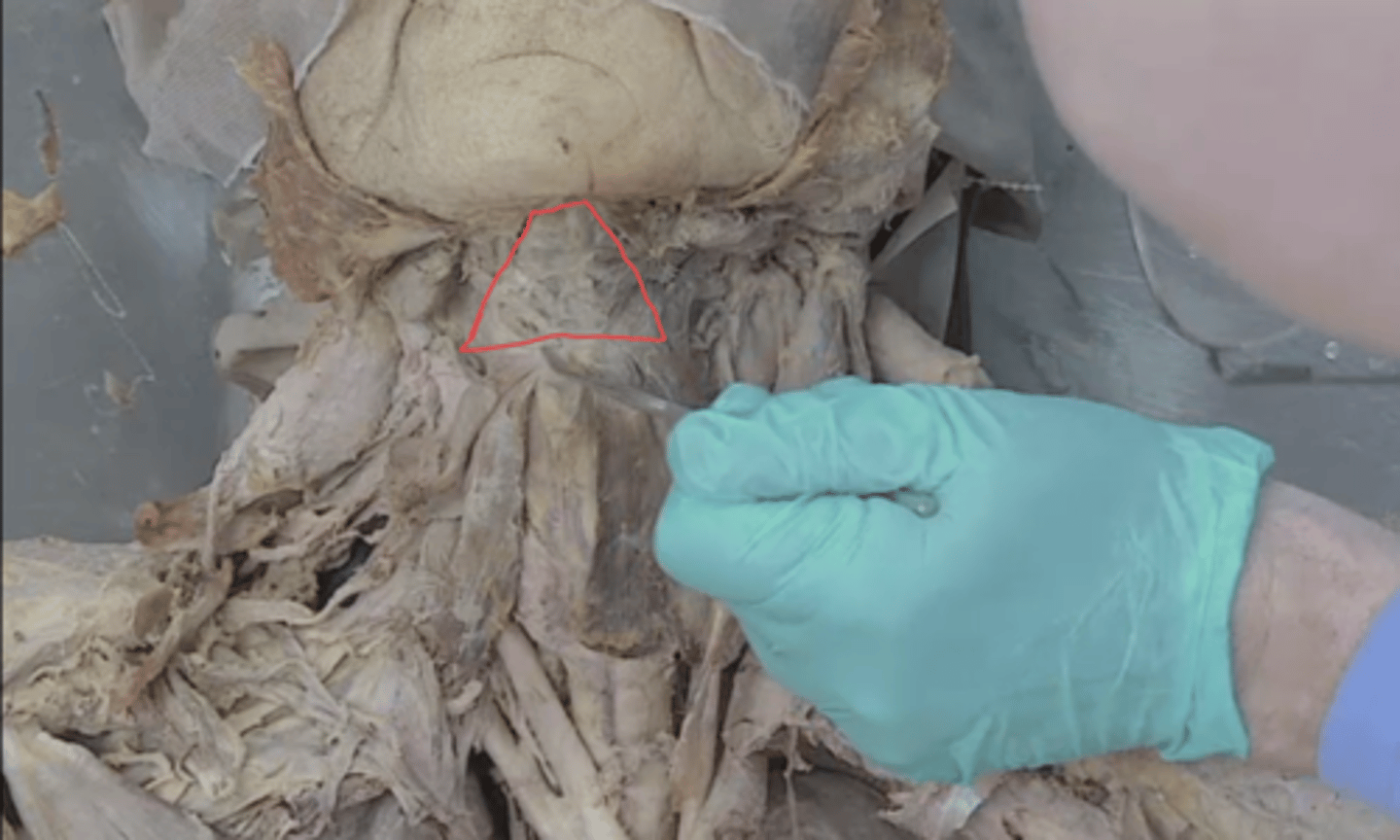
submental triangle
identify this triangle
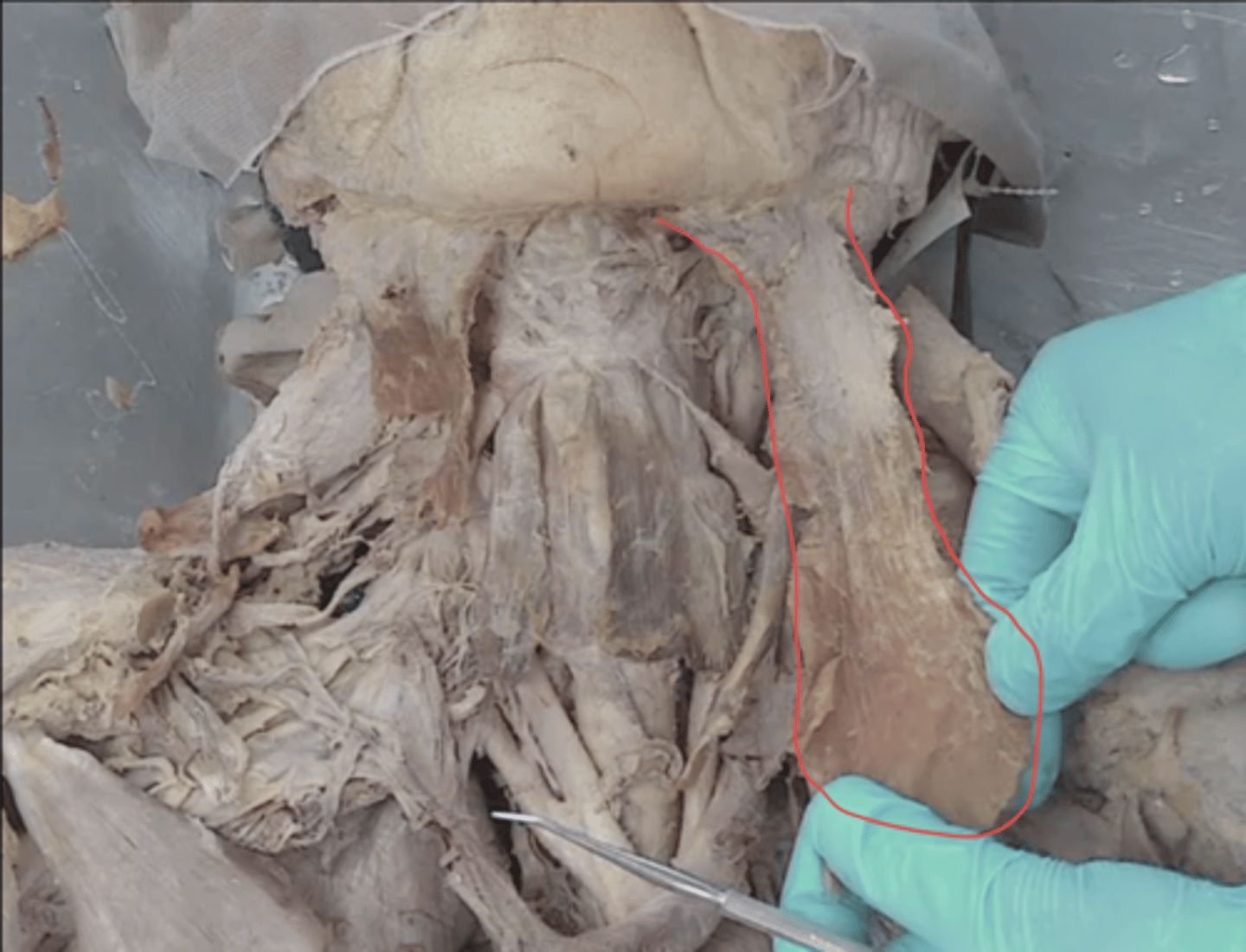
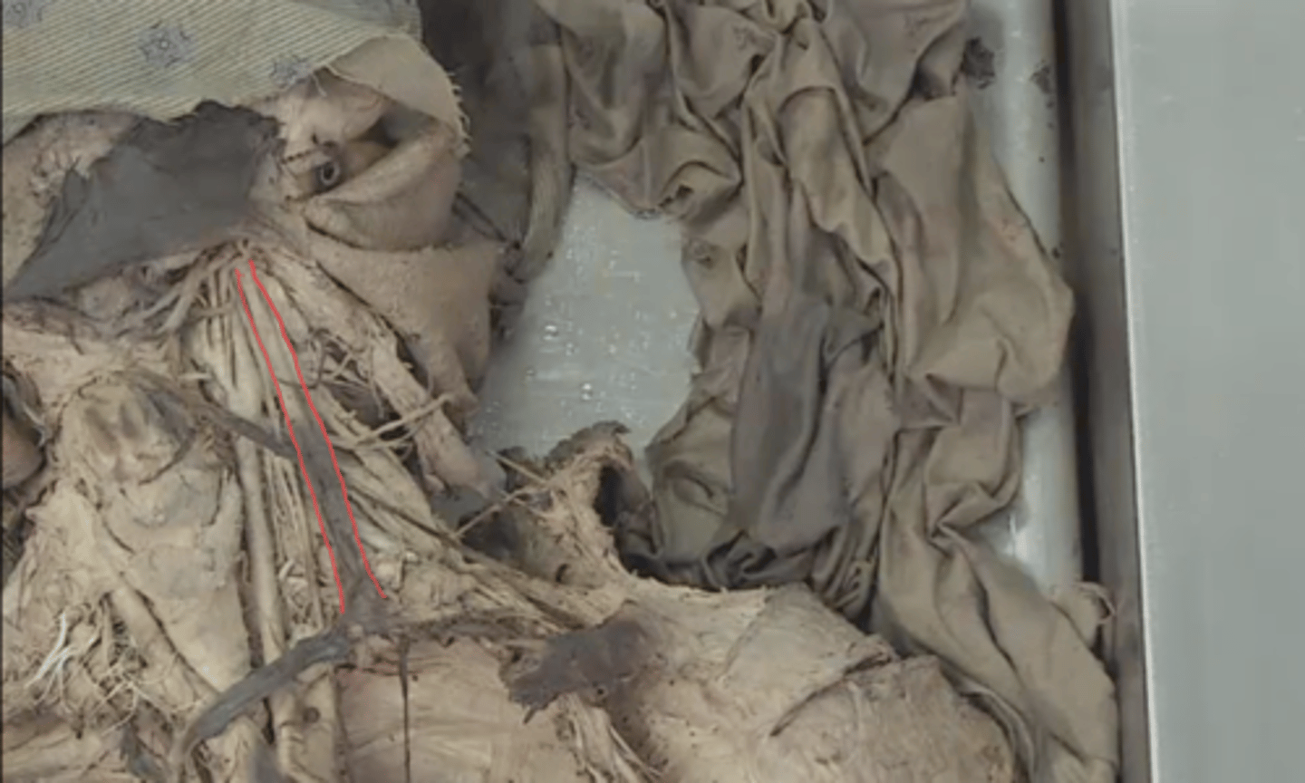
superior belly of omohyoid muscle
identify this muscle
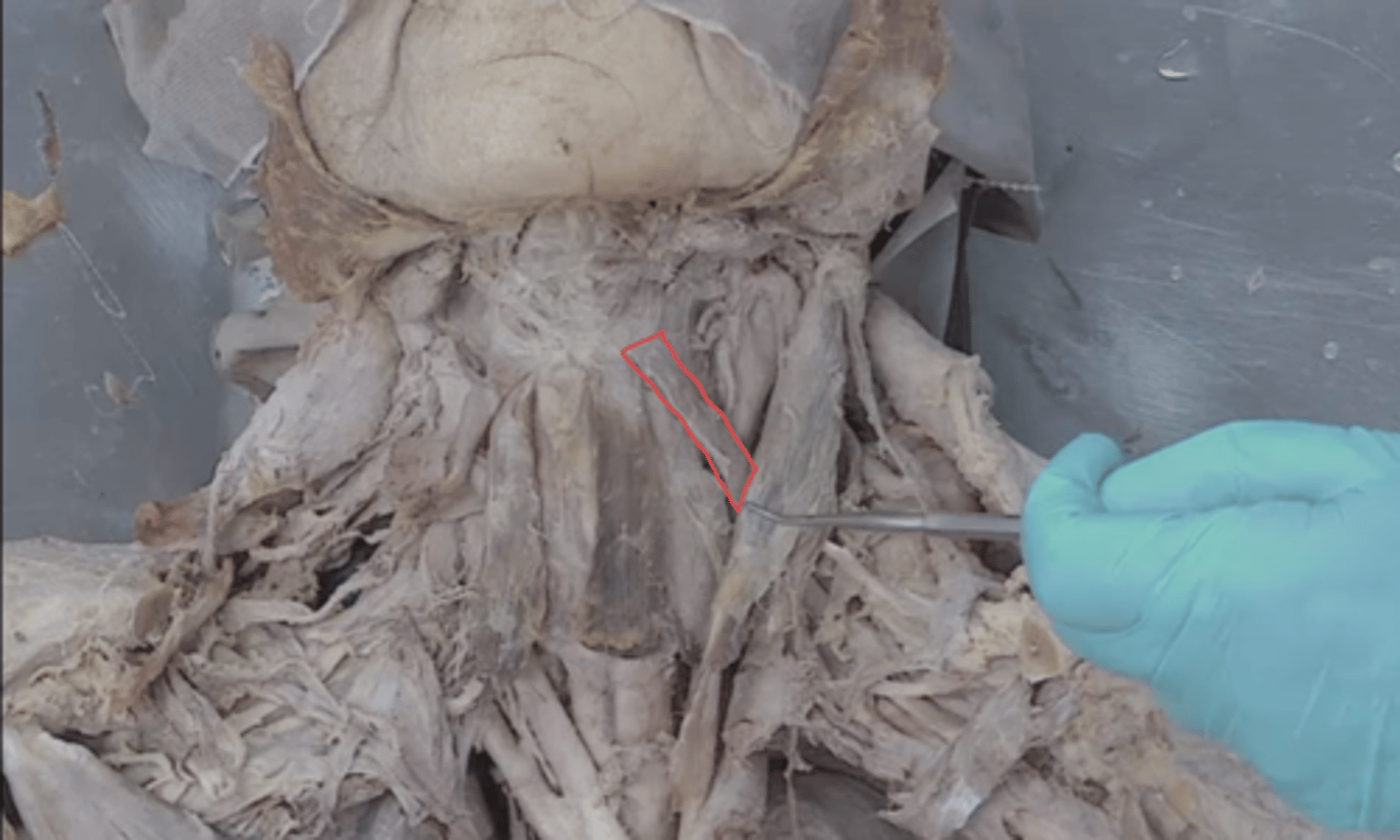
sternohyoid muscle
identify this muscle
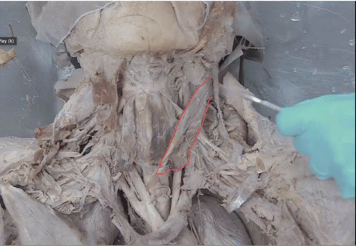
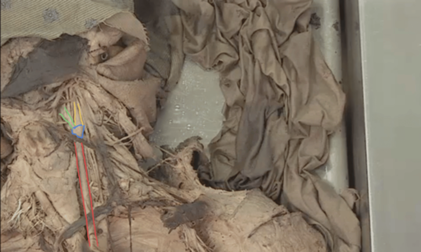
superior belly of omohyoid muscle
identify this muscle in red
intermediate tendon of omohyoid muscle
identify this structure in blue

inferior belly of omohyoid muscle
identify this muscle in green

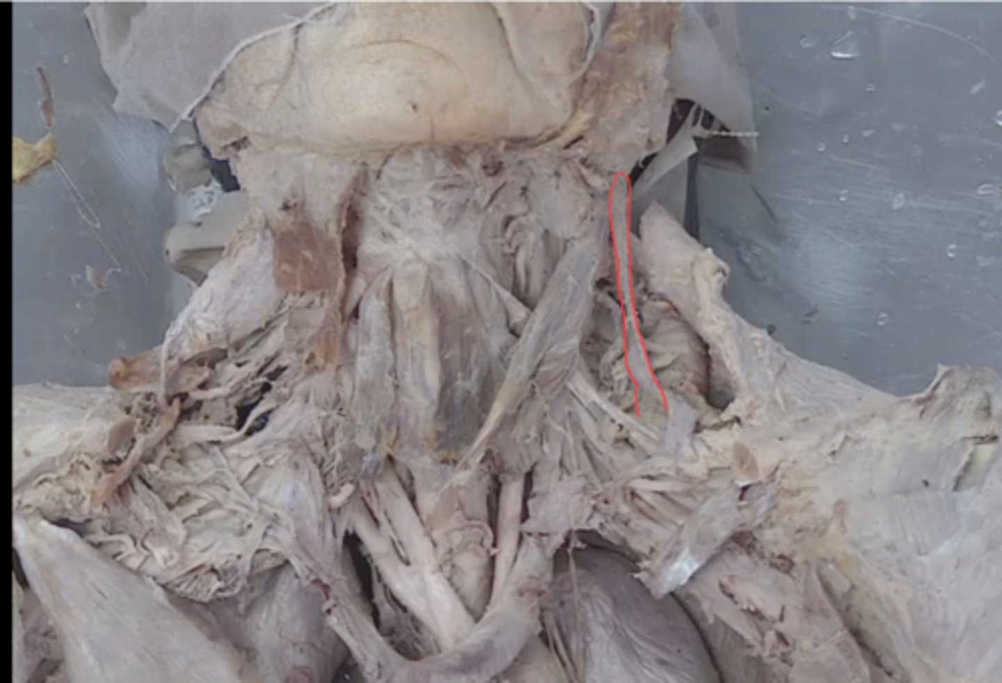
sternothyroid muscle
identify this muscle (sternohyoid muscle reflected)
thyrohyoid muscle
identify this muscle (sternohyoid muscle reflected)
right lobe of the thyroid gland
identify this structure in red

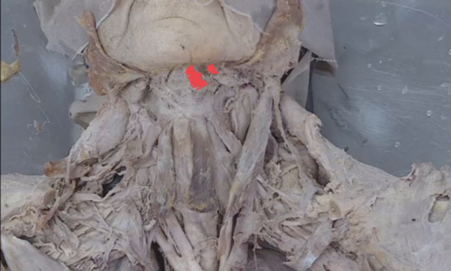
isthmus of the thyroid gland
identify this structure in blue

left lobe of the thyroid gland
identify this structure in green
pyramidal lobe of the thyroid gland
identify this structure (50% have it)
carotid sheath
identify this fascia
hypoglossal nerve
identify this structure
ansa cervicalis
identify structure in blue (off of hypoglossal nerve)
internal jugular vein
identify this structure
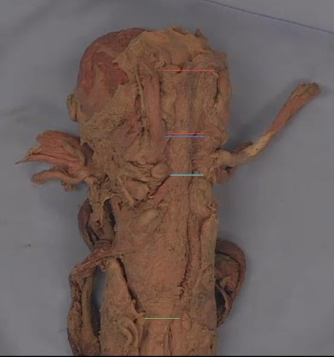
common carotid artery
identify structure in red

carotid sinus
identify structure in blue

external carotid artery
identify structure in green
internal carotid artery
identify structure in yellow

superior thyroid artery
identify structure in red

superior laryngeal artery
identify structure in blue
lingual artery
identify this structure
facial artery
identify this structure
vagus nerve
identify this structure
sympathetic trunk
identify this structure (behind internal jugular vein and vagus nerve)
superior cervical ganglion
identify this structure in red (very top of sympathetic trunk)
superior laryngeal nerve
identify this nerve (behind external carotid artery)
phrenic nerve
identify this nerve
mylohyoid muscle
identify this muscle (behind anterior belly of digastric) Duray didn't show it in videos but this is where it will be)
mylohyoid muscle
identify this structure
geniohyoid muscle
identify this structure
genioglossus muscle
identify this structure
lingual nerve
identify this structure (tongue reflected)

submandibular duct
identify this structure (tongue reflected)
submandibular gland
identify structure that lies here (tongue reflected, deep to lingual nerve)
sublingual gland
identify structure that lies in this fossa (tongue reflected)
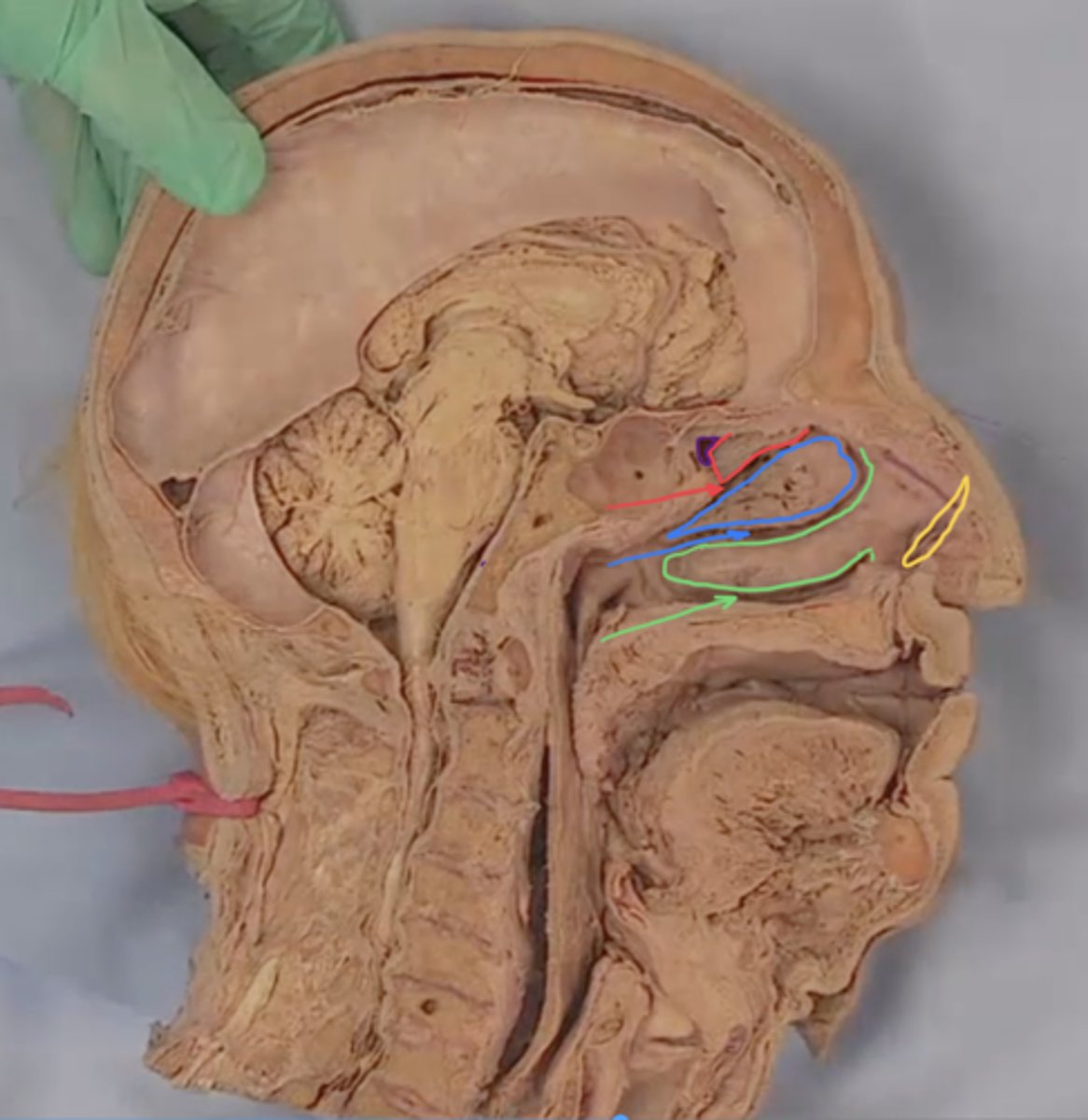
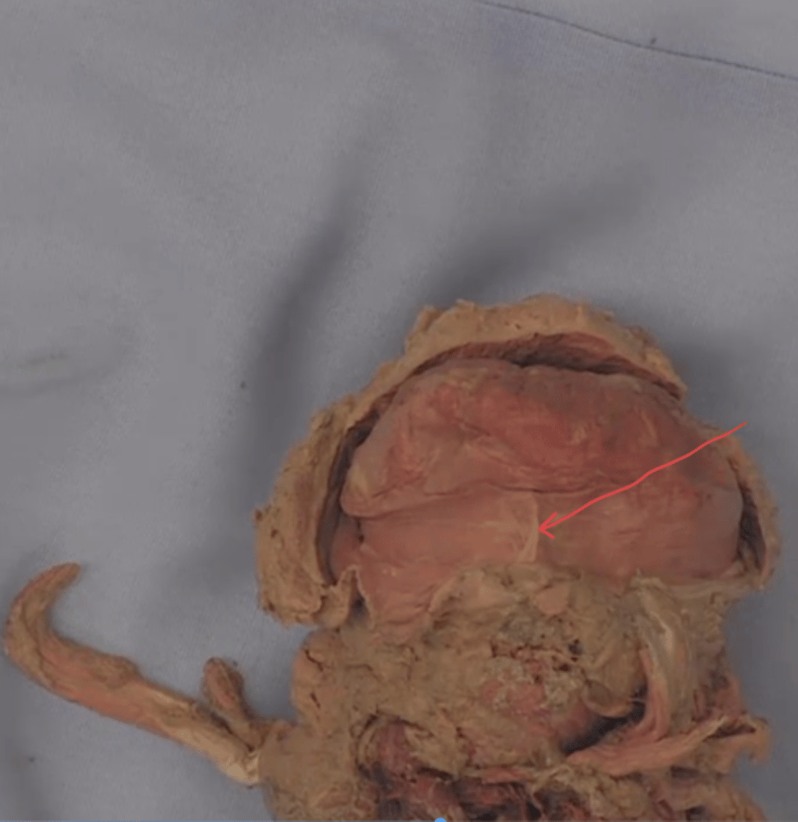
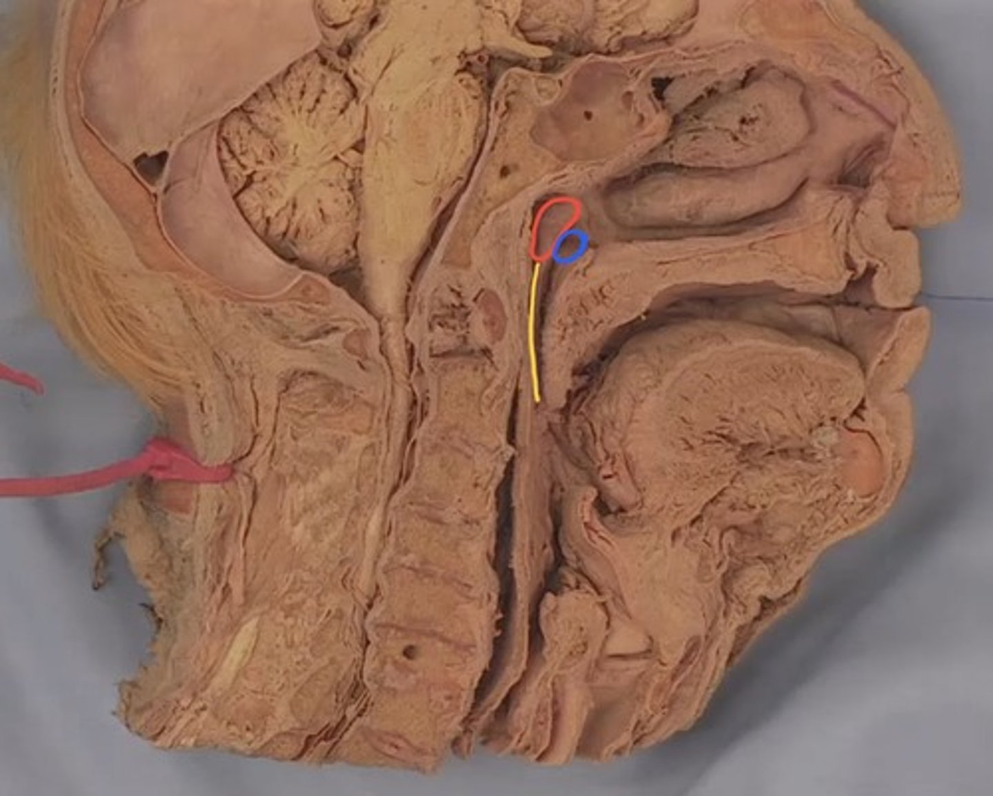
superior nasal concha
identify structure outlined in red

middle nasal concha
identify structure outlined in blue

inferior nasal concha
identify structure outlined in green

superior meatus
identify red arrow

middle meatus
identify blue arrow

inferior meatus
identify green arrow

nasal vestibule
identify structure outlined in yellow

sphenoethmoidal recess
identify structure outlined in purple (very small superior to superior nasal concha)
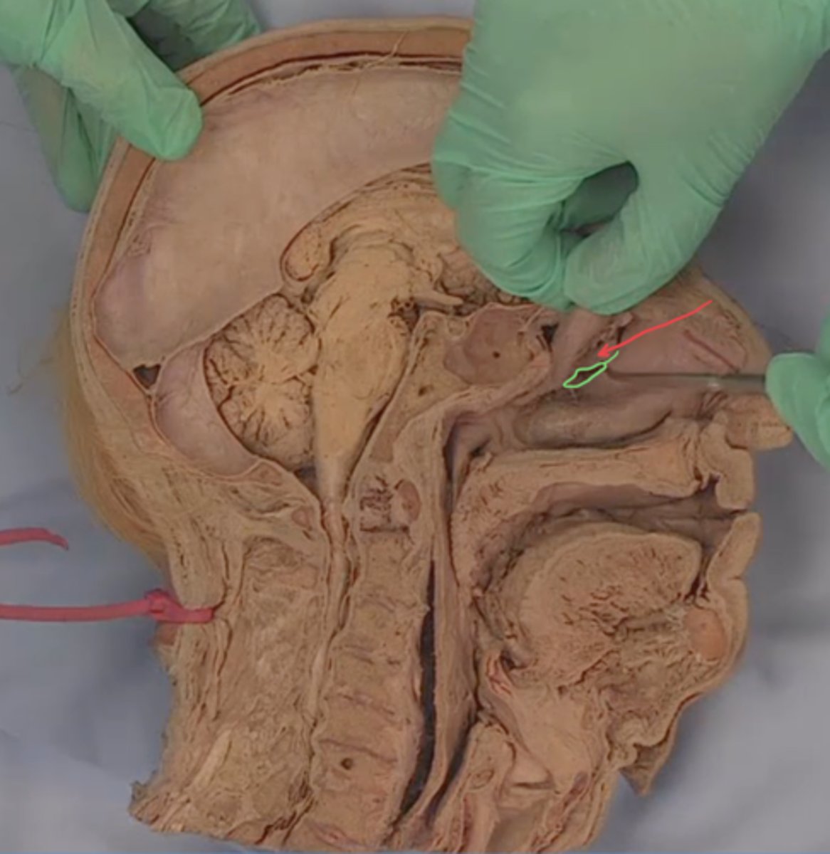
ethmoidal bulla
identify red arrow (middle nasal conch reflected)

hiatus semilunaris
identify structure outlined in green (middle nasal concha reflected below ethmoidal bulla)
sphenoid sinus
identify structure outlined in red
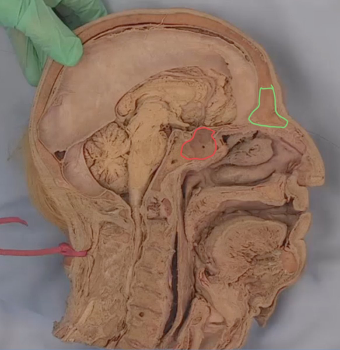
frontal sinus
identify structure outlined in green

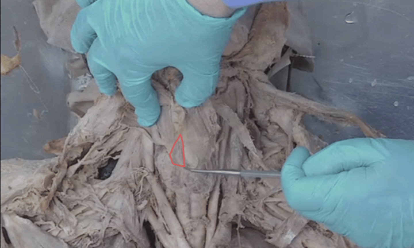
anterior belly of digastric muscle
identify structure outlined in red

intermediate tendon of digastric muscle
identify structure outlined in blue
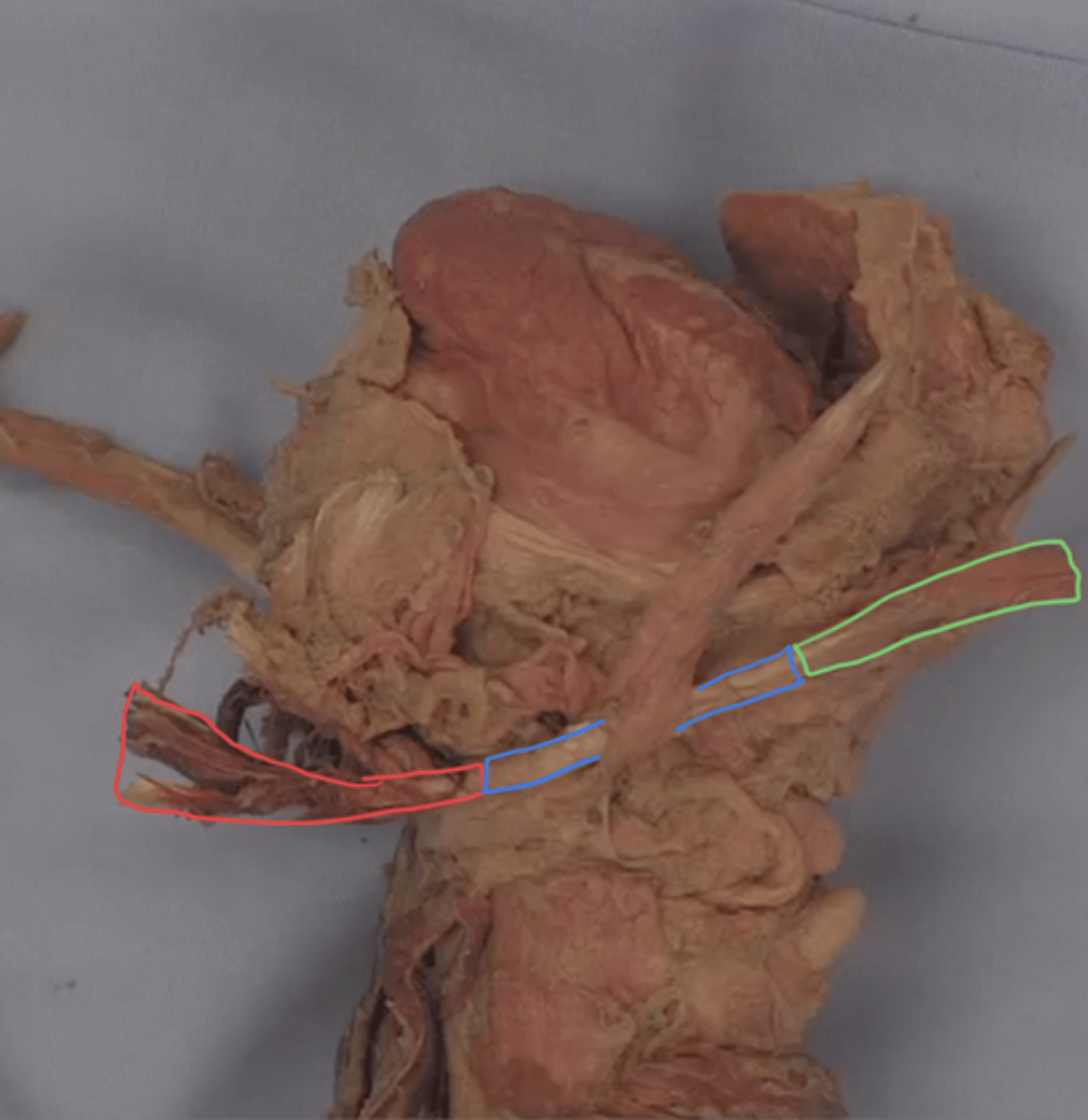
posterior belly of digastric muscle
identify structure outlined in green

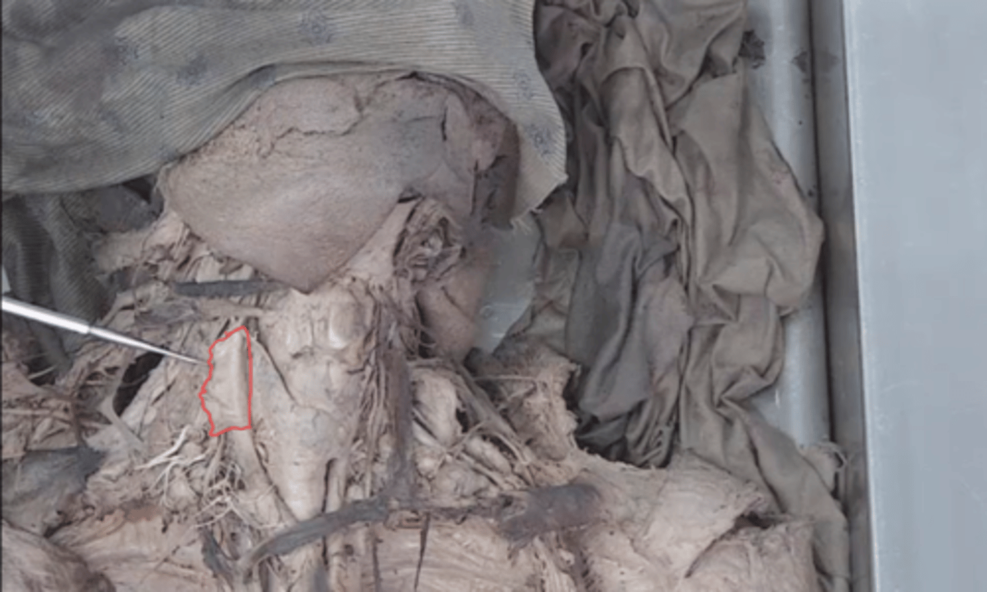
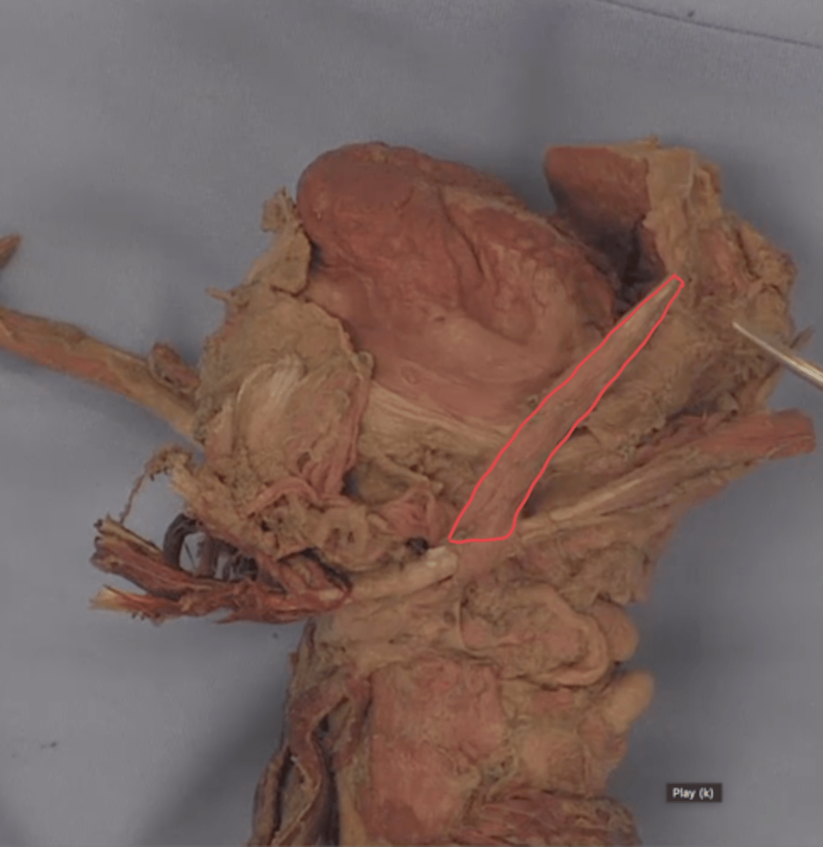
stylohyoid muscle
identify this structure
styloglossus muscle
identify structure outlined in red
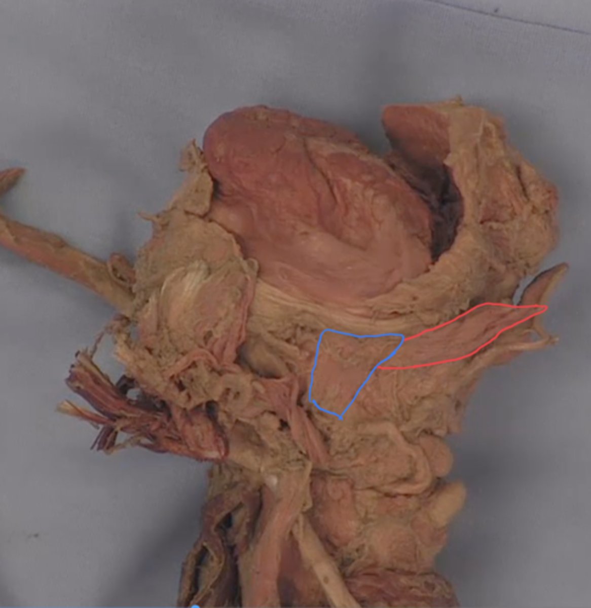
hyoglossus muscle
identify structure outlined in blue

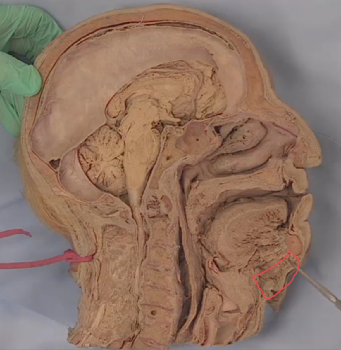
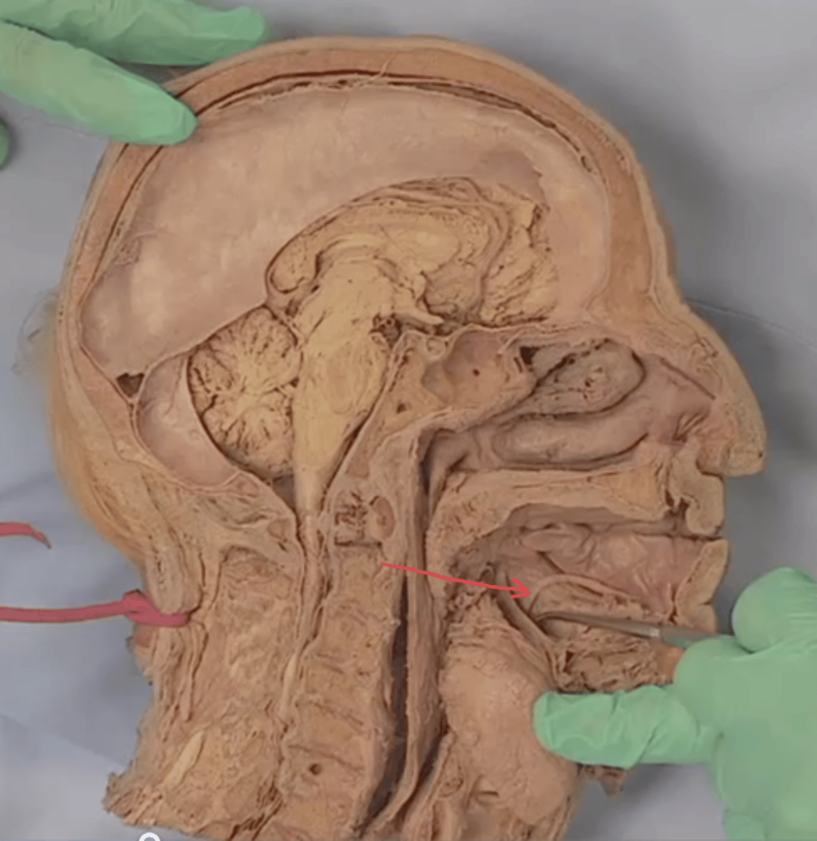
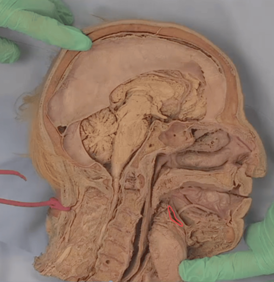
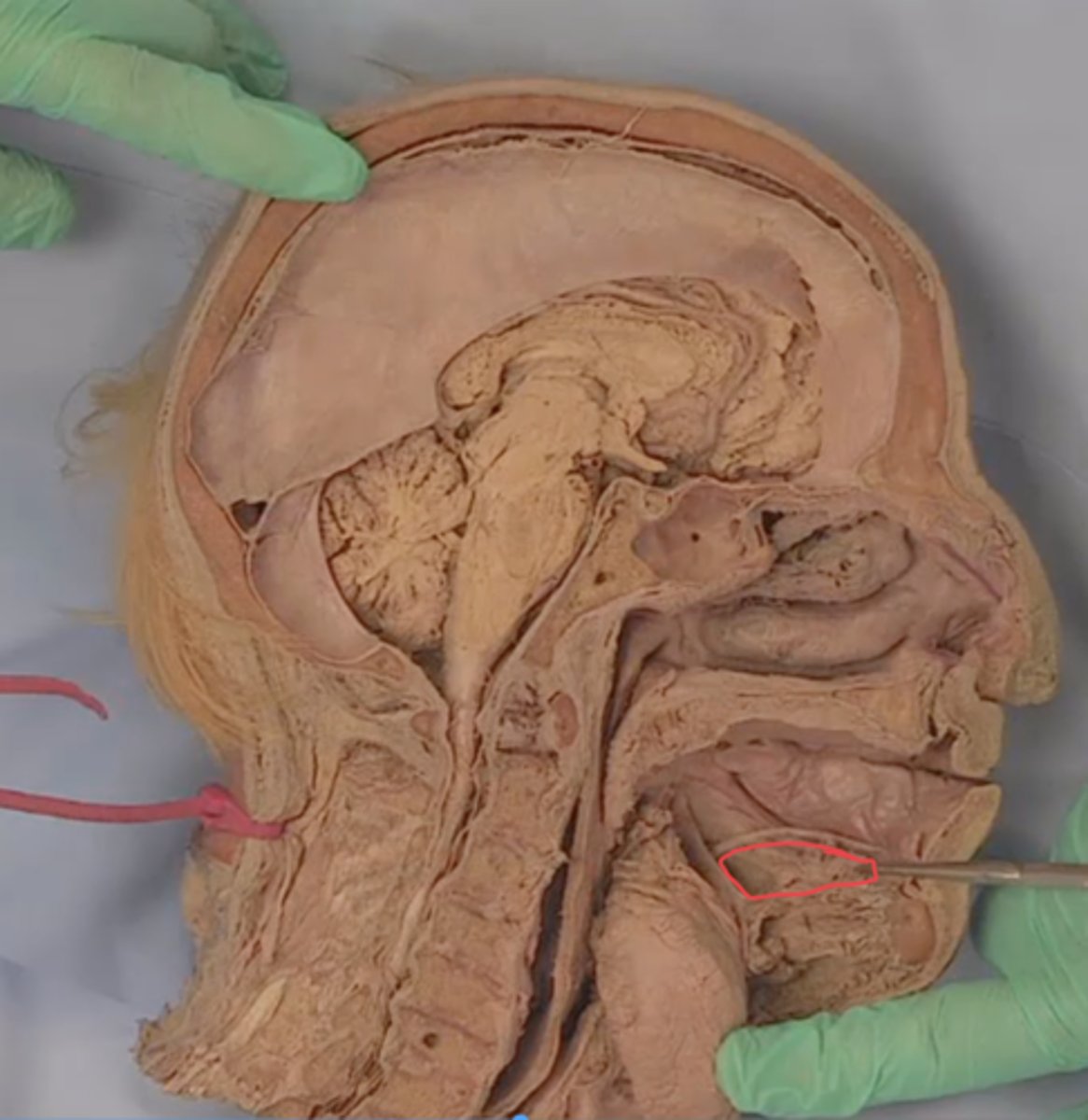
frenulum of the tongue
identify this structure
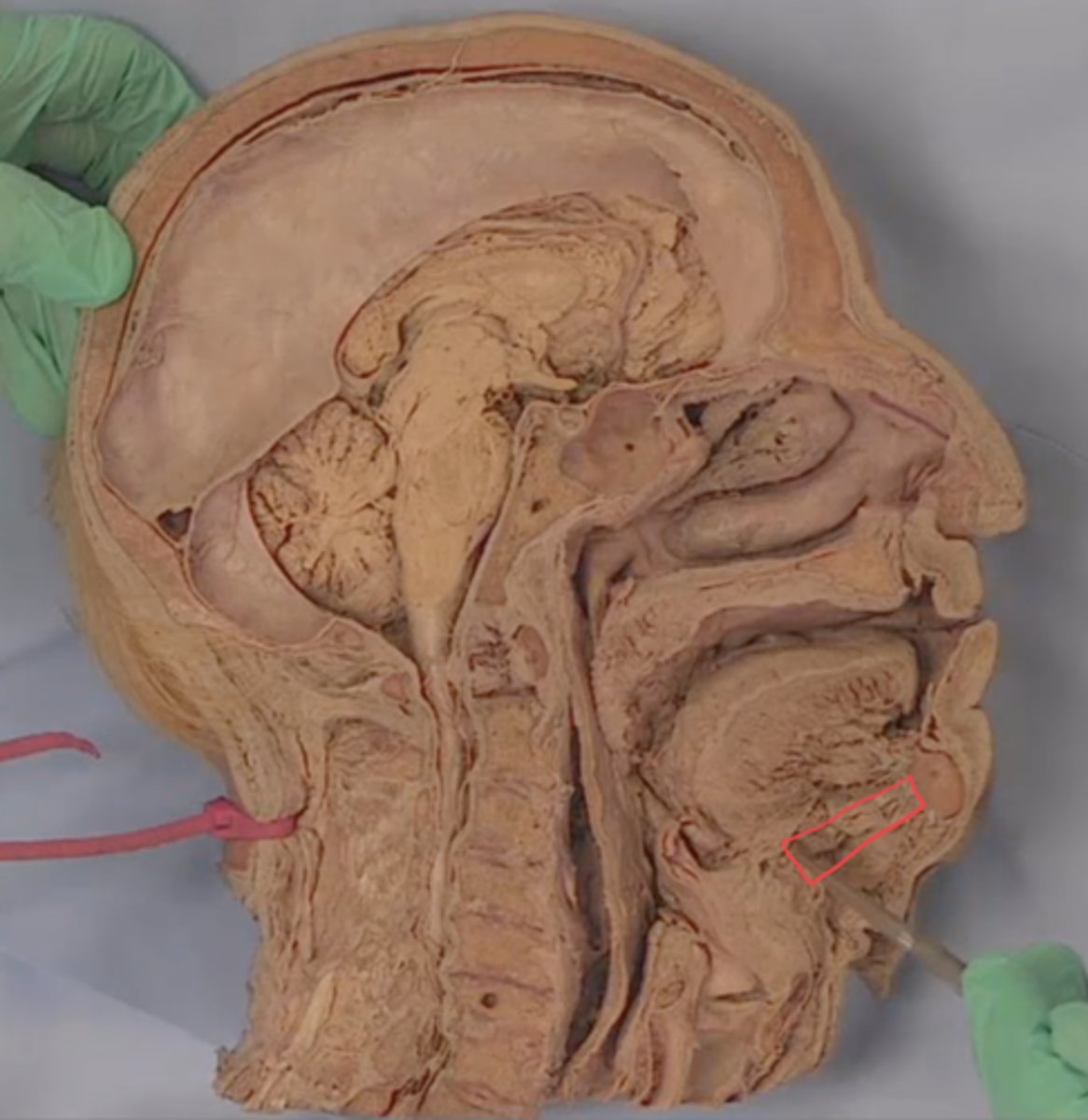
sulcus terminalis
identify structure outlined in red

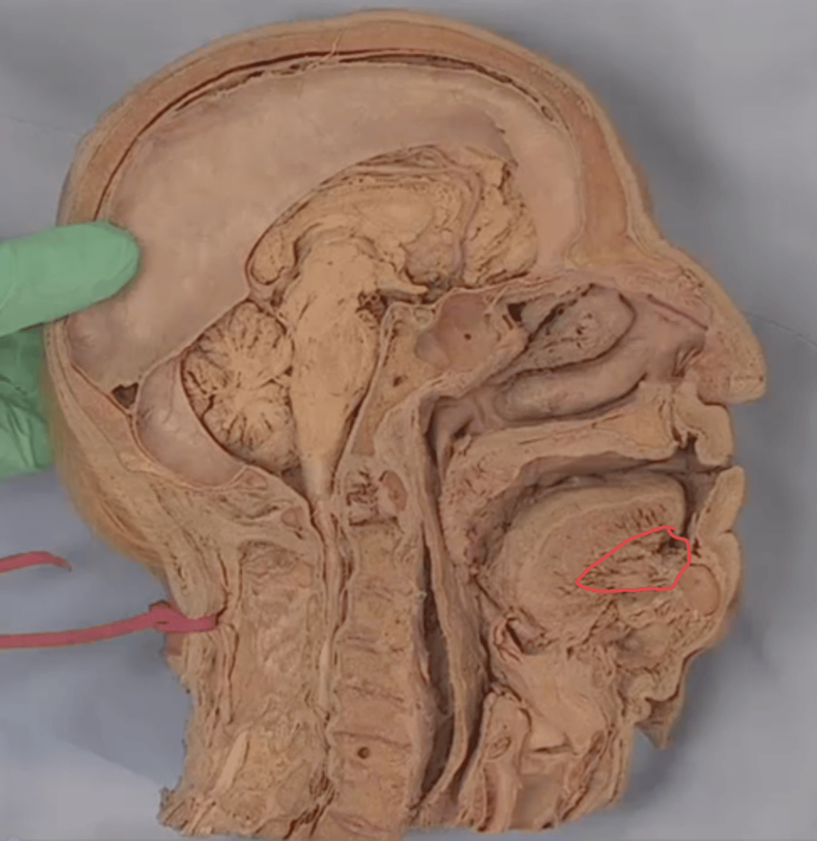
foramen cecum
identify structure outlined in purple

vallate papillae
identify structures in green
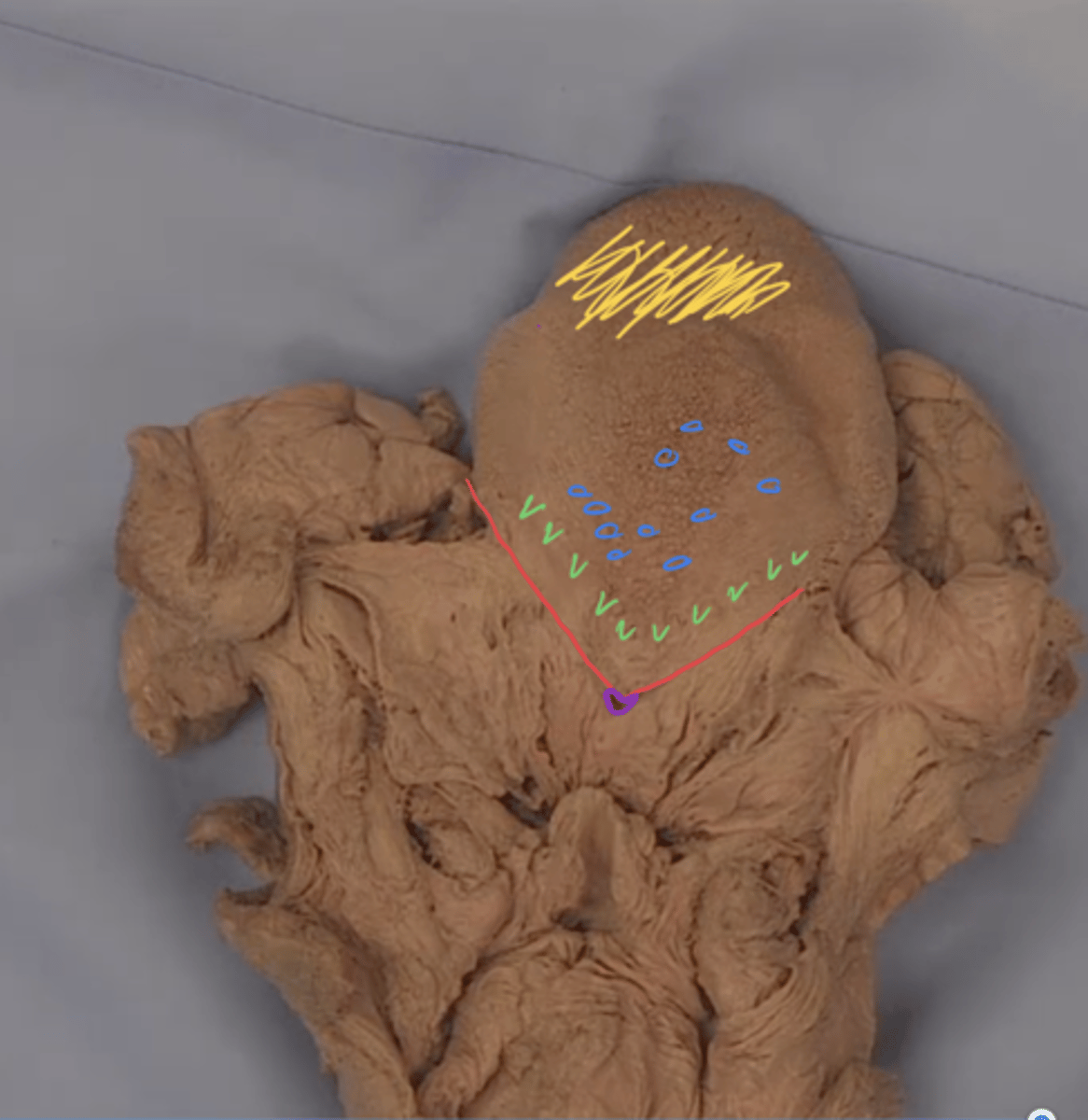
fungiform papillae
identify structures in blue

filiform papillae
identify structures in yellow

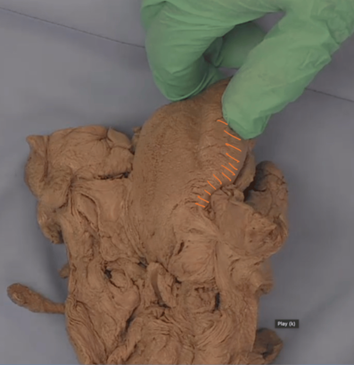
foliate papillae
identify structures in orange
Stylopharyngeus muscle
What is this structure?
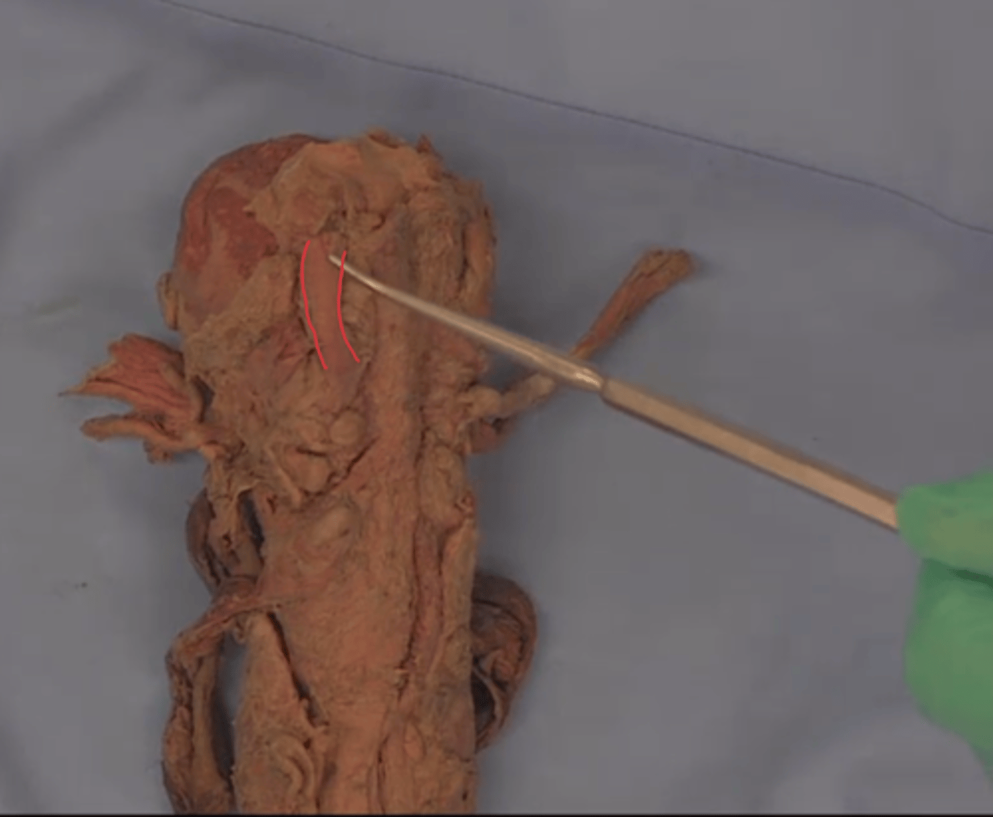
Glossopharyngeal nerve (CN IX)
What is this structure?
Superior pharyngeal constrictor muscle
What is the structure outlined in red?

Middle pharyngeal constrictor muscle
What is the structure outlined in blue?

Inferior pharyngeal constrictor muscle
What is the structure outlined in green?
Pharyngeal raphe
What is this structure?
Retropharyngeal space
What is the space outlined in green?
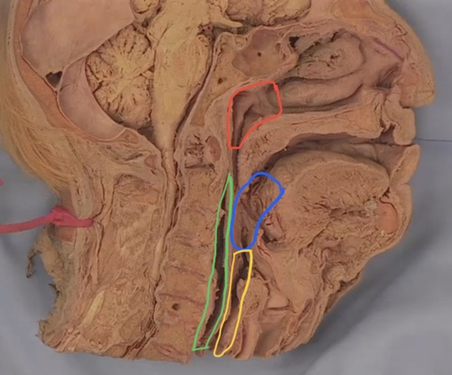
Nasopharynx
What part of the pharynx is outlined in red?

Oropharynx
What part of the pharynx is outlined in blue?

Laryngopharynx
What part of the pharynx is outlined in yellow?

Piriform fossa
What is the structure colored green? (constrictor muscles are reflected)

Laryngeal inlet
What is this opening colored white? (constrictor muscles are reflected)
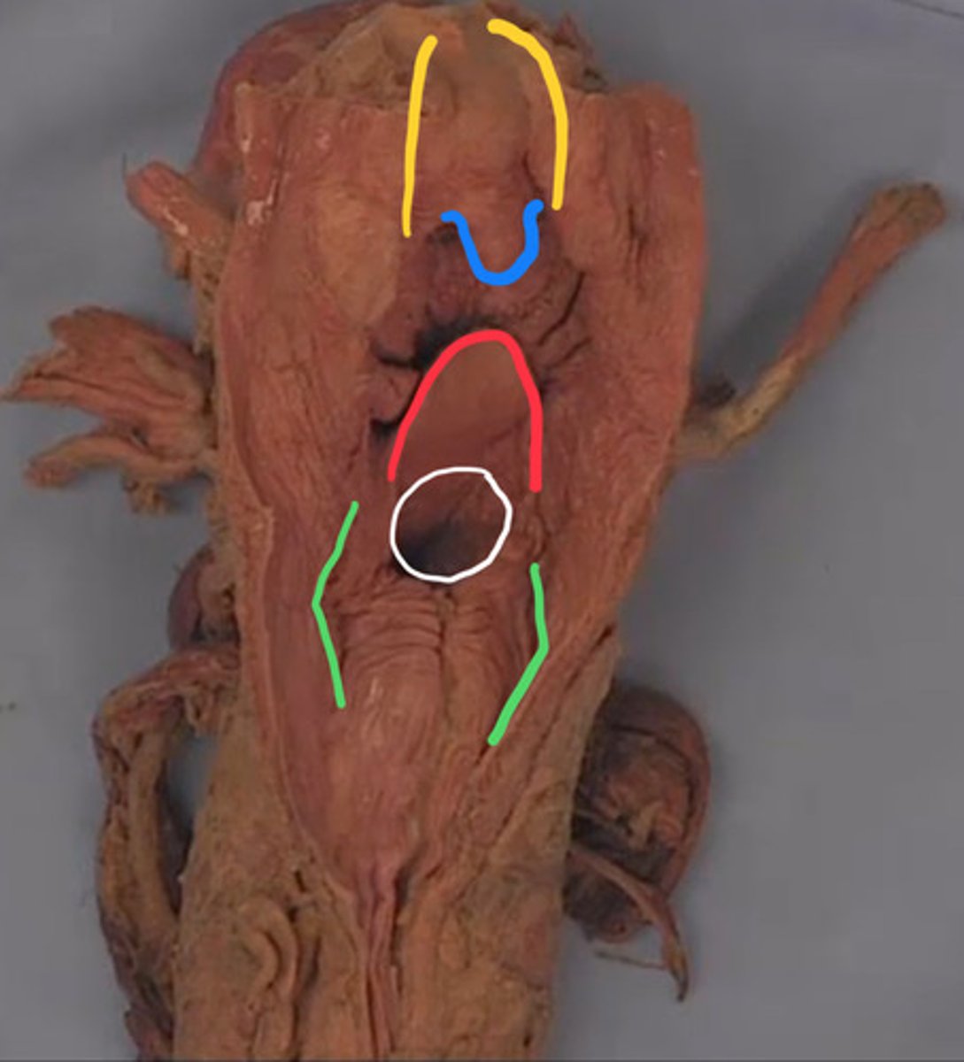
Epiglottis
What is the structure outlined in red? (constrictor muscles are reflected)

Opening of pharyngotympanic tube
What is the opening outlined in blue?

Torus tubarius
What is the structure outlined in red?
Salpingopharyngeal fold
What is the fold colored yellow?

Soft palate
What is the structure outlined in yellow? (constrictor muscles are reflected)

Uvula
What is the structure outlined in blue? (constrictor muscles are reflected)

Palatopharyngeal fold
What is the fold colored blue? (constrictor muscles are reflected)
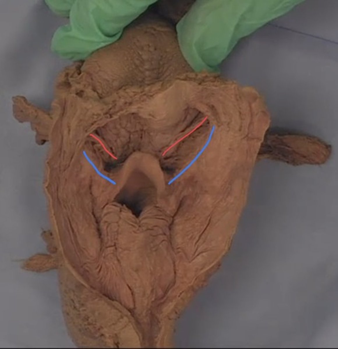
Palatoglossal fold
What is the fold colored red? (constrictor muscles are reflected)

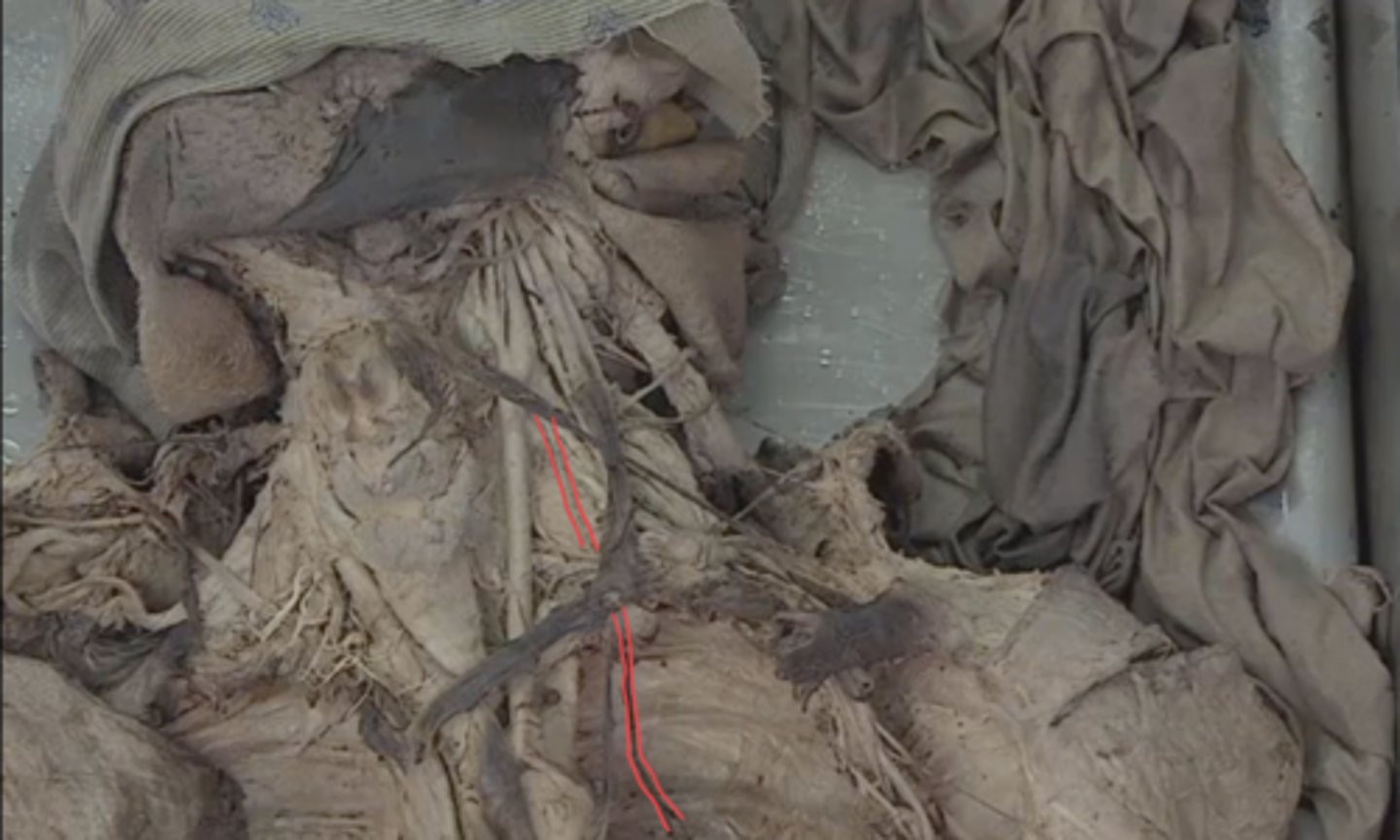
Tonsillar fossa
What is the structure between the two lines? (constrictor muscles are reflected)

Median glossoepiglottic fold
What is the fold colored red? (constrictor muscles are reflected, epiglottis is pushed down)

Lateral glossoepiglottic fold
What is the fold colored blue? (constrictor muscles are reflected, epiglottis is pushed down)

Velleculae
What is between the yellow lines? (constrictor muscles are reflected, epiglottis is pushed down)