Bio 222 Week 8: Animal Nutrient Homeostasis
1/43
There's no tags or description
Looks like no tags are added yet.
Name | Mastery | Learn | Test | Matching | Spaced | Call with Kai |
|---|
No analytics yet
Send a link to your students to track their progress
44 Terms
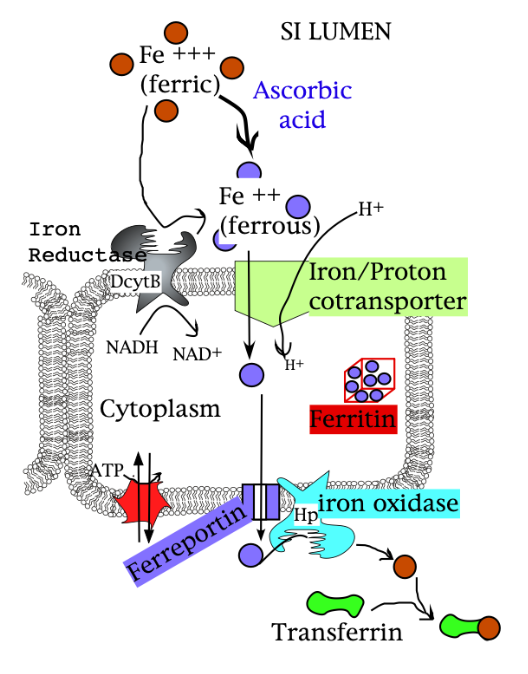
Animal Iron Uptake into the Brush Border Cell (in the small intestine)
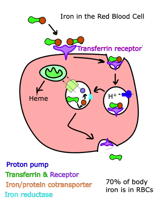
Animal Iron Uptake in the Red Blood Cell
This diagram must be a baby red blood cell (an erythrocyte) because mature RBCs don’t have organelles like the mitochondria.
Notice that iron does not leave the red blood cell!!!
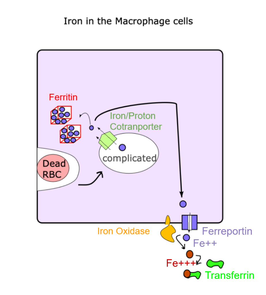
Animal Iron Uptake in the Macrophage Cells
Animal Iron Uptake in Liver Cells
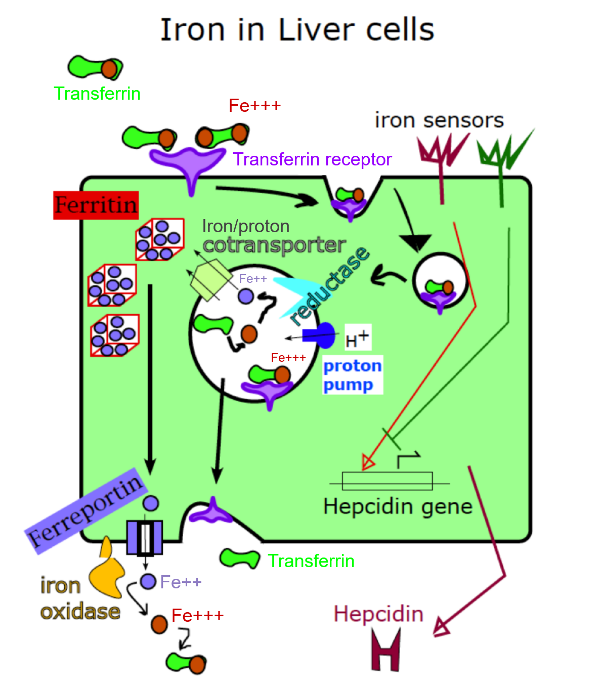
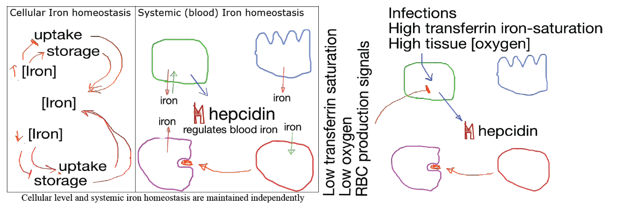
Cellular homeostasis and blood/body iron homeostasis are maintained by…
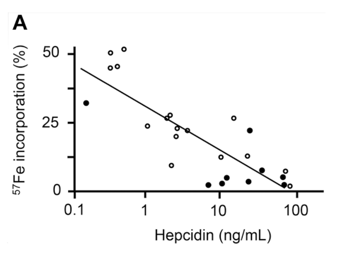
Hepcidin
Decreases blood iron levels
Is made when [iron] is high in the blood
Decreases ferroportin (iron exit channel) activity in brush border cells, liver cells, and macrophages
Hepcidin Regulation:
Activated by…
Infections
High transferrin-iron saturation
High tissue [oxygen']
Inhibited by…
RBC production signals
Low transferrin-iron saturation
Low tissue [oxygen]
![<p>Hepcidin</p><ul><li><p>Decreases blood iron levels</p></li><li><p>Is made when [iron] is high in the blood</p></li><li><p>Decreases ferroportin (iron exit channel) activity in brush border cells, liver cells, and macrophages</p></li></ul><p></p><p>Hepcidin Regulation:</p><ul><li><p>Activated by…</p><ul><li><p>Infections</p></li><li><p>High transferrin-iron saturation</p></li><li><p>High tissue [oxygen']</p></li></ul></li><li><p>Inhibited by…</p><ul><li><p>RBC production signals</p></li><li><p>Low transferrin-iron saturation</p></li><li><p>Low tissue [oxygen]</p></li></ul></li></ul><p></p>](https://assets.knowt.com/user-attachments/a4958704-c004-4830-833b-b31277430a12.png)
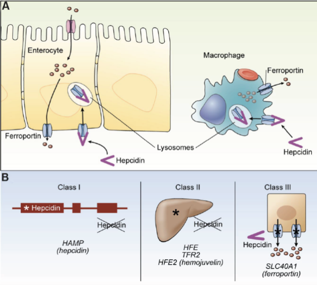
MIGHT BE ON MIDTERM (Epistasis)
Hepcidin blocks export through ferroportin, but it also causes these channels to be endocytosed and digested.
Genetic diseases that lead to excessive systemic iron include:
Non-functional genes for hepcidin itself (class I)
Non-functional genes for proteins that regulate hepcidin gene regulation(class II)
Ferroportin alleles that code for a protein (ferroportin) that fails to be bound and stopped by hepcidin (class III).
Excess iron symptoms are called hemochromatosis.
If you have excess iron and lack hepcidin, you can use leeches to relieve your symptoms!
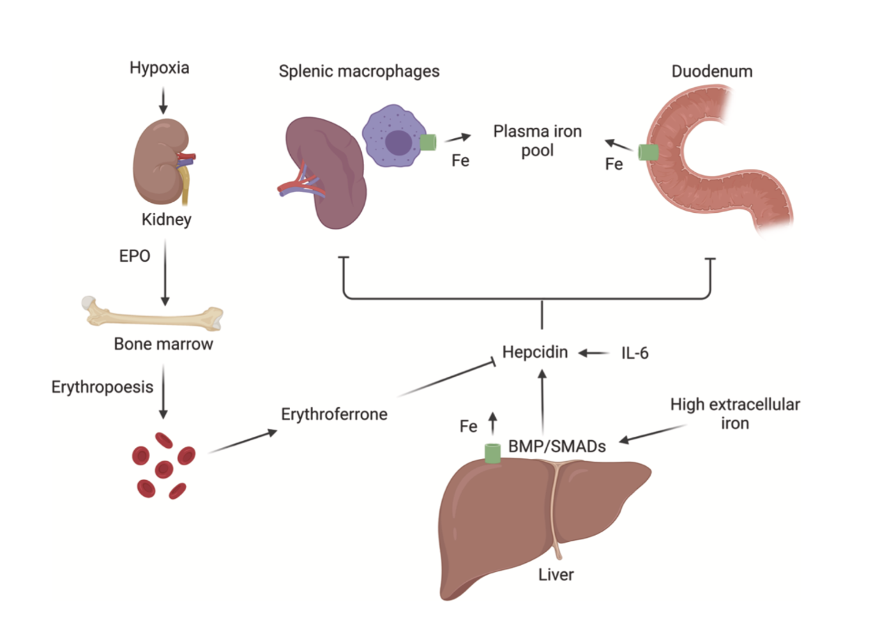
Hepcidin Regulation Diagram
It’s important to know that hepcidin ALSO inhibits export from the liver itself to the plasma iron pool. The blood cell’s arrows represent developing RBC stem cells sending a signal that stops hepcidin expression.
Iron deficiency is an example of malnutrition.
Although there are genetic and infectious diseases that cause anemia, the most common cause is a lack of dietary supply.
Meat and fish contain large quantities of very bioavailable iron. Meat is muscle, and myoglobin is a great source of iron.
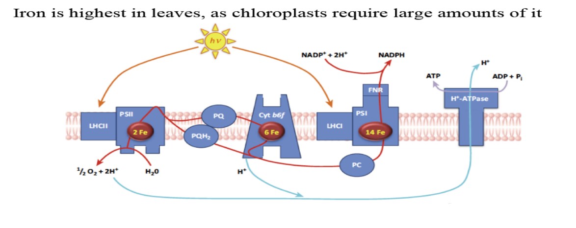
Leafy vegetables also contain lots of iron.
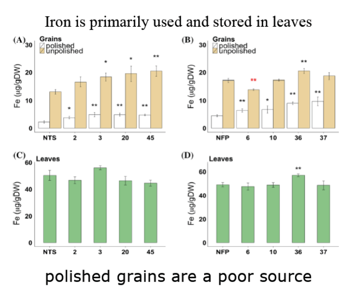
IRON IS PRIMARILY STORED IN LEAVES
Polished grains however typically contain quite low quantities of iron.
This is why it is legally mandated that packaged grain foods in the united states are fortified with iron (among other things).
However, there is a reasonable amount of iron in whole grains.
With rice the preference for polished grains, or “white rice” stems in very large part from the storage life it has when compared to whole grain rice.
The bran (bits of the floral organs that make up the seed) and the germ (the embryo) are the locations of seed iron, but they are also the parts that “go bad” (become rancid quickly).
Genetically modified rice:
Increased phytase (gets rid of phytate, the thing so good at storing iron that if you eat it it can take iron out of you!)
Increased ferritin (the other means of iron storage)
Increased Vitamin B
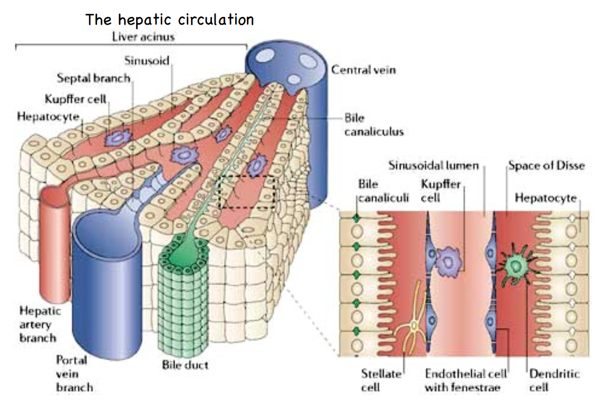
The hepatic circulation (liver addendum)
The liver is the organ responsible for adding and subtracting material to and from the blood, including nutrients.
Fuels and building blocks are maintained within homeostatic levels in the blood in this way.
Blood leaving the intestine is delivered to the liver. The veins deliver blood to sinusoids. These look and act a lot like capillaries. The liver extracts excesses and adds what is lacking to the blood, then delivers it to the central vein on its way back to the heart for the next round of circulation.
Another function of the liver is the production of bile.
This doesn’t enter the blood, not ever, but as you can see from the diagram the bile ducts derive their material to carry from the opposite side of the same set of cells that are intimately associated with blood in the sinusoids.
The space of Disse is the location of interstitial fluid between sinusoid and liver cells. Don’t be confused by the shape of the liver cells (hepatocytes). That surface area increasing finger shaped projection motif is seen over and over again. Those are not brush border cells.
Essentially: the point is that the liver is a massively redundant collection of blood adjusting cells. The portal vein moves blood from collection in the guts directly to the liver, which can then alter the content of the blood by taking up much of the nutrients.

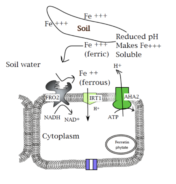
Plant Iron Uptake Review
Iron uptake in plants involves something akin to (but not really) extra-cellular digestion. The proton pump is hyperactivated to acidify the environment to make Fe+++ soluble. FRO2 (iron reductase (oxidase???)) converts ferric (Fe+++) to ferrous iron (Fe++).
That reduction isn’t a cleavage, so this isn’t digestion.
Ferrous iron is taken up by IRT1. This is a high affinity system, so it is a cotransporter (HATS).
Iron is stored by ferritin in many plant cells.
This is the major vegetable source of iron in our diets.
But when stored linked to phytate we can’t get the iron, and in fact lose iron to the phytate as it passes through our gut.
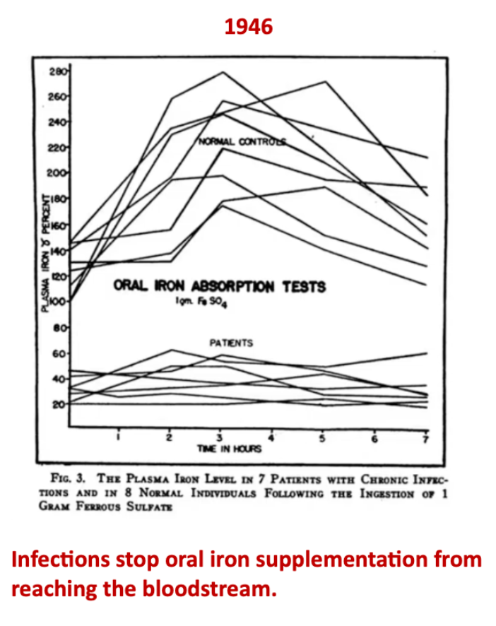
Infections stop oral iron supplementation from reaching the bloodstream because…
Iron cannot be absorbed properly due to increased circulating hepcidin (inhibition of brush border cell ferroportin).
Form of iron taken up by brush border cells: Fe++ (ferrous)
Form of iron taken up by liver cells: Fe+++ (ferric)
Ferroportin activity is regulated directly by interaction with: Hepcidin
Hepcidin levels are increased by: High transferrin saturation
Form of iron that exits liver cells before enzymatic conversion: Fe++ (ferrous)
Form of iron that exits brush border cells before enzymatic conversion: Fe++ (ferrous)
Form of iron that is bound by or passes through transferrin: Fe+++ (ferric)
Form of iron that is bound by or passes through ferroportin: Fe++ (ferrous)
Form of iron that is bound by or passes through hepcidin: This protein does not interact with iron
Mechanism of iron uptake in juvenile red blood cells: Endocytosis
Mechanism of iron uptake in brush border cells: Cotransport with protons
Protein required for release of iron from proteins in endosomes: Proton pump
Iron exits macrophages through: Ferroportin channels
More hepcidin activity leads to which? increased or reduced blood iron levels: Reduced
More ferroportin activity leads to which? increased or reduced blood iron levels: Increased
Mechanism of iron uptake in liver cells: Endocytosis
In class I asked you to draw iron uptake, processing and delivery to the blood after seeing the set of machinery that participates in these processes in the context of blood, macrophage and brush border cells. Some of the key facts I highlighted as I shared the cell diagrams with you included the fact that iron is delivered to the blood only through ferroportin, that iron is converted into iron 3+ form so that proteins called transferrin can grab it and move it through the blood.
I hoped that you would recognize that since that's how iron travels in the blood, uptake of that structure must be the mechanism of iron import by liver cells.
True or False:
Upregulation of the hepcidin gene is likely during infection and inflammatory responses, and leads to increased iron to the blood.
False
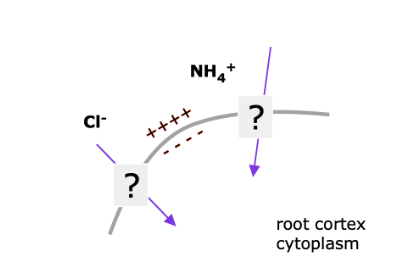
Which ion must be moving through HATS given greater concentration in the soil water?
Neither??????????????
During cholesterol regulation which protein interacts with nucleic acids to accomplish its role within the regulatory pathway?
SREBP
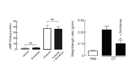
Given only the data from the graphs…
Diclofenac may be an inhibitor of PKA (cAMP works even with diclofenac, so it must be further downstream)
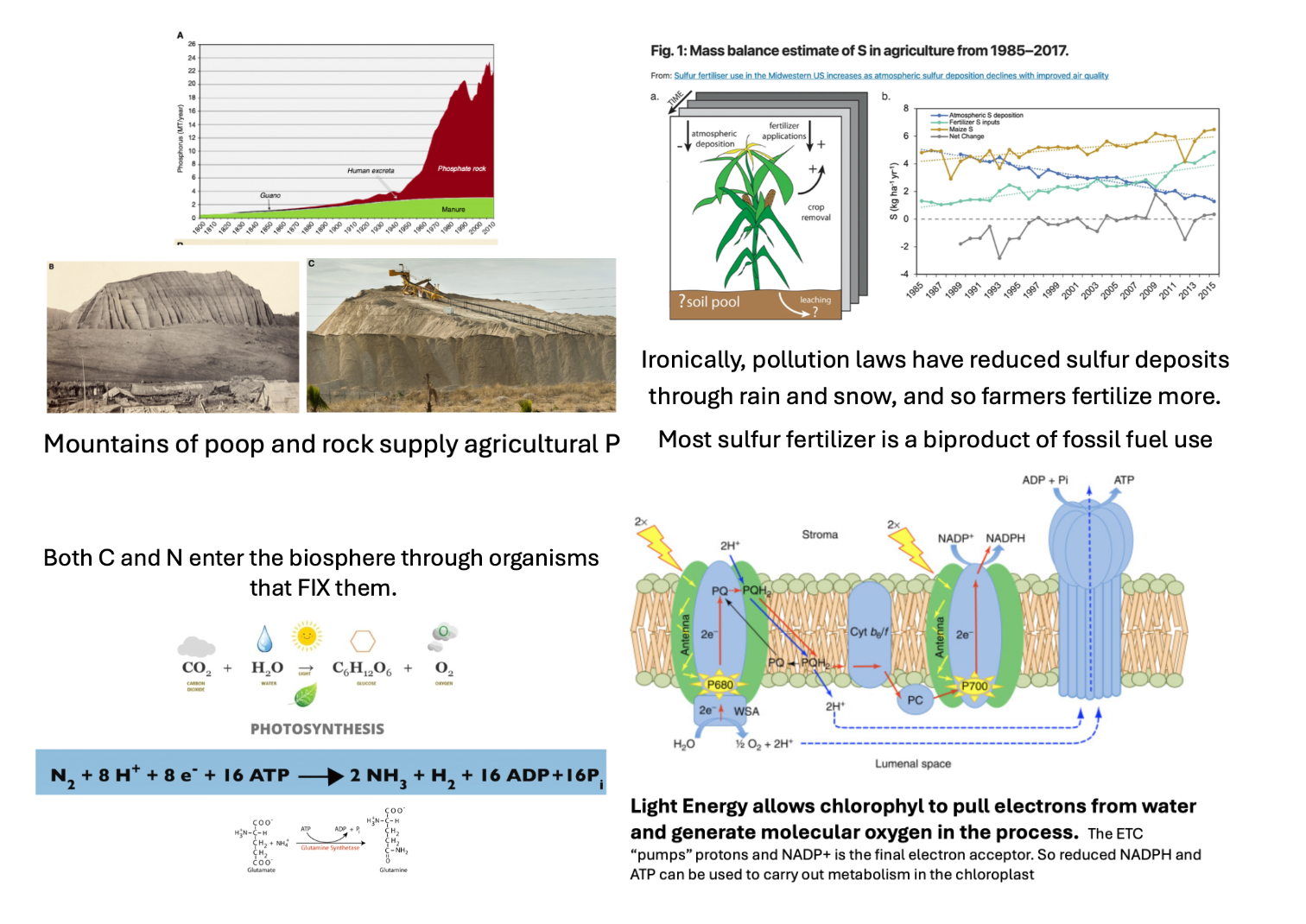
NCHOPS revisited:
Mountains of poop and rock supply agricultural phosphorus (P)
Both carbon (C) and nitrogen (N) enter the biosphere through organisms that FIX them
Pollution laws have reduced sulfur (S) deposits through rain and snow, and so farmers fertilize with it more (most sulfur fertilizer is a byproduct of fossil fuel use).
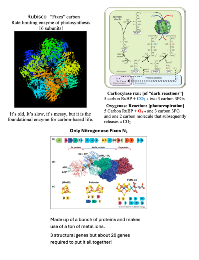
Rubisco “fixes” carbon. It is the rate limiting step of photosynthesis, and because it is so old it cannot tell the difference between CO2 and O2. Because of this, it can “fix” both of these in carboxylase (“dark”) reactions and oxygenase (photorespiration) reactions..
In a similar vein, only nitrogenase fixes N2 (an anaerobic process). However, it cannot tell the difference between N2 and O2 (actually binding O2 better than N2), and this creates problems since is O2 binds the nitrogenase will denature/unfold in a case of irreversible competitive inhibition. However, in many cases aerobic respiration is required to provide the fuel (ATP AND NADH) required to carry out the nitrogen fixation.
Note: the active site of nitrogenase has lots of iron in it. Iron binds to N2.
A brief history lesson:
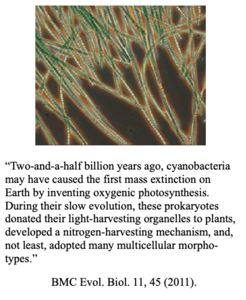
Once upon a time earth had no oxygen in the atmosphere. The organisms that “invented” oxygenic photosynthesis through nitrogen (N2) fixation are known as the cyanobacteria.
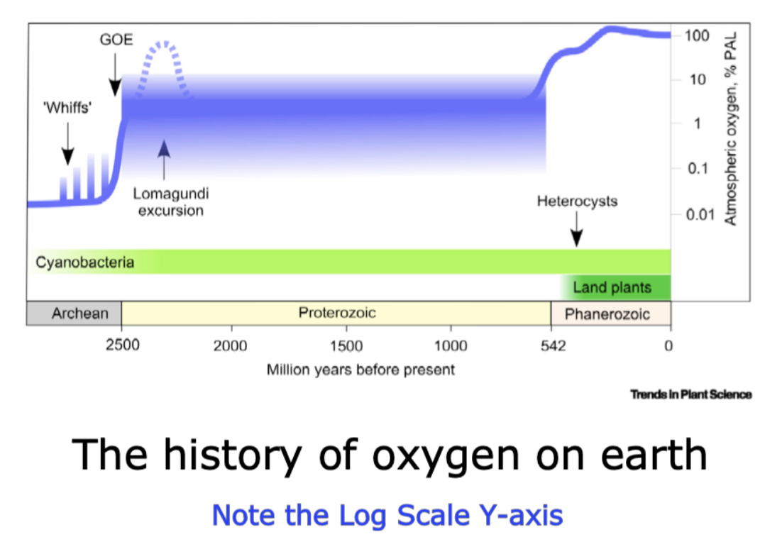
The levels of oxygen in the atmosphere were low and seemingly consistent for a couple of billion years. The shaded area indicates the uncertainty of our data within the Proterozoic, but the scientific community is confident that levels never exceeded 10% of our current oxygen levels throughout that majority of life history.
Atmospheric oxygen comes from photosynthesis, and it obviously predates aerobic respiration. A recent paper makes an elegant argument that the quite recent (in a geological time frame, so over 500 million years) increases in oxygen come from land plants evolving to allow photosynthesis to occur above the ground, at a distance from where nitrogen fixation occurs.
The logic they propose is that while unicellular organisms were carrying out both nitrogen fixation and photosynthesis the oxygen generated by photosynthesis created a negative feedback of biomass accumulation (the amount of life happening) because oxygen stops nitrogenase.
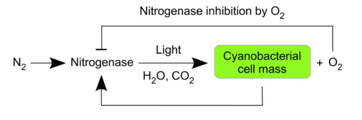
Nitrogenase Inhibition by O2
This is a key component of the argument – the evolution of heterocysts, the cyanobacteria cells that are impervious to oxygen, became widespread AFTER land plants were prevalent. Before that cyanobacteria were happy to live with the amount of oxygen they were generating.
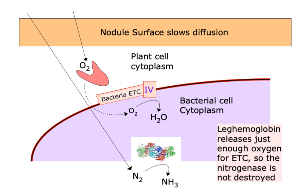
Symbiotic Nitrogen Fixation
Nodules are condos for N2 fixing bacteria.
Plant gets NH4+ (the product of N fixation) from the bacteria. Most often, these are Rhizobium bacteria, but also cyanobacteria in association with cycads, and others.
Bacterium gets an O2-controlled environment and carbohydrates from the plant
Oxygen affinity relationships:
Cytochrome c Oxidase > Leghemoglobin > Nitrogenase
(Leghemogloben releases just enough oxygen for ETC so the nitrogenase doesn’t get destroyed)
Location:
Cytochrome c Oxidase = in rhizobial cell membrane
Leghemoglobin = in the cytoplasm of host cell
Nitrogenase = inside the rhizobium (bacterioid)
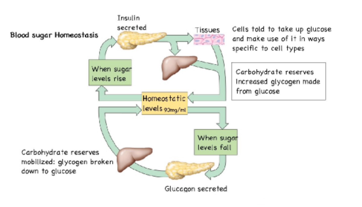
Blood Sugar Homeostasis
Pancreatic alpha cells secrete glucagon in response to low blood sugar.
Pancreatic beta cells secrete insulin in response to high blood sugar
Glucagon:
Increases blood sugar levels by…
Signalling the liver to break down glycogen and release the breakdown product (glucose) or make gluose from stored reserves (lipids and AAs).
^ The latter is GLUCONEOGENESIS.
Clearly liver cells must have receptors for glucagon, as liver cells also stop making glycogen when glucagon arrives. So these are supply, but not demand changing mechanisms.
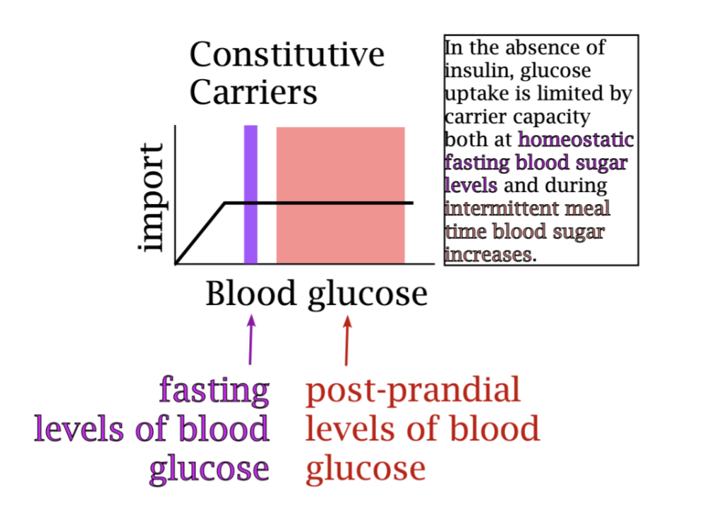
In the absence of insulin, glucose uptake is limited by carrier capacity both at homeostatic fasting blood sugar levels and during intermittent meal time blood glucose levels.
Note: BOTH ARE AT VMAX! The machinery present in your cells when you are at fasting levels of blood sugar could not accommodate more intake. The import carriers present on the surface of the cells are working as fast as they can even at fasting glucose levels.
Postprandial = After a meal
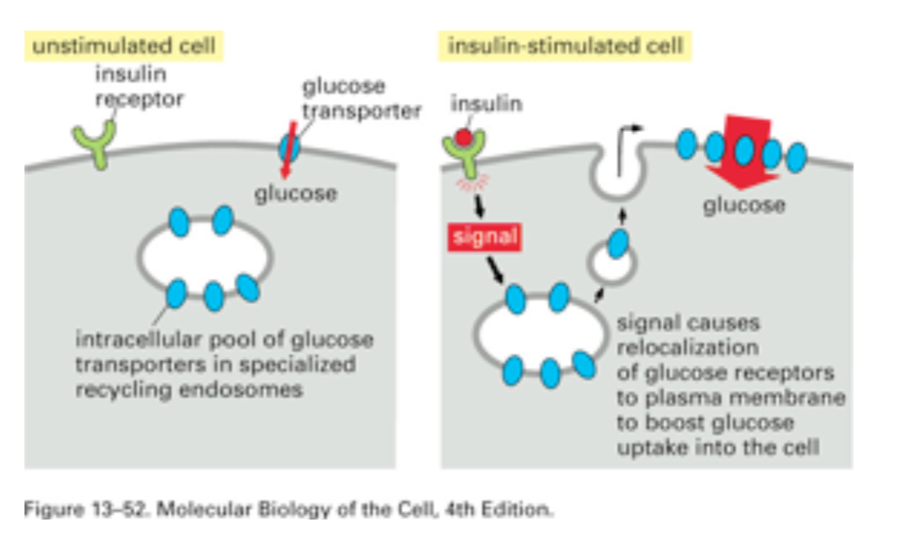
In the presence of insulin, glucose uptake increases to allow glucose uptake proportional to insulin levels.
Note: VMAX HAS INCREASED BUT KM IS THE SAME. From this you might think you see a noncompetitive activator of insulin uptake carriers, but in fact you are seeing the result of the addition of carriers, akin to the addition of enzyme in a michaelis menten plot.
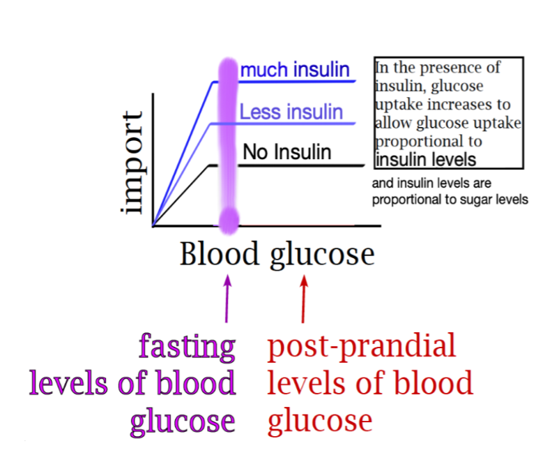
As blood sugar increases above fasting…
Both circulating insulin levels and cellular glucose uptake levels increase.

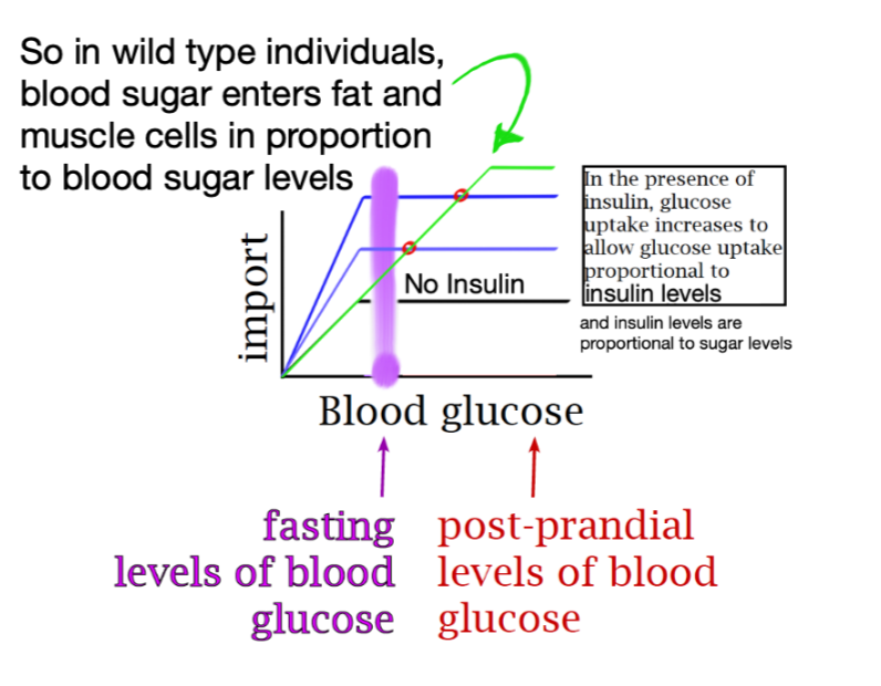
In WT individuals, blood sugar enters fat and muscle cells in proportion to blood sugar levels.
Essentially:
Tissues take up sugar from blood at a rate that is independent of blood sugar unless insulin signals otherwise.
Iron uptake in the small intestine is accomplished via proton cotransport. It should not surprise you then to discover that iron uptake is limited to the upper end of the intestine, before bicarbonate action has accomplish buffering. Iron is taken up in ferrous form and exits the brush border cells through ferroportin, which is tightly integrated with hephaestin, the enzyme that returns iron to the less toxic form that is then carried through the blood by transferrin.
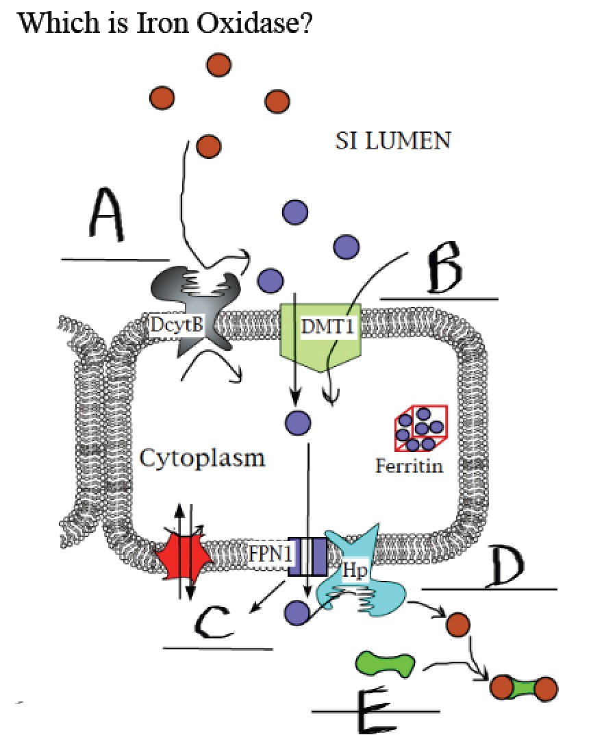
Which is iron oxidase?
D
Uptake of iron form the small intestine lumen occurs via proton cotransport. Uptake of iron from the blood occurs via transferrin endocytosis. In all cells that grab iron from the blood the iron and iron carrier and iron carrier receptor are pulled apart by pH denaturation of the proteins. All iron released into the blood is exported through ferroportin.
Hepcidin function is to decrease blood iron levels.
Ferroportin mutants that fail to bind with hepcidin lead to excess circulating iron.
A hepcidin loss of function mutation would lead to excess blood iron (same thing).
How do cells export copper?
Copper pumps! (not tested on)
Over the course of billions of years atmospheric oxygen levels increased from essentially non-existant as a result of accumulated photosynthesis using water as the electron donor. This in turn led to a dramatic decrease in the amount of biologically available iron in the oceans.
Cyanobacteria were responsible for this accumulation. They were also also available for much of the biologically available nitrogen on the planet.
What molecule reduces the activity of both nitrogenase and rubisco?
Oxygen
The Km of leghemoglobin is greater than that of rhizobium cytochrome c oxidase but lower than that of nitrogenase.
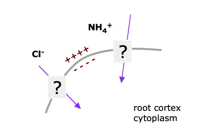
Which ion could be moving through HATS, given greater concentration in the soil water (outside)?
Chloride (????????????????????????????????)
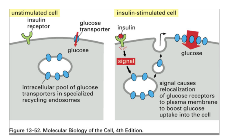
Watch sugar into muscle cell video?
Glucose receptors exocytosed to the membrane of the cell, and then endocytosed back into the cell.
What does insulin do?
Insulin signals target tissues to make things
Upregulates fuel consumption (often via PFK)
Upregulates synthesis of fuel storage molecules (fat, glycogen)
Upregulates protein synthesis in all target cells
Insulin signals target cells not to mobilize fuel reserves
Downregulates breakdown of fuel storage molecules (fat, glycogen, protein)
Downregulates release and distribution of fuel molecules (glucose, fatty acids, amino acids)
Liver is major target in terms of tissue that will deliver nutrients into the blood, but other cells too.
Insulin acts in these ways on target cells throughout the body, but not the brain. Insulin acts primarily on fat and muscle and the liver.
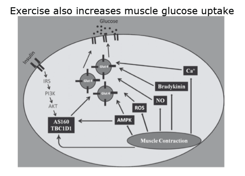
Exercise also increases muscle glucose uptake.
(Muscle cells exercised intensely also have a 48 hour period of increased insulin sensitivity post workout).
How Pancreatic b-cells respond to blood glucose levels
Glucose enters b-cells at rate proportional to blood glucose
(unlike in typical tissues, compare the kinetics!)
Beta cells of the pancreas secrete insulin at a rate determined by glucose concentration of the blood.
So glucose must be imported at a rate proportional to blood sugar.
Glucokinase is the rate-limiting step.
Glucokinase kinetics allow the rate of ATP production to match the blood sugar concentration (PFK not limiting here)

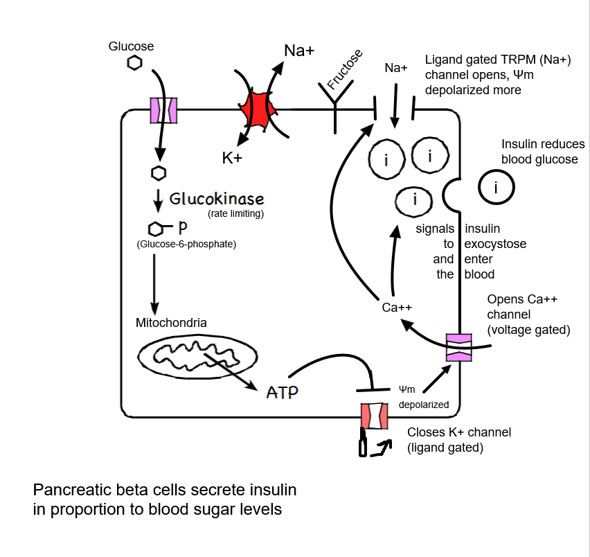
Beta Cell Pathway:
Glucose
↓ Glucokinase (rate limiting)
Glucose-6-phosphate
↓ Mitochondria
ATP
↓
Closes ligand gated K+ channel
↓
Membrane depolarized
↓
Voltage gated Ca++ channel opens
↓
Ca++ signals insulin to exocytose and enter the blood AND opens the ligand gated TRPM (Na+) channel (COINCIDENCE DETECTOR!)
↓
Na+ entering depolarizes the membrane more
↓
Blood glucose reduced!
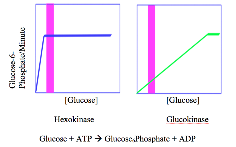
Hexokinase vs Glucokinase
Hexokinase
High affinity
Low km
Glucokinase
Low affinity
High km
Better at signalling a need for things (like cytochrome c oxidase!)
Measures how much sugar is around, processing sugar in proportion to how much sugar is in the cell
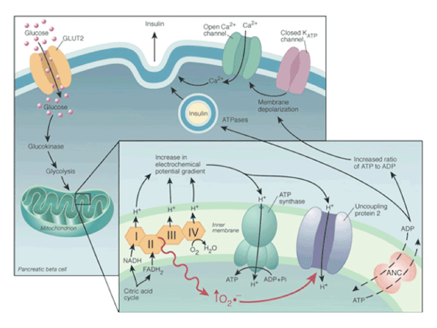
Uncoupler activity has a role in cellular ATP concentration in beta cells. This is in extreme contrast to the other cells in your body, that have a paired negative feedback loop based around the enzyme PFK. Because in these cells PFK isn’t rate limiting, the feedback system doesn’t compensate for uncoupler activity. A significant fraction of people who have lived to 100 years old have a high uncoupler activity in beta cells, suggesting to many scientists that this contributes to longevity. Note this is a correlation, and I am not telling you that the evidence for a causal relationship is sufficent for me to teach that this is so.
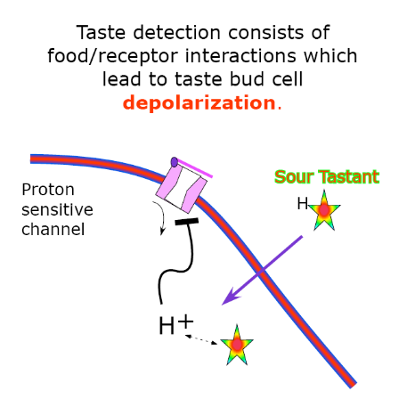
Taste review:
Which ion moves through the proton sensitive channel when protons are not closing it?
K+ (depolarizes the cell)
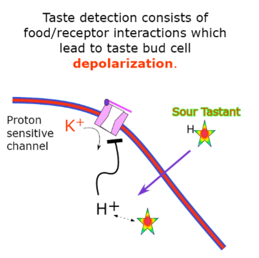
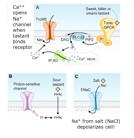
Taste review:
The process of flavor detection is a function of recognition of a particular chemical leading to depolarization of the cell that recognized it. This cell sends the message to the brain that it has detected that chemical as a result of this depolarization.
Sour tastes led to a mechanism of depolarization similar to that of b cell responses to glucose, namely the closure of a K+ channel.
Salt detecting cells use the Na+ of NaCl to depolarize the cell.
Other flavors use interaction with a receptor molecule leading to signal transduction leading to the opening of Na+ channels. These are called the sweet channels because they were discovered in experiments with sweet detection.
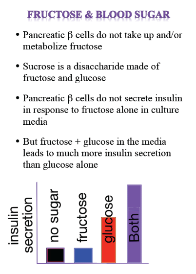
The odd case of fructose response in animals:
Pancreatic beta cells do NOT take up/metabolise fructose.
Sucrose is a disaccharide made of glucose and fructose monomers. It’s the most abundant dietary sugar (think plants!).
Pancreatic beta cells do not secrete insulin when fructose is used at low or high levels in culture media.
Glucose + fructose increases insulin levels more than glucose alone.
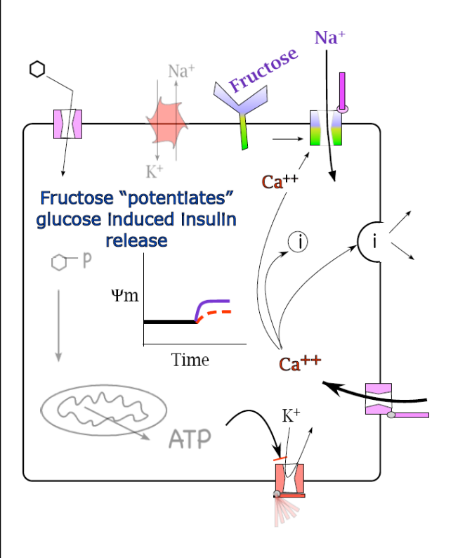
Fructose “potentiates” (makes stronger/more effective) beta cell response to glucose.
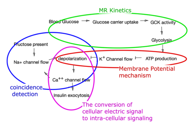
A sweet detection receptor with a much higher affinity for fructose than glucose is bound by fructose. When this happens at the same time that Ca++ is present (due to high levels of glucose metabolism) a channel opens that allows Na+ in. This adds to the depolarization of the membrane and so the amount of insulin released.
Note that without glucose, no Ca++ is present, and this explains why fructose alone doesn’t lead to insulin release.
This is the coincidence detection mechanism!!!!! (Ca++ & Fructose = stronger depolarization)
Beta Cell Pathway
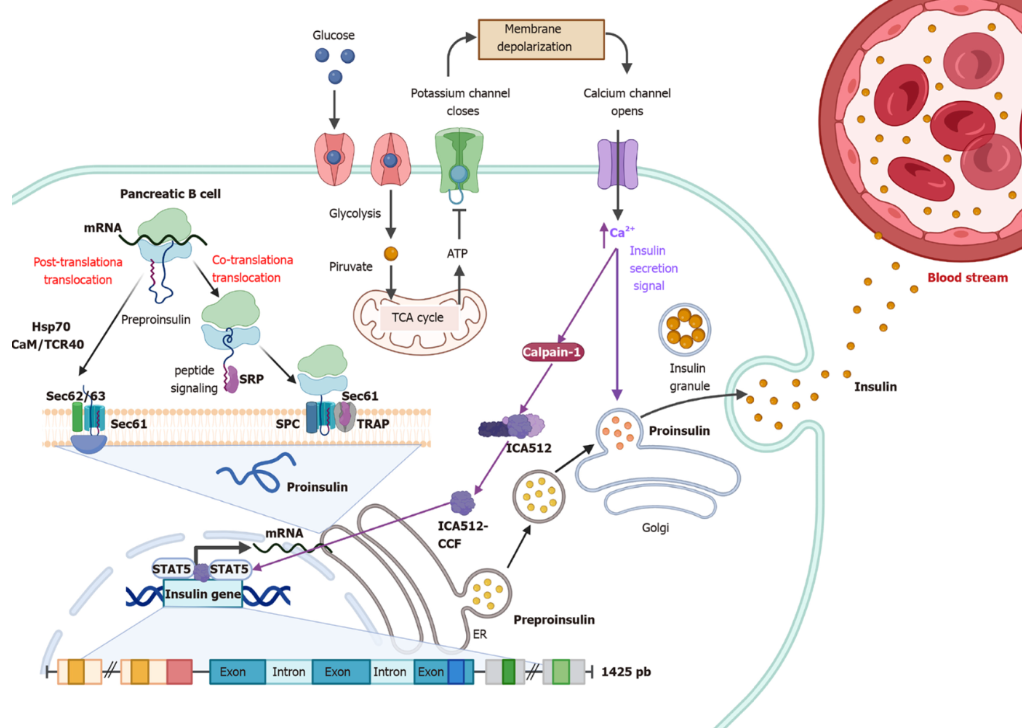
In this recent review of glucose stimulated insulin secretion we see that the process both causes release of insulin containing vesicles and promotes their replacement through upregulation of the insulin gene itself.
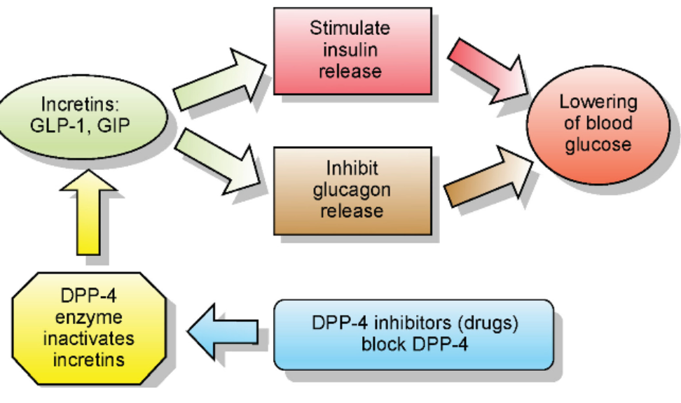
GLP1 Pathway Response:
GLP1 is made by cells located in the intestines and brain. It is made during nutrient uptake. It is very short lived and probably is released by these cells to preserve beta cells when blood sugar levels are not being well maintained. Our GLP1 doesn’t survive long in circulation, hence the large impact of the pharmaceutical version.
The GLP-1 agonists are medications that activate the GLP-1 receptor and have much longer lifespans in circulation. They were created to treat blood sugar problems, but seem to play a role in a weirdly large number of systems.
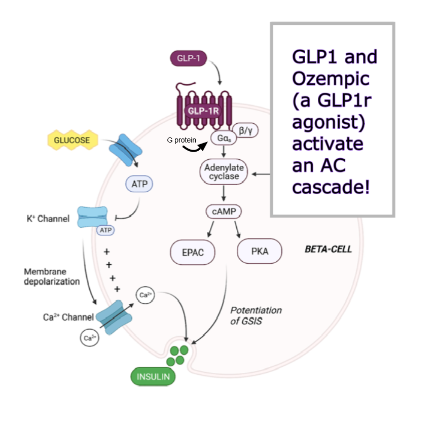
The potentiation of Glucose stimulated insulin secretion must be a form of coincidence detection, in that the hormone increases the response, rather than replacing it.
GLP1 leads to upregulation of the protein activity that is required for Ca++ dependent insulin exocytosis.
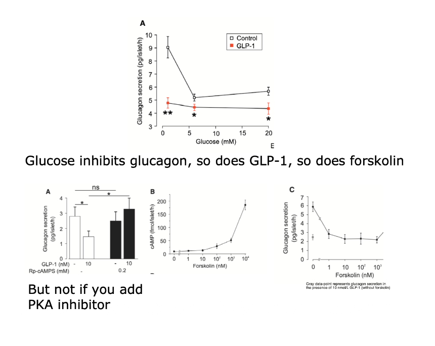
Glucose inhibits glucagon, so does GLP1, so does forskolin…
But not if you add PKA inhibitor!