Practicals
1/76
There's no tags or description
Looks like no tags are added yet.
Name | Mastery | Learn | Test | Matching | Spaced | Call with Kai |
|---|
No analytics yet
Send a link to your students to track their progress
77 Terms
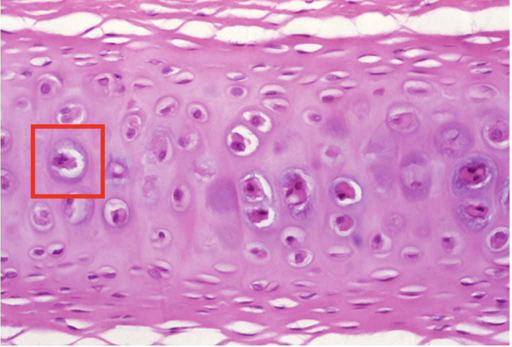
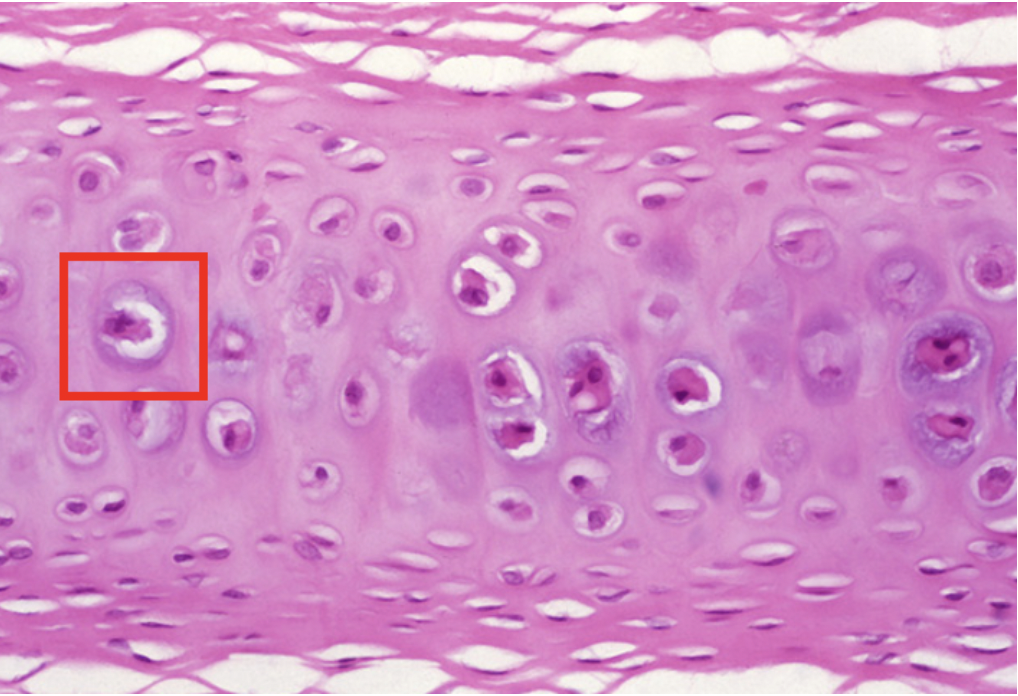
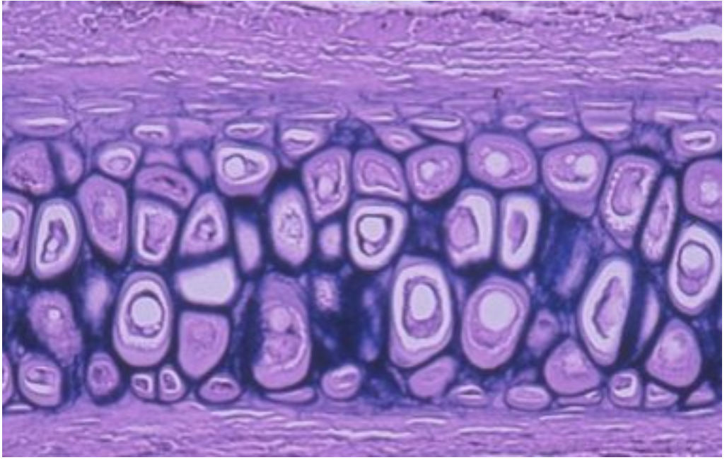
Identify the CELL seen in the red box
chondrocyte
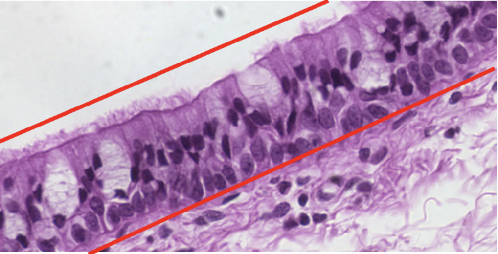
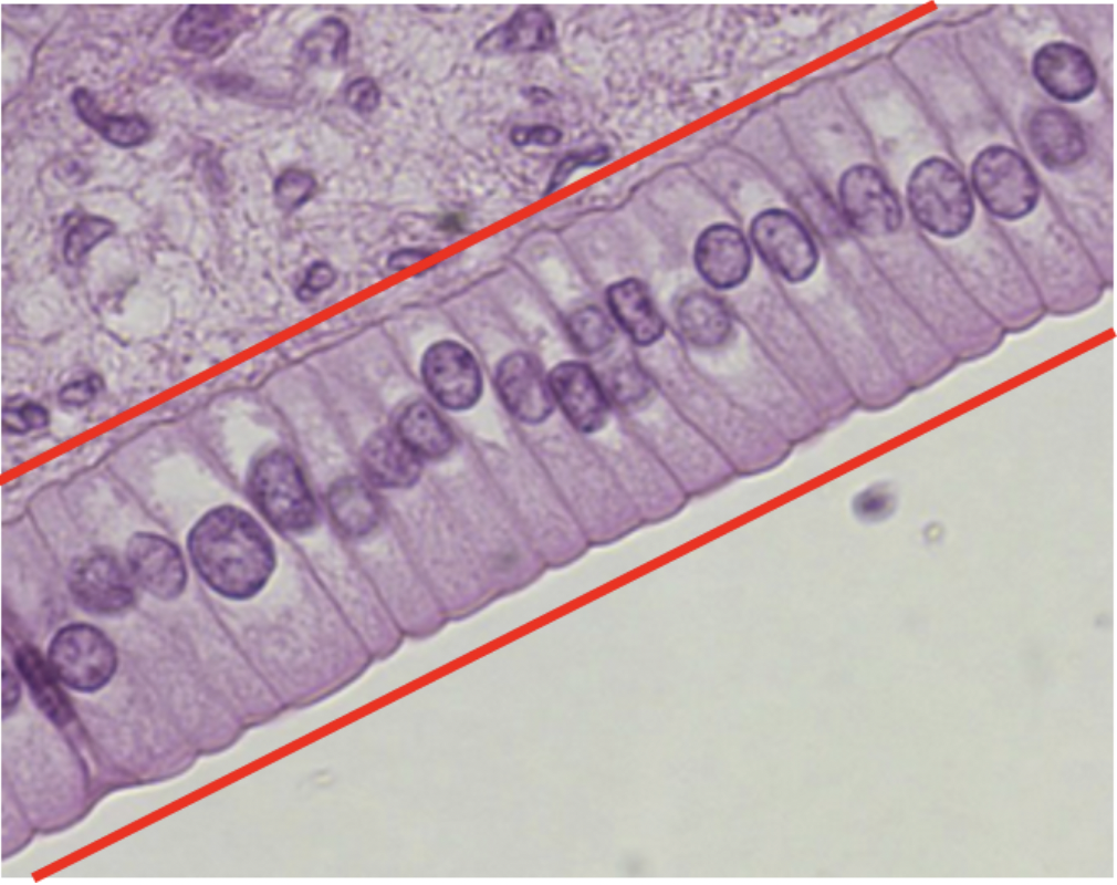
Identify the tissue seen between the red lines
pseudostratified epithelium
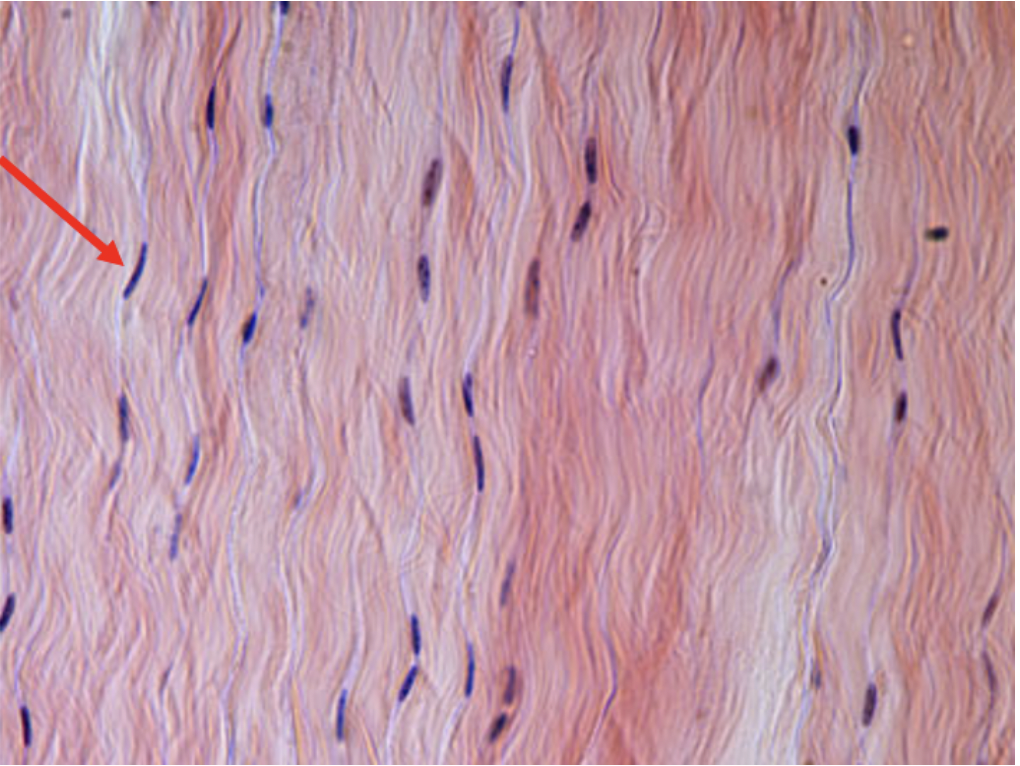
Identify the fiber seen in this photo
collagen fiber
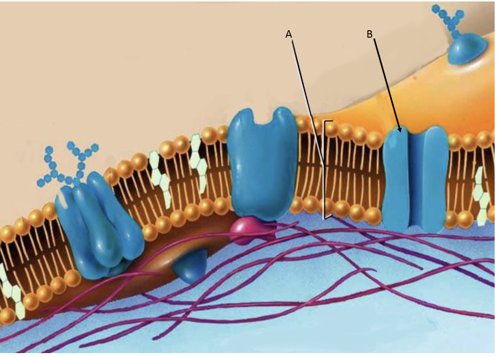
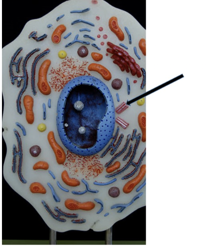
Identify the molecules/structures bracketed by “A”
phospholipid bilayer
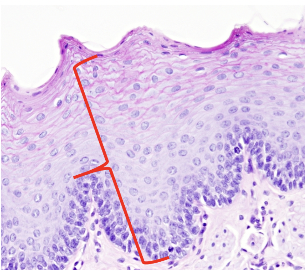
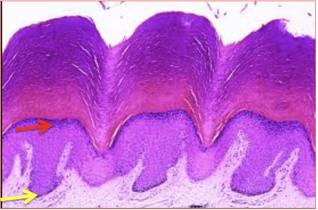
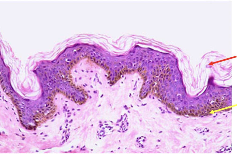
Identify tissue type indicated by the red bracket
stratified squamous epithelium
identify the tissue seen between the red lines
pseudostratified columnar epithelium
Name a LOCATION for this tissue
ends of long bones
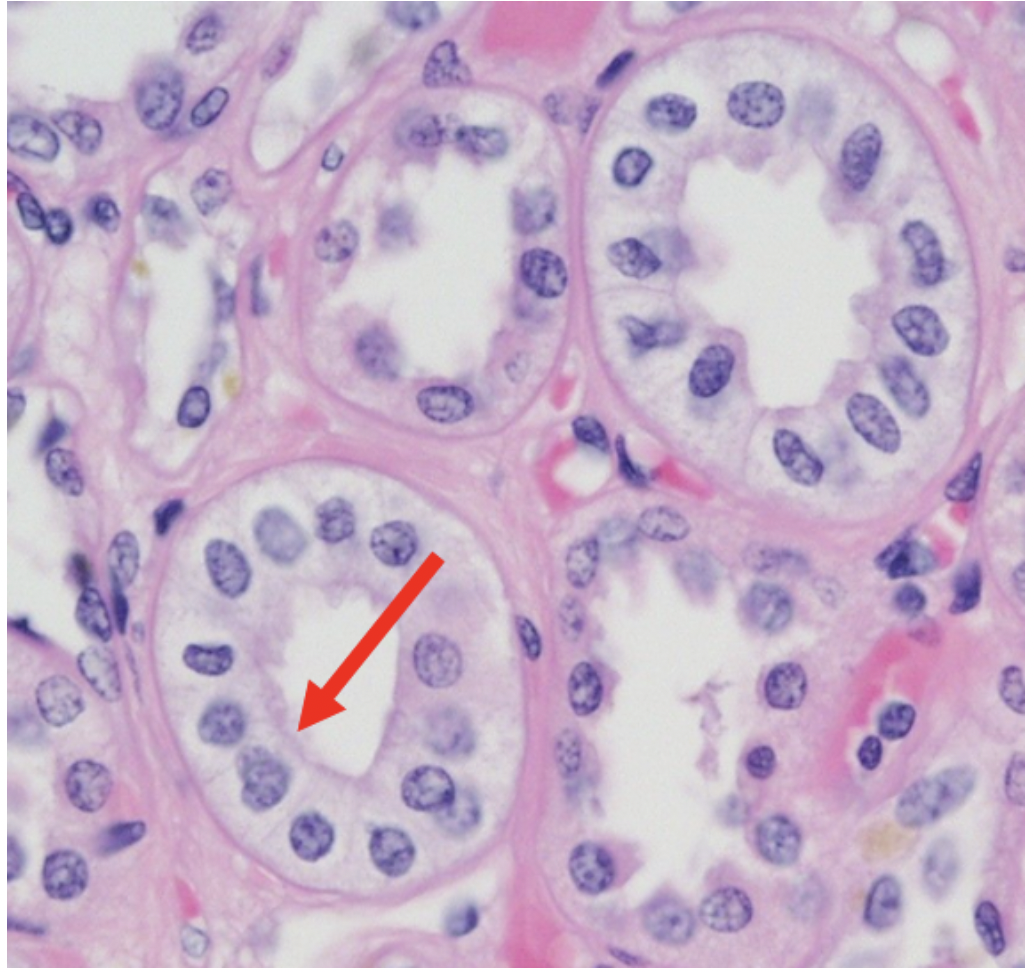
Identify the tissue type at the tip of the red arrow
simple cuboidal epithelium
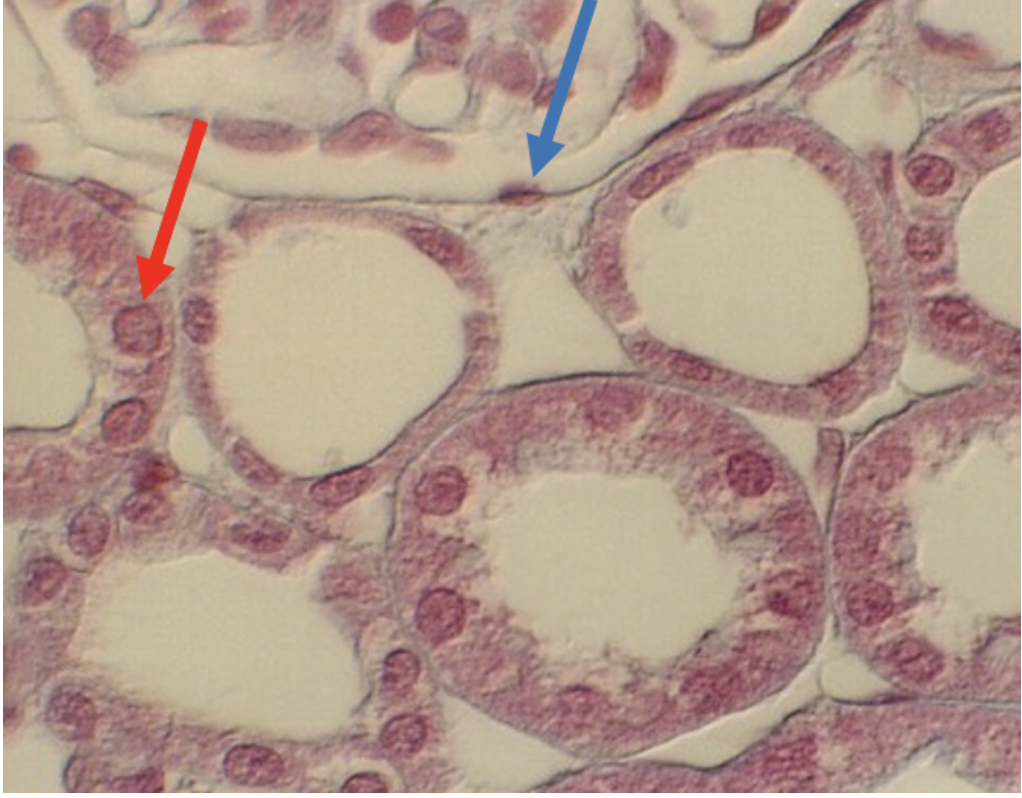
Identify the epithelial type seen at the tip of the blue arrow
simple squamous epithelium
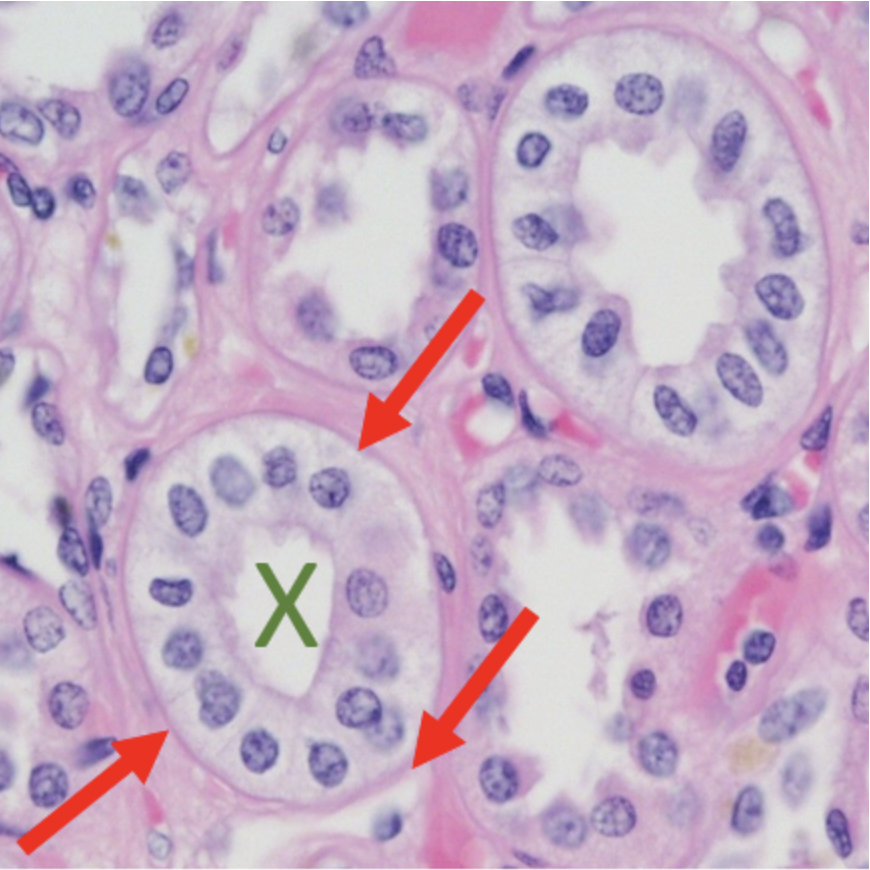
Identify the STRUCTURE at the tip of the red arrows (both photos are the same, photo below is only to see without markups)
basement membrane
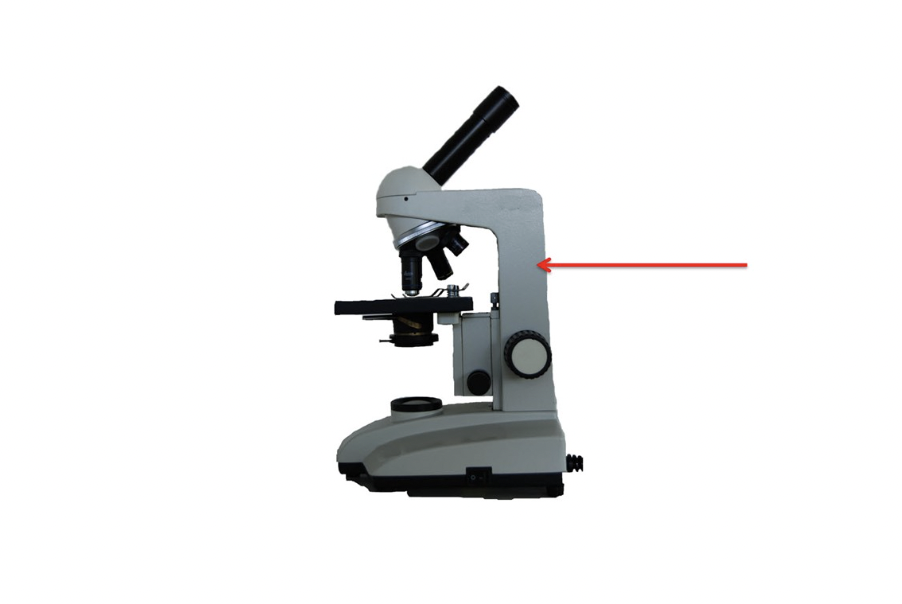
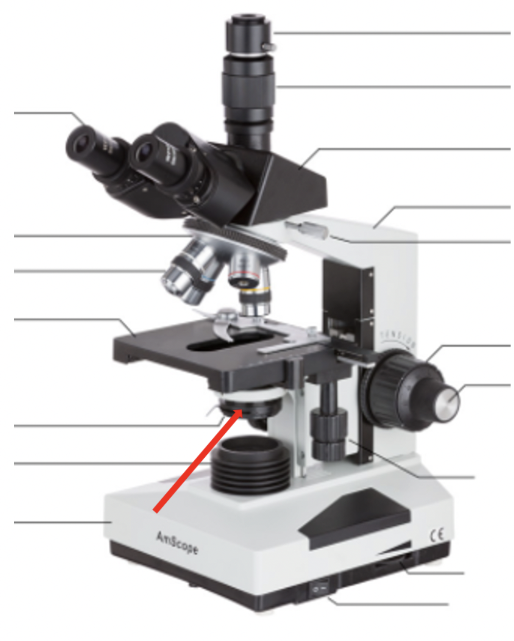
Identify the microscope part at the red arrow
arm
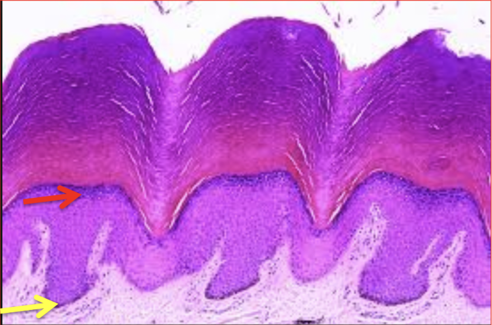
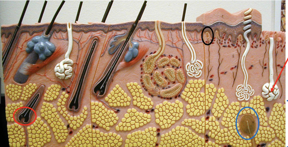
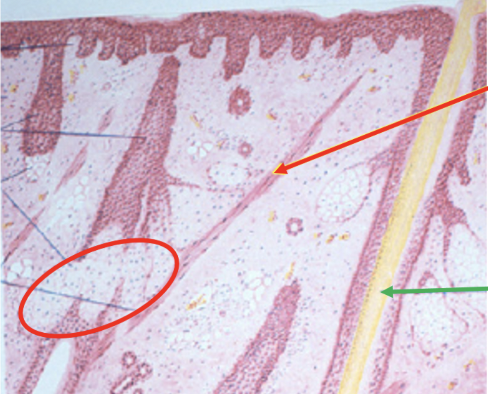
Identify the organ seen here. Be specific.
thick skin
Identify the structure at the tip of the black arrow
centriole
Identify the microscope part at the red arrow
condenser
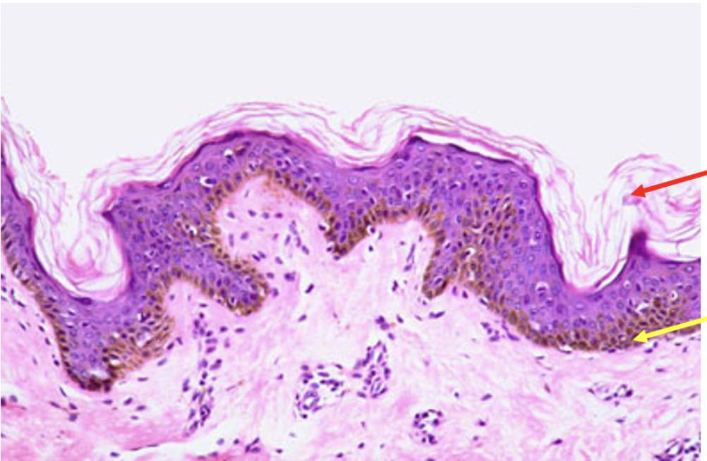
Identify the ORGAN seen in this photo. Be Specific
thin skin
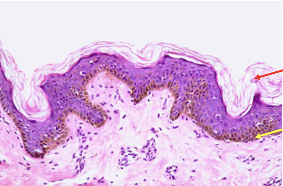
Identify the epithelial layer at the tip of the red arrow
stratum corneum
Identify the structure at the tip of the red arrow
eccrine sweat gland
Name a location for this tissue
soles of feet or palms of hands
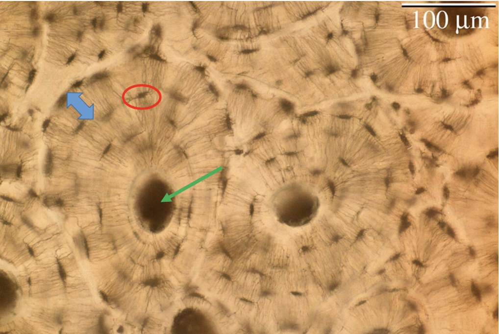
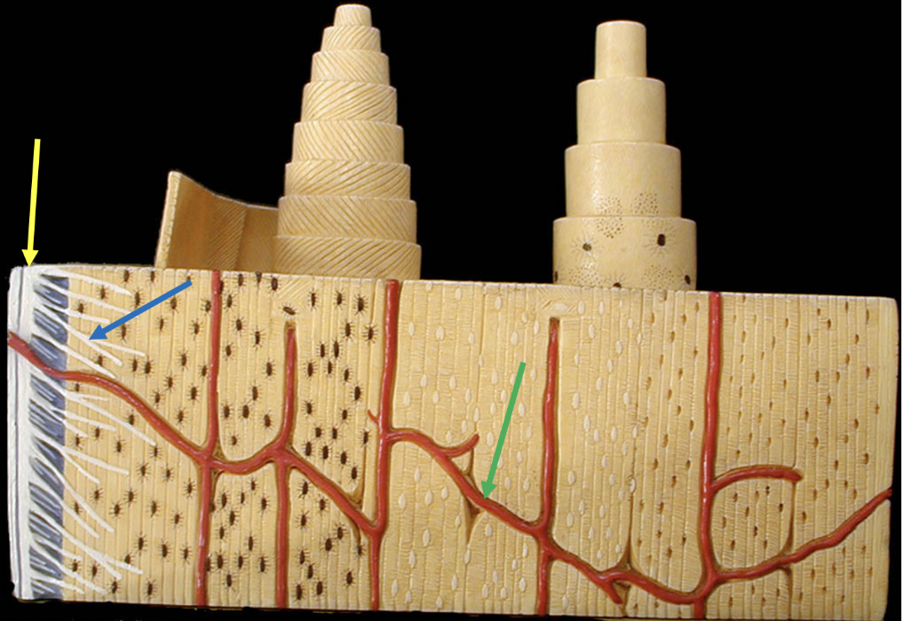
Identify the type of tissue on this slide
compact bone
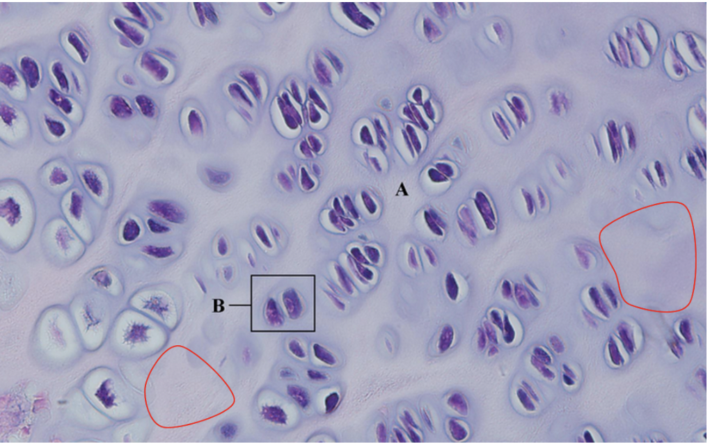
Identify the tissue part seen in the red outlined areas
matrix
Name a location for this tissue
ear
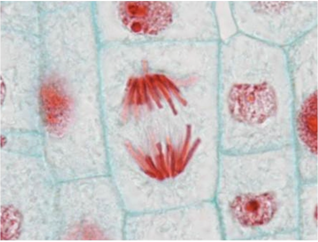
Identify the stage of mitosis seen here in the center of the photo
anaphase
Name the gland circled in red
sebaceous gland
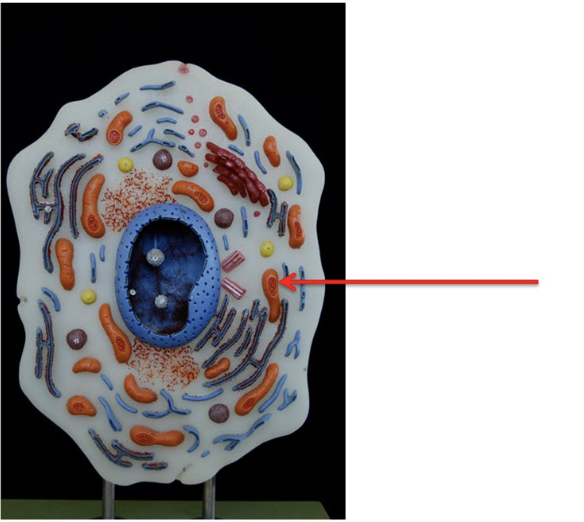
Identify the structure at the tip of the red arrow
mitochondria
Identify the dark/brownish cell at the tip of the yellow arrow
melanocyte
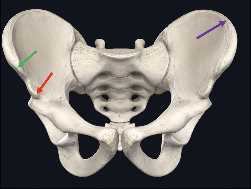
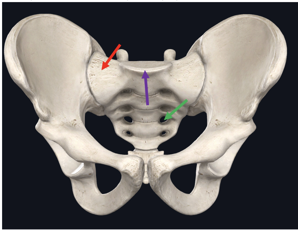
Identify the structure at the tip of the purple arrow
iliac crest
Identify the indentation at the tip of the red arrow
olecranon fossa
Identify the structure at the tip of the red arrow (be specific)
medial condyle of femur
Identify the connective tissue at the tip of the yellow arrow
periosteum
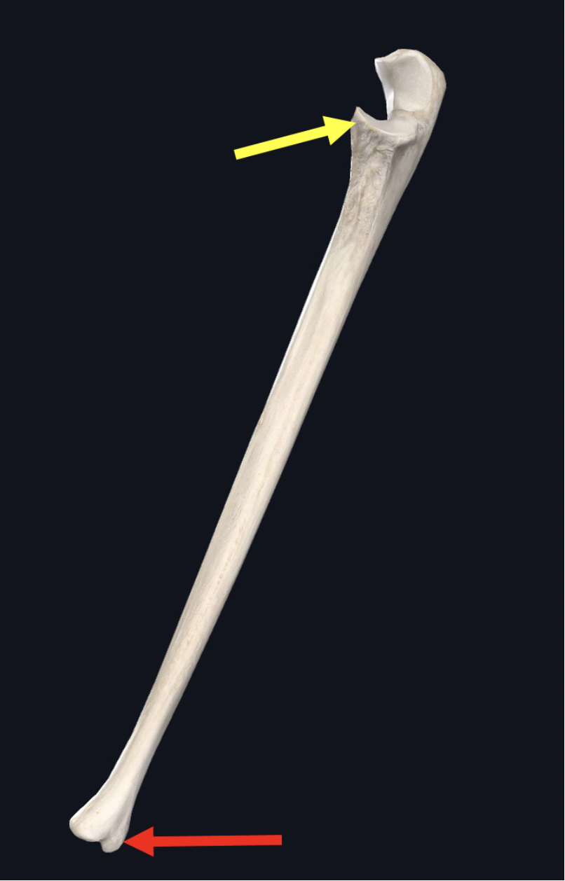
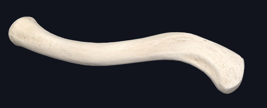
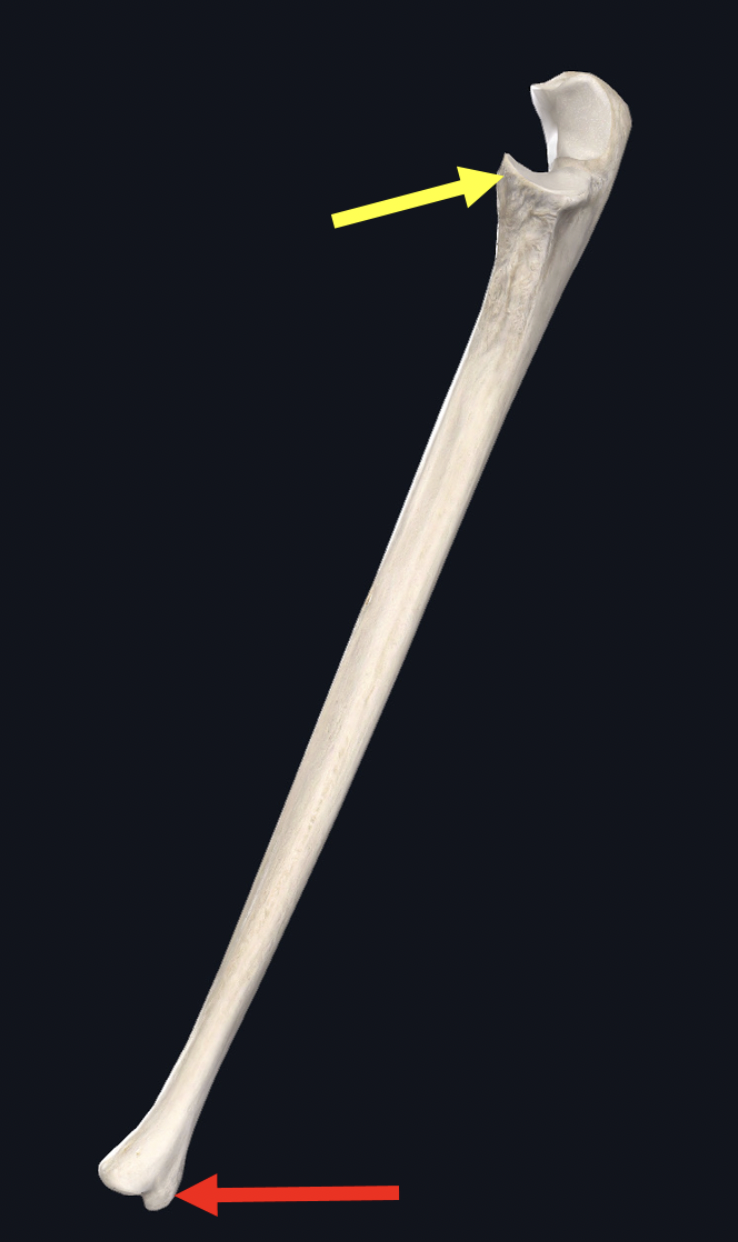
Identify this bone
ulna
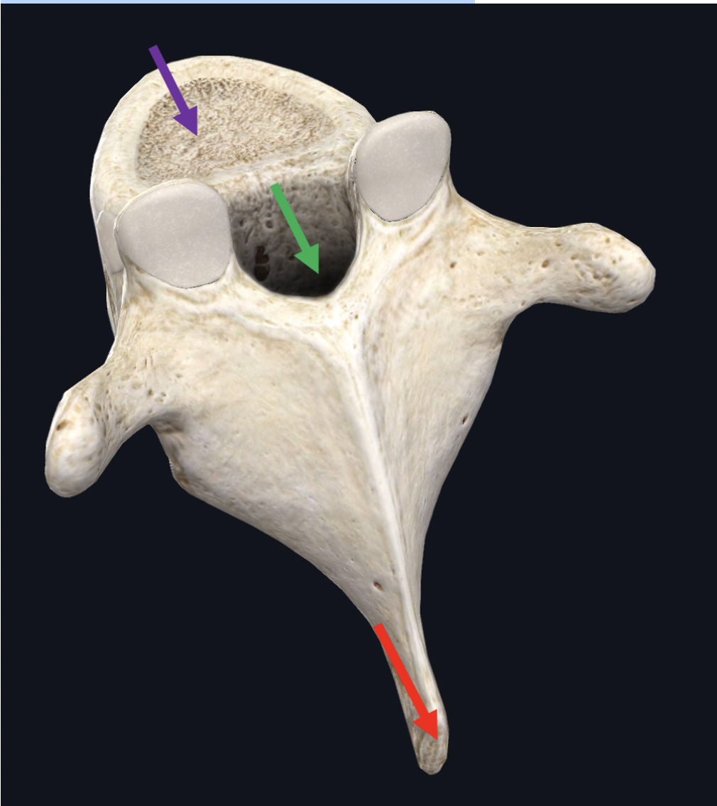
Identify the structure at the tip of the red arrow
spinous process
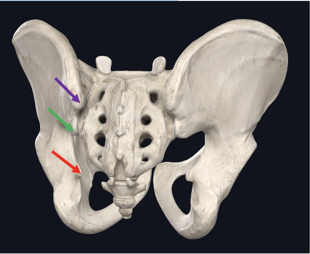
Identify the bone with the top two arrows
ilium
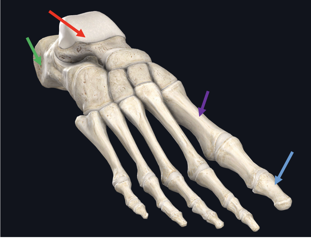
Identify the bone at the tip of the purple arrow
metatarsal
Identify this bone
clavicle
What bone connects at the tip of the red arrow
rib
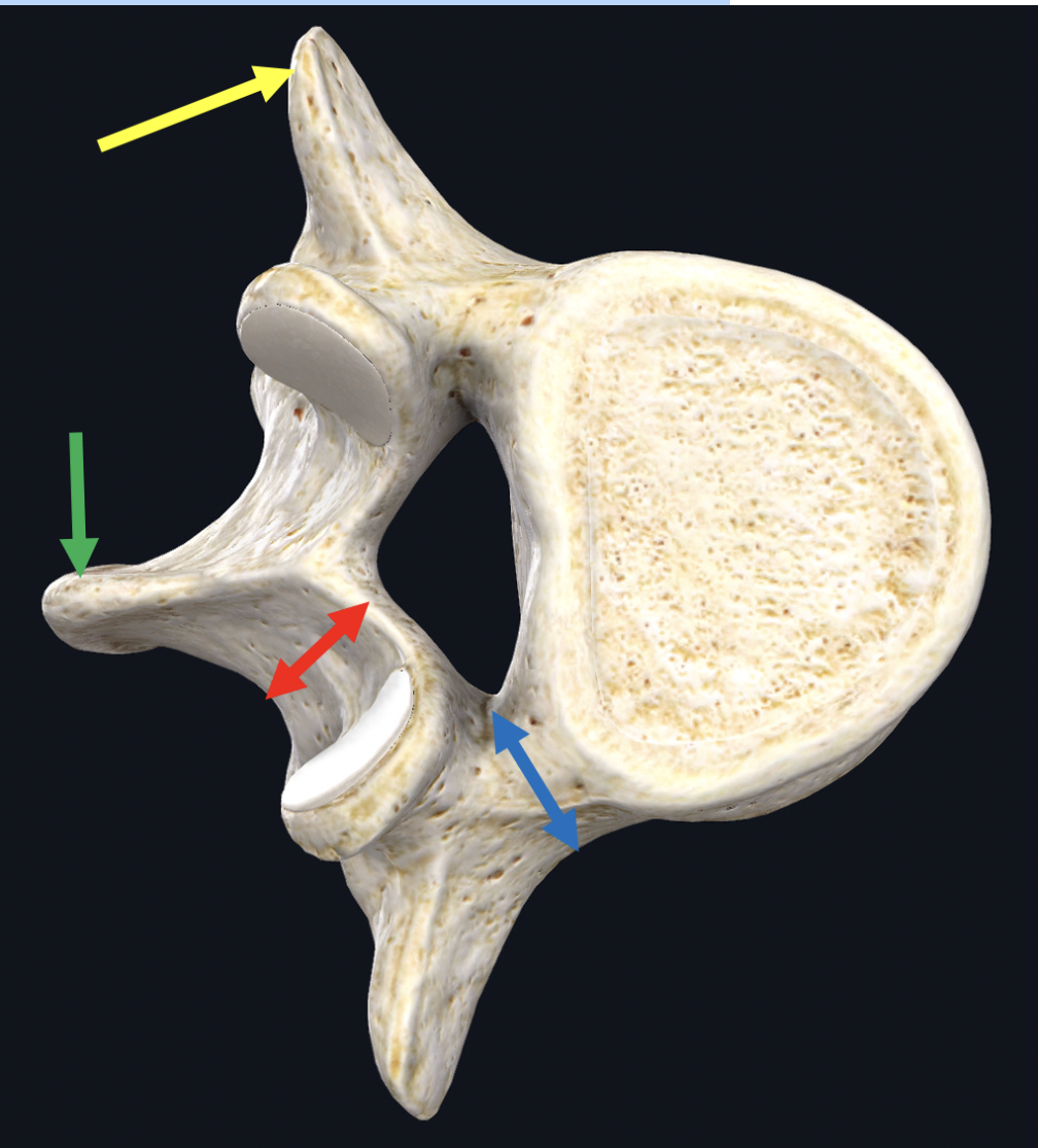
Identify the structure at the tip of the yellow arrow
transverse process
Identify the structure at the tip of the red arrow
spinous process
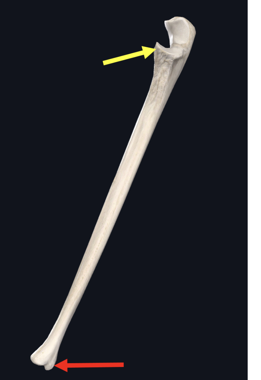
identify the structure at the tip of the yellow arrow
radial notch

Identify the group of bones within the blue circle
carpals
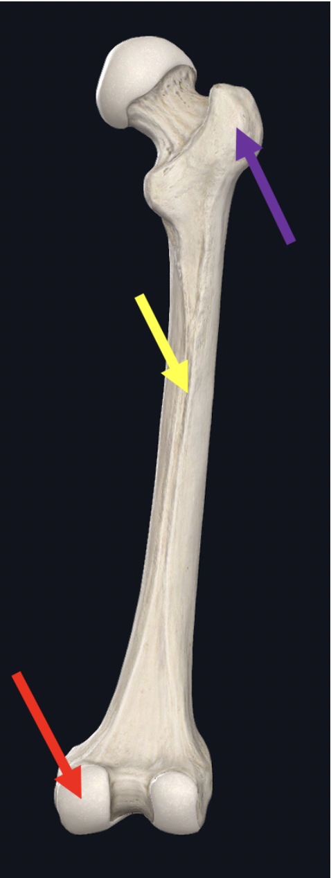
Identify the structure at the yellow arrow
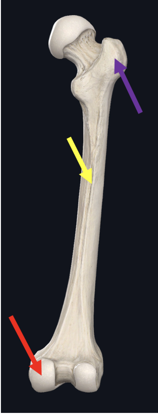
linea aspera
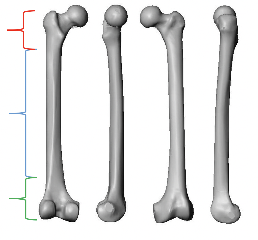
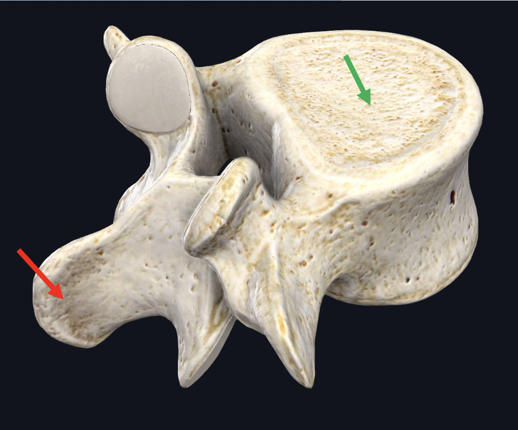
Identify the REGION of the bone indicated by the red bracket. Be specific.
proximal epiphysis
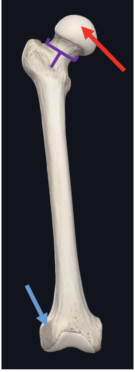
identify the structure indicated by the purple bracket
neck of the femur
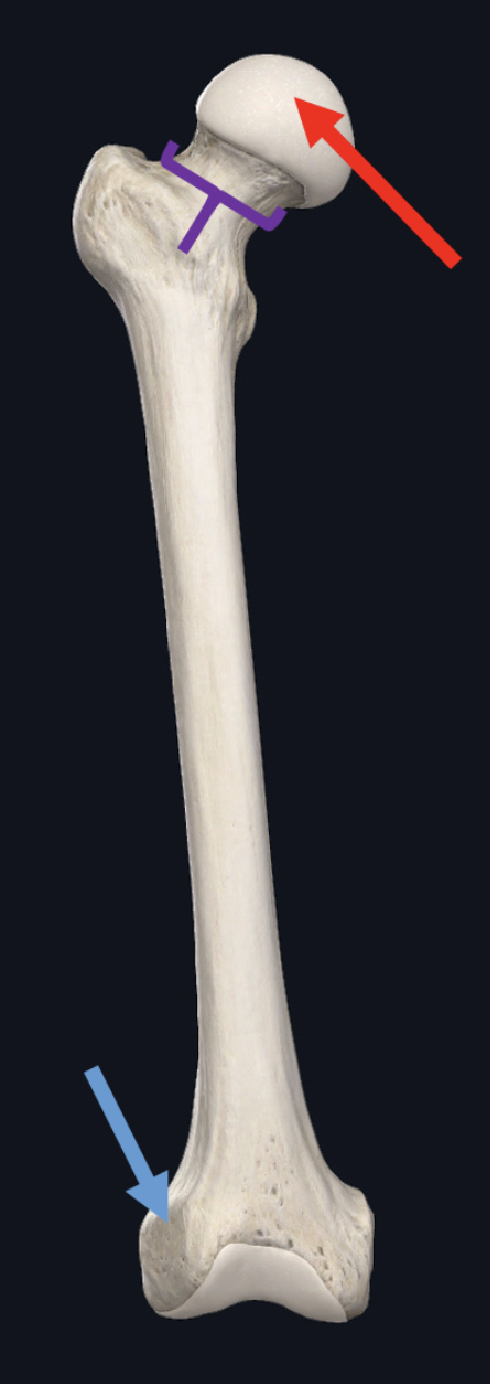
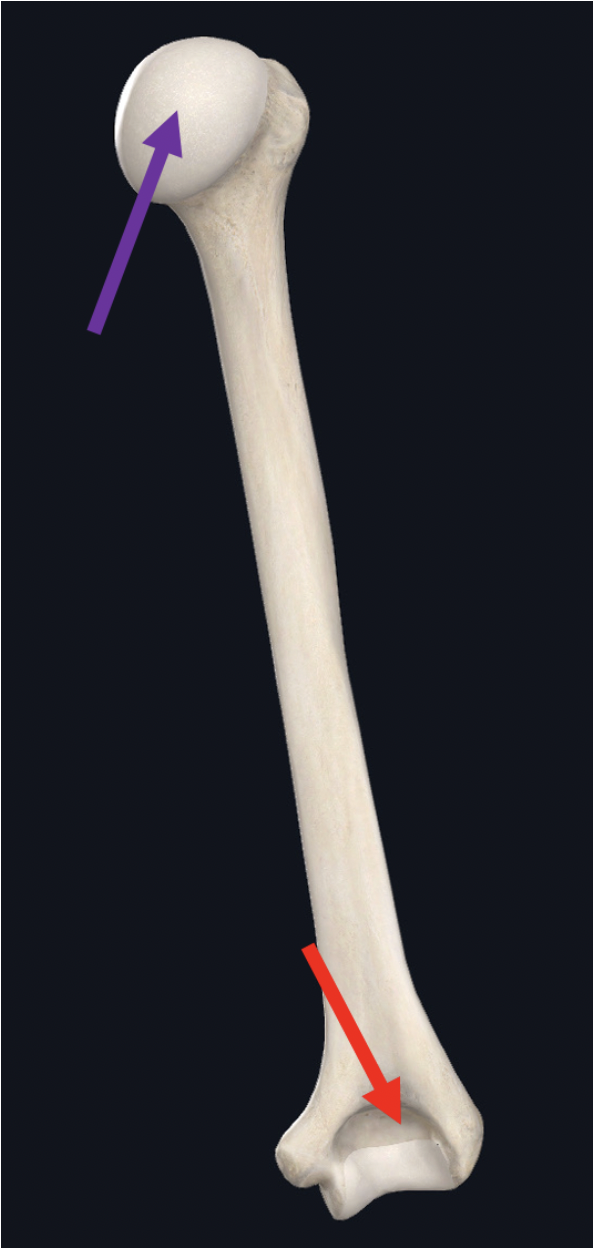
Identify the structure at the red arrow
head of femur
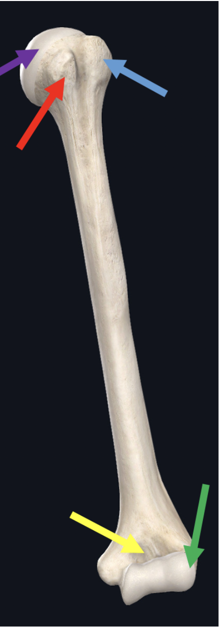
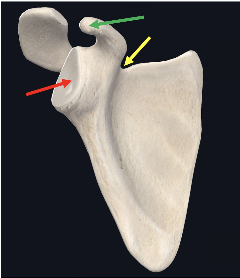
Identify the structure at the tip of the green arrow
lateral epicondyle of humerus
Identify the structure at the tip of the red arrow
styloid process
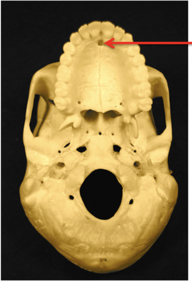
Identify the arrowed structure. Be specific.
incisive foramen
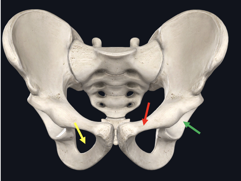
Identify the BONE at the tip of the red arrow
pubis
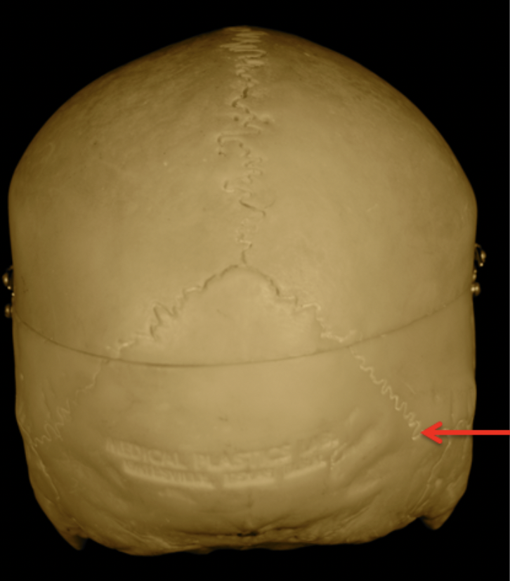
Identify the arrowed structure. Be specific.
lambdoidal suture
Identify which (left or right) this bone is
right
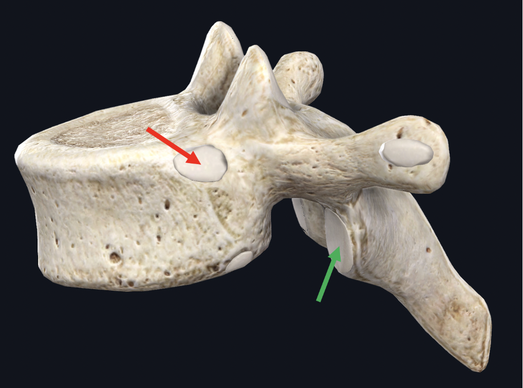
Identify the structure at the tip of the purple arrow
sacral promontory, red arrow is sacral ala, green arrow is sacral foramen

Identify the structure circled in blue
pacinian corpuscle
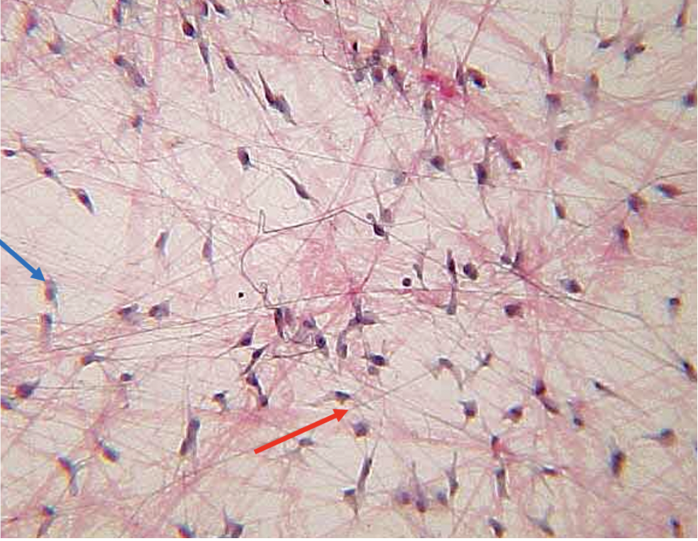
Identify this tissue
areolar connective tissue
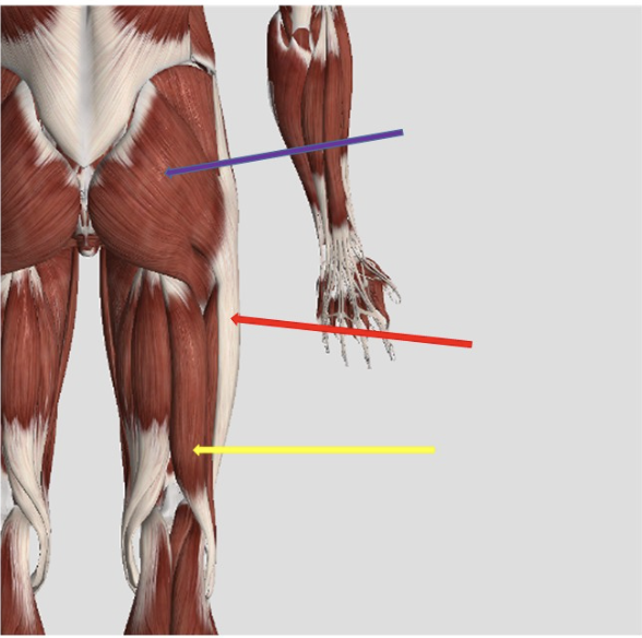
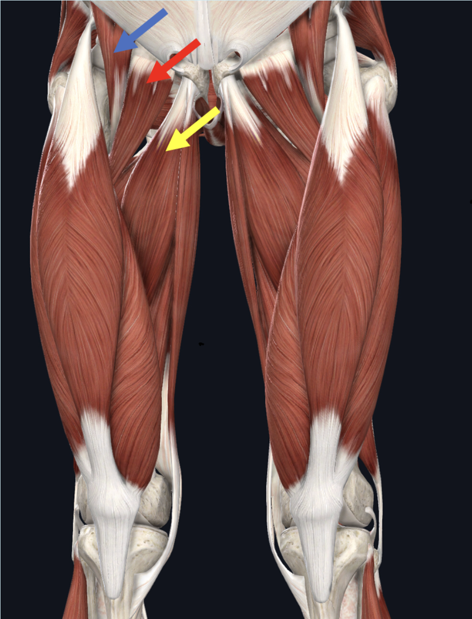
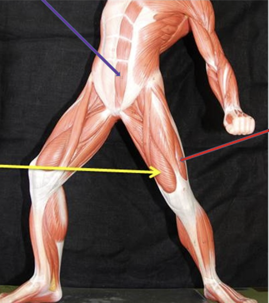
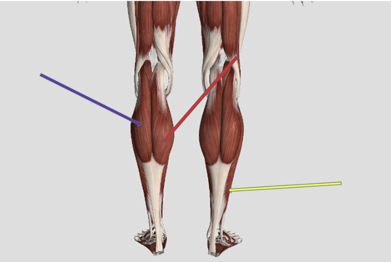
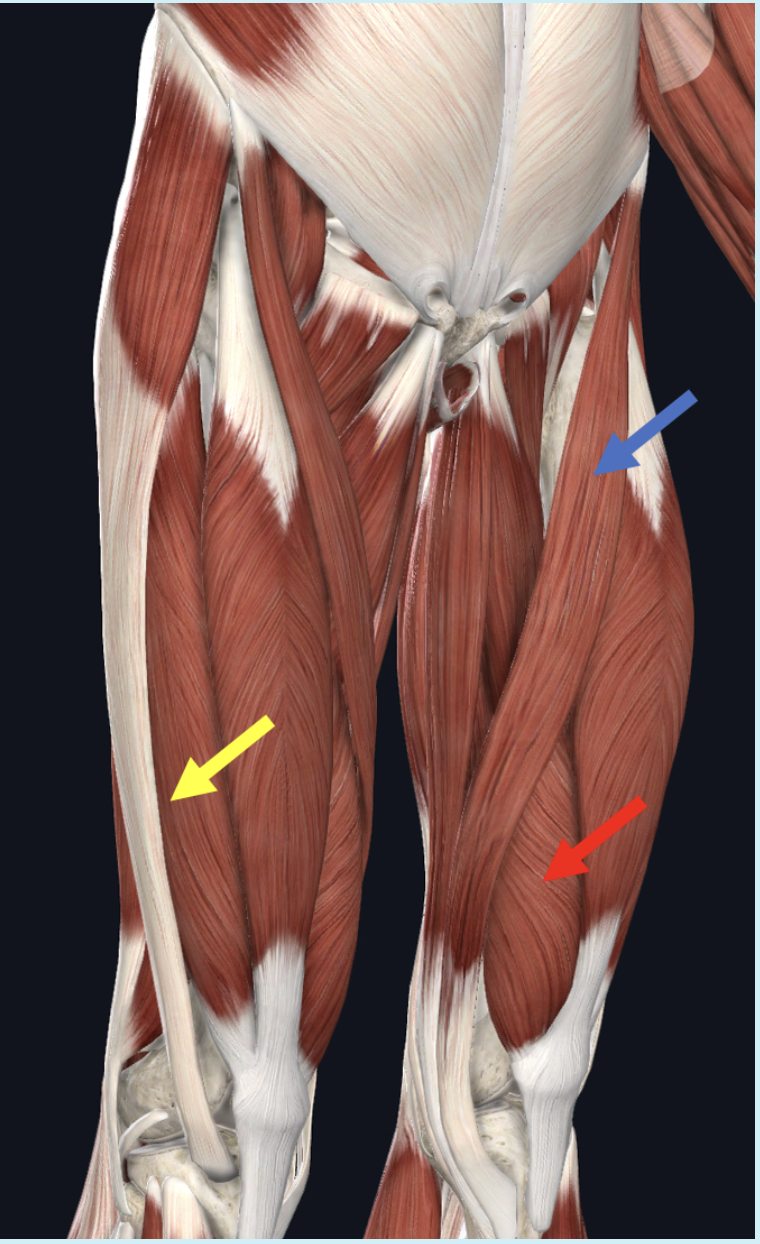
Identify the muscle at the tip of the blue arrow
short head of biceps femoris
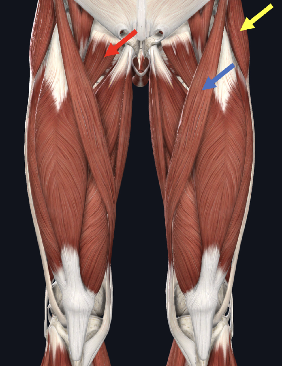
Identify the muscle at the tip of the yellow arrow
adductor longus
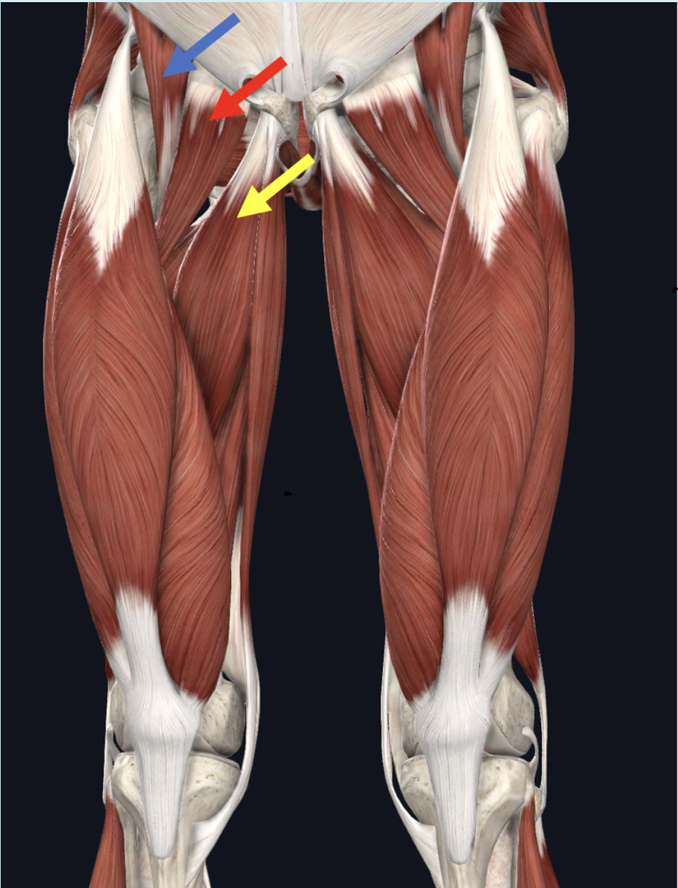
Identify the muscle at the tip of the red arrow
pectineus
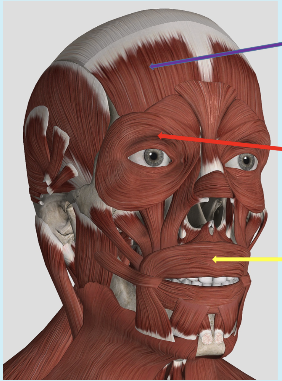
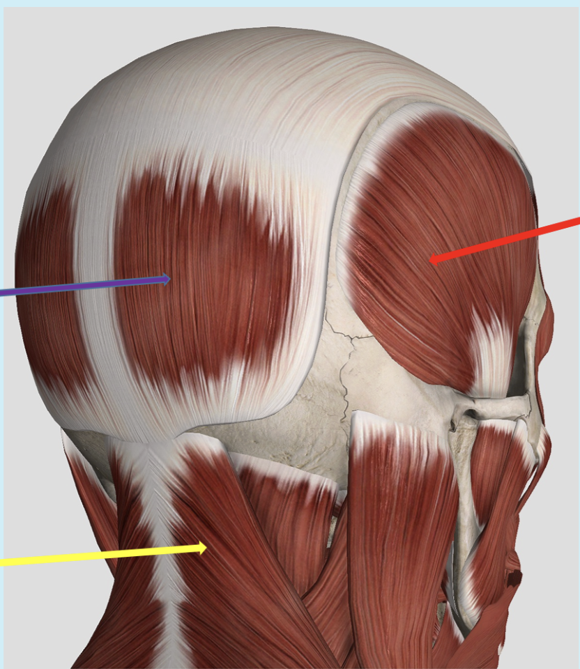
Identify the muscle at the tip of the purple arrow
frontalis, red arrow orbicularis oculii, yellow arrow orbicularis oris
Identify the muscle at the tip of the yellow arrow
external oblique
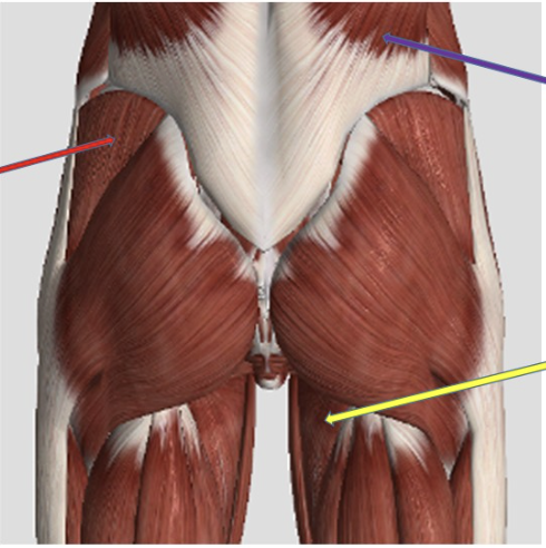
Identify the muscle at the tip of the red arrow
gluteus medius
Identify the muscle at the tip of the yellow arrow. BE SPECIFIC
brachialis
Identify the muscle at the tip of the yellow arrow
trapezius
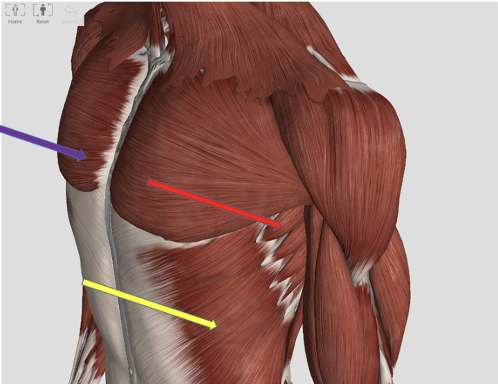
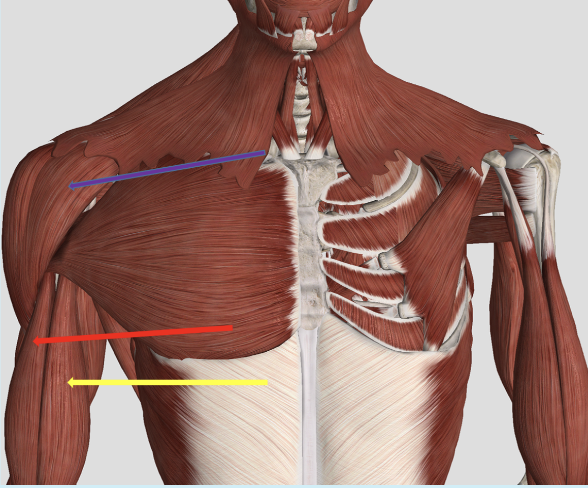
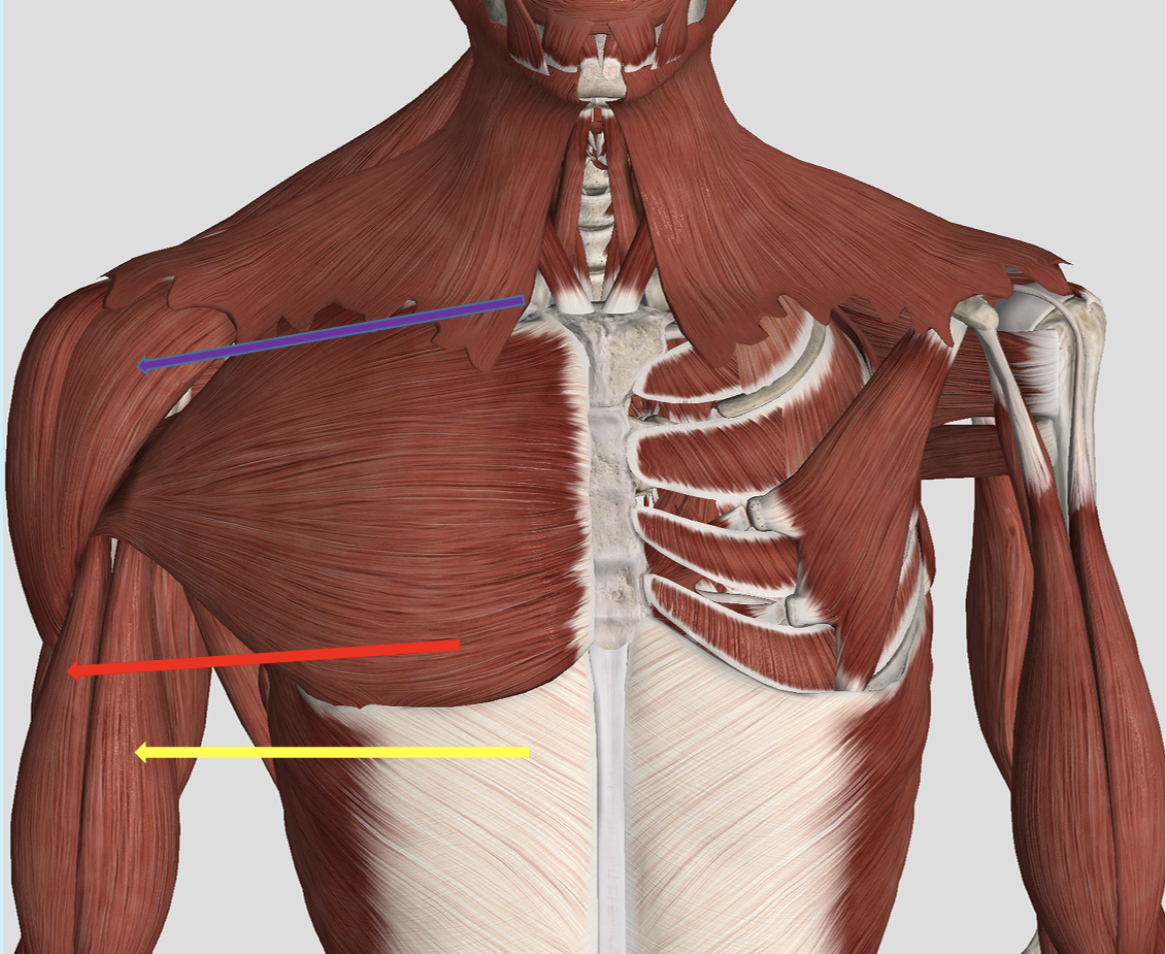
Identify the muscle at the tip of the purple arrow
pectoralis minor
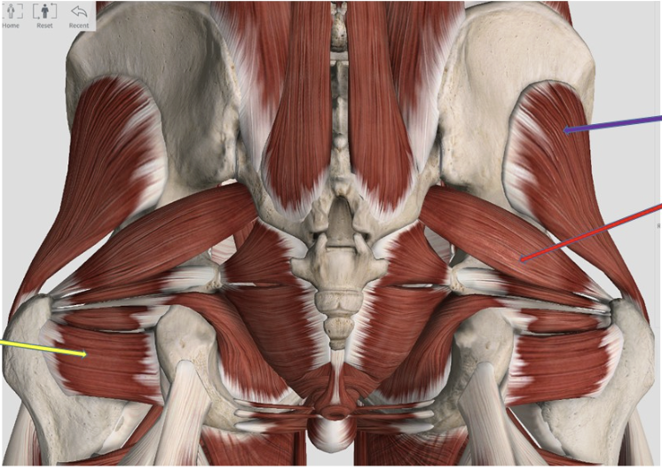
Identify the muscle at the tip of the purple arrow
gluteus minimus
Identify the muscle at the tip of the yellow arrow
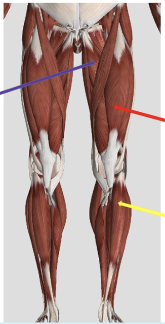
semitendinosus
Identify the muscle at the tip of the red arrow
rectus femoris
Identify the muscle at the tip of the red arrow
semitendinosus
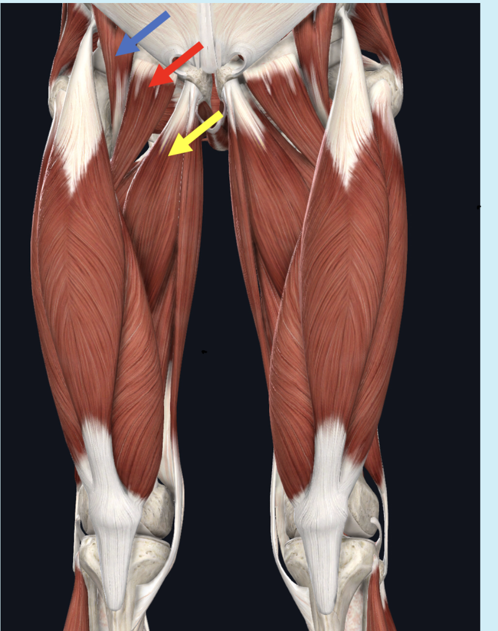
Identify the muscle at the tip of the blue arrow
iliopsoas
Identify the muscle at the tip of the purple arrow
rectus abdominis
Identify the muscle at the tip of the blue arrow
fibularis longus
Identify the muscle at the tip of the purple arrow
deltoid
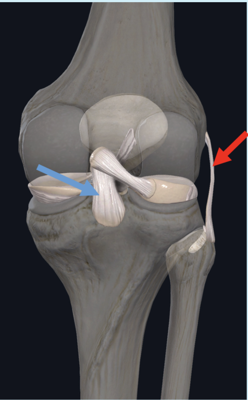
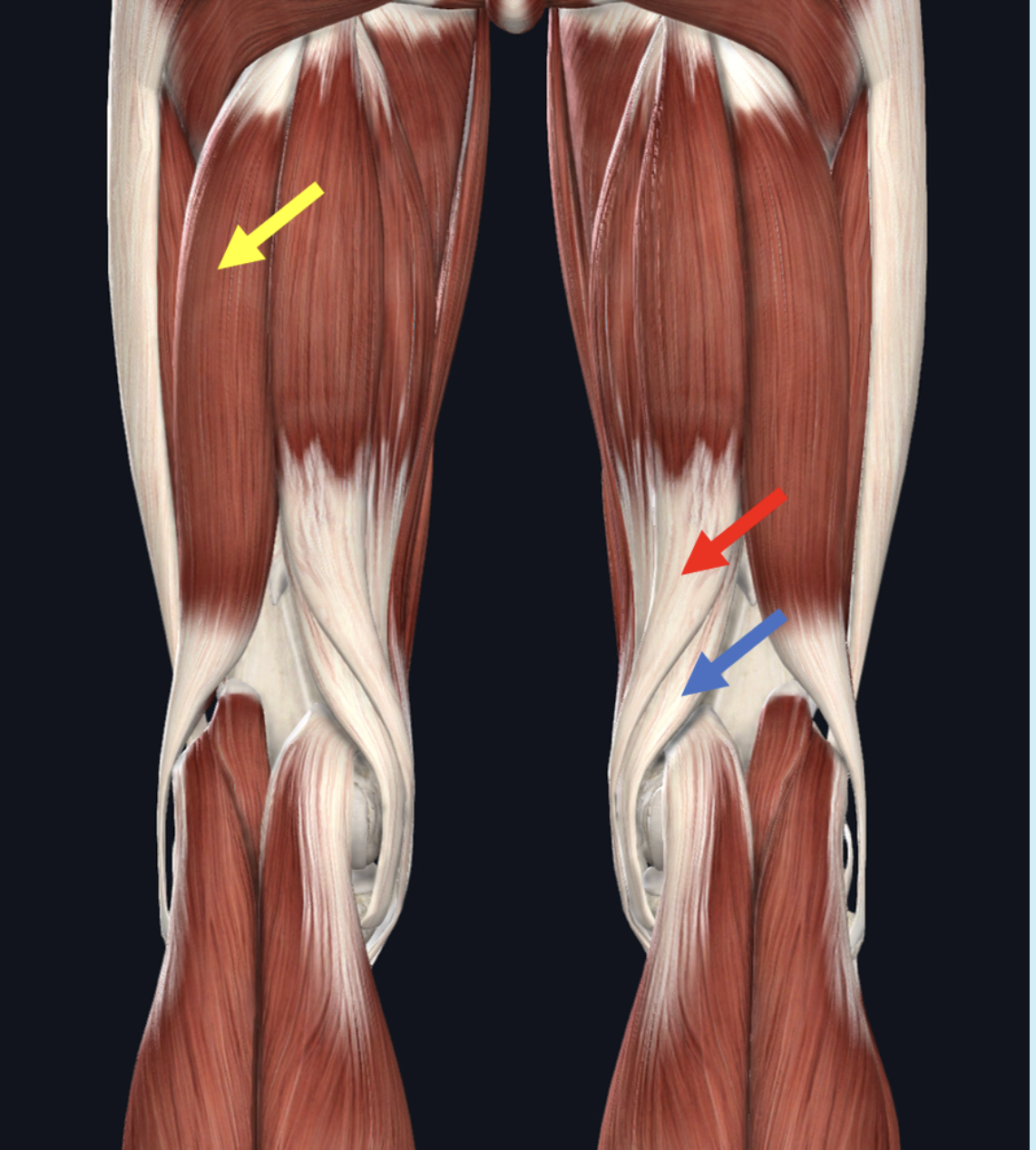
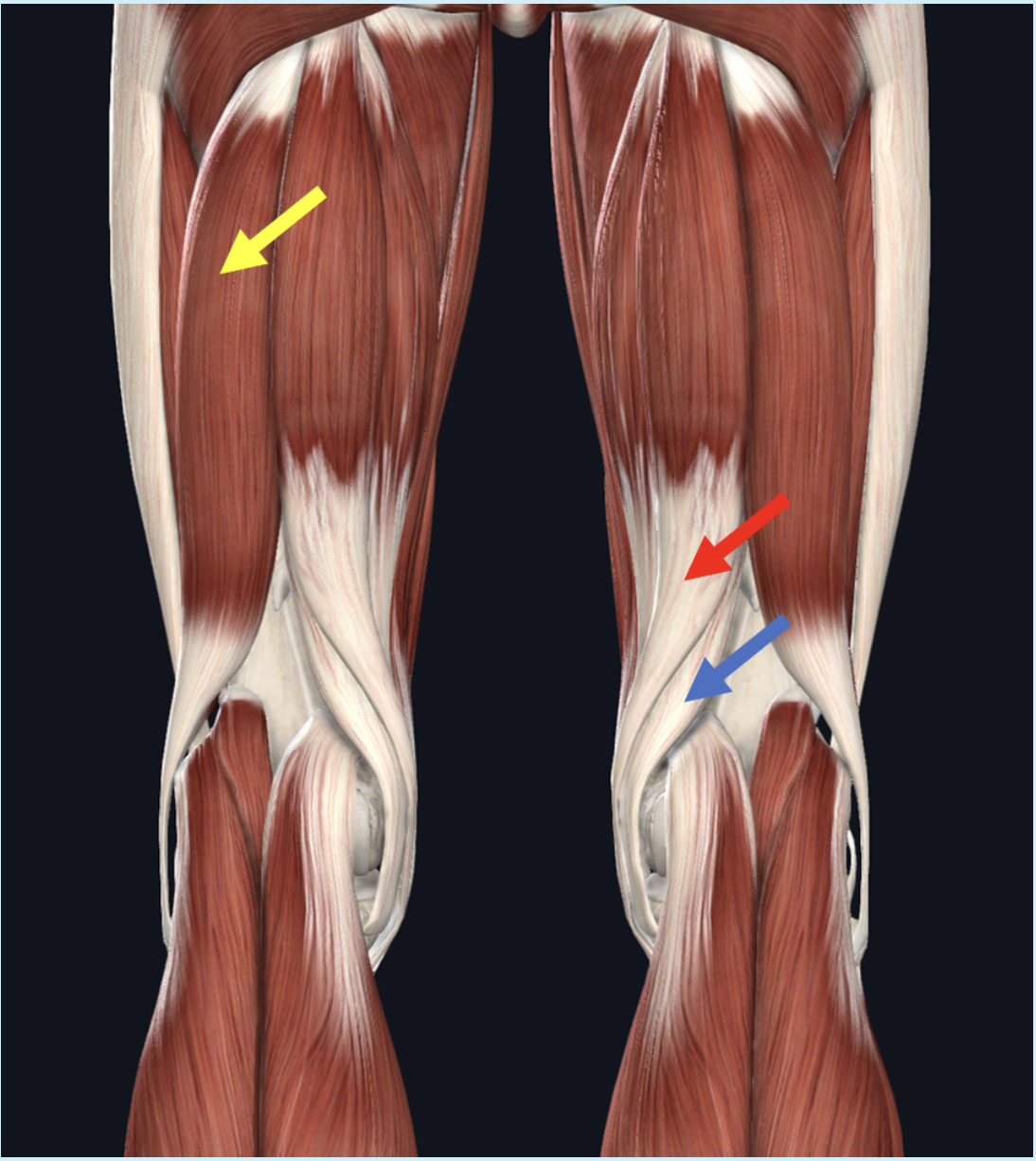
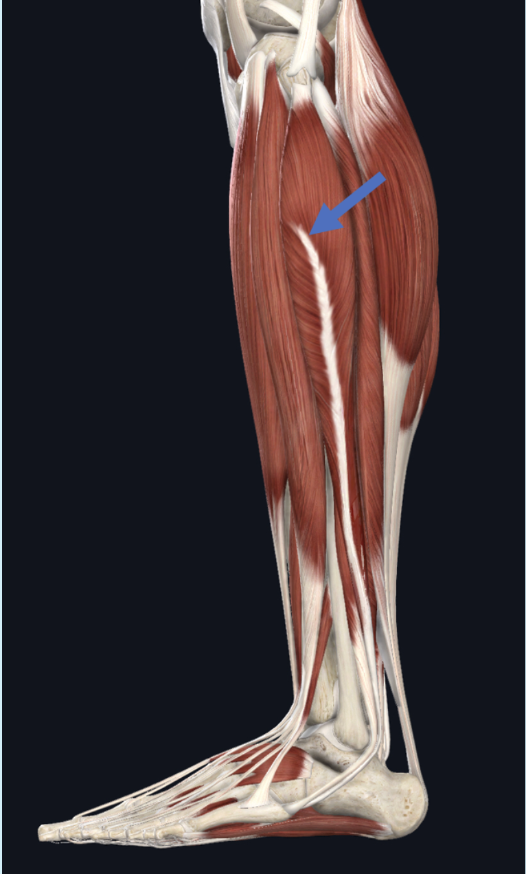
Identify the connective tissue structure at the tip of the red arrow
red arrow; fibular collateral ligament
blue arrow: patellar ligament
Identify the muscle at the tip of the yellow arrow
fibularis brevis
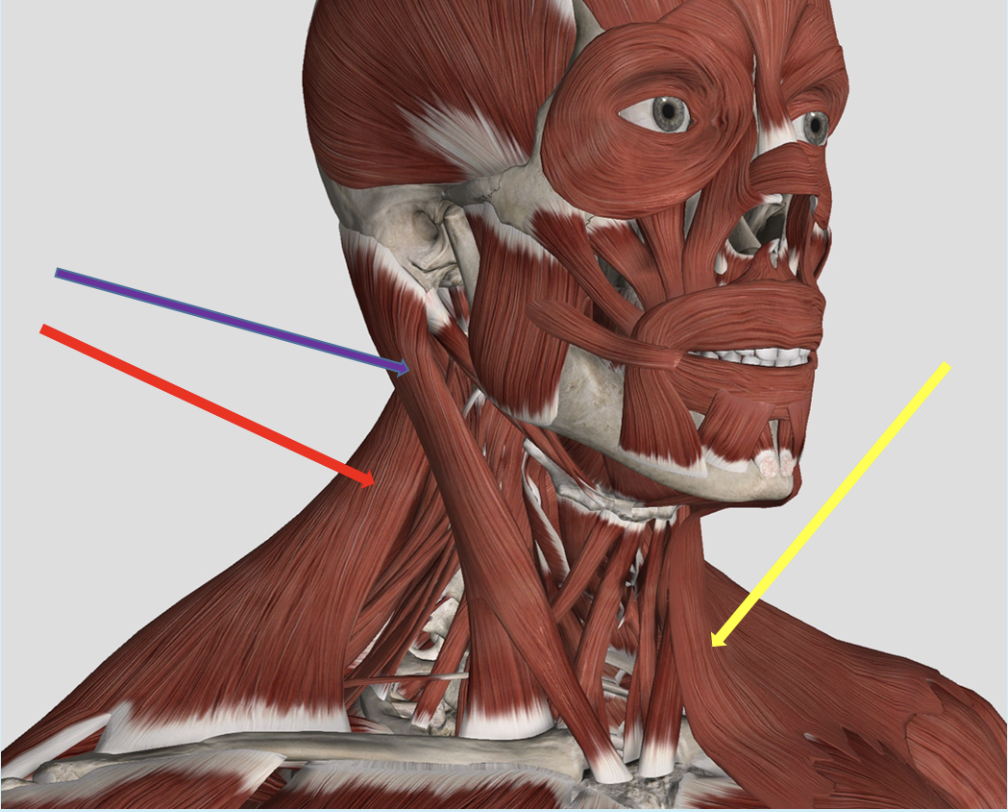
Identify the muscle at the tip of the purple arrow
sternocleidomastoid
Identify the muscle at the tip of the yellow arrow
tensor fascia lata
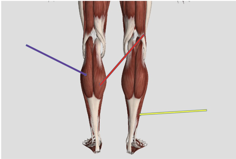
Identify the muscle at the tip of the purple arrow.
gastrocnemius
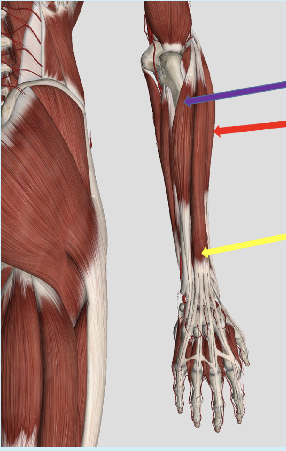
Identify the muscle at the tip of the purple arrow
purple arrow: extensor carpi radialis brevis
red arrow: extensor digitorum
yellow arrow: flexor pollicis longus
Identify the muscle at the tip of the red arrow
vastus medialis
Identify the muscle at the tip of the yellow arrow
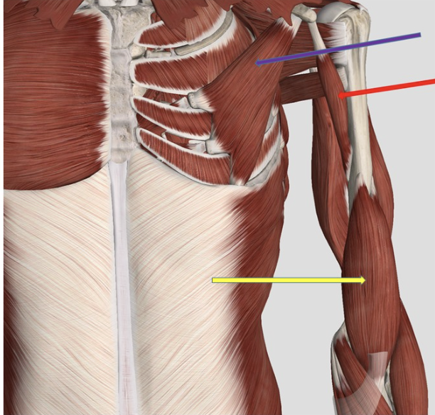
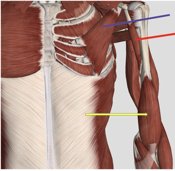
yellow arrow: brachialis
purple arrow: pectoralis minor
red arrow: coracobrachialis