3.3 pelvis, perineum, gluteal region, hip, thigh
1/90
There's no tags or description
Looks like no tags are added yet.
Name | Mastery | Learn | Test | Matching | Spaced | Call with Kai |
|---|
No analytics yet
Send a link to your students to track their progress
91 Terms
What is the pelvis inlet formed by?
sacral promontory
sacral ala
arcuate line
pecten pubis (pectineal line)
superior pubic ramus
what area is considered the false pelvis?
lower abdomnal cavity → lower parts of abdomincal viscera
what area is considered a true pelvis
terminal digestive tract, urinary and reproductive organs
what divides the false and true pelvis?
the pelvic inlet / superior pelvic aperture
what is known as the “doorway” into the pelvic cavity and also serves as a superior boundary?
the pelvic inlet!
what are the bones that makeup the anterior, lateral, and posterior boundaries of the pelvic cavity?
os coxae
sacrum
What are the muscles that make up the anterior, lateral, and posterior boundaries of the pelvic cavity
obturator internus
piriformis
what creates the inferior boundary (floor) of the pelvic cavity?
pelvic diaphragm
levator ani
coccygeus
What is the function of the pelvic diaphragm?
support abdominopelvic viscera
involved with control over urination and defecation
what’s the difference between the pelvic diaphragm in females?
floor of pelvic cavity has an additional opening for vaginal canal
what is the roof of the perineum?
The pelvic diaphragm
what is the perineum?
the area between the pubic symphysis and coccyx
what is the area where we find external genitalia called?
perineum
where is the perineum in relation to the pelvic diaphragm?
superficial to the pelvic diaphrahm
what are the boundaries of the pelvic OUTLET / perineum
pubic symphysis
ischiopubic rami
ischial tuberosities
sacrotuberance ligaments
coccyx
what are the two triangles of the pelvic outlet / perineum?
urogenital triangle
anal triangle
what are the urogenital and anal triangles of the pelvic OUTELT bound by?
ischial tuberosities
pubic symphysis
coccyx
where are muscles of the perineum primarily found in?
urogenital and anal triangles
what nerve are the muscles of the perineum innervated by?
pudendal nerve
what nerve exists the pelvic cavity through the greater sciatic foramen/notch and then enters the lesser sciatic foramen/notch?
pudendal nerve!
finish this phrase: pee, poop, …
PUDENDAL
what bony landmarks is the greater sciatic notch found between?
Posterior inferior iliac spine ( PIIS) and the ischial spine
What bony landmarks is the lesser sciatic notch found between?
ischial spine and ischial tuberosity
what ligament turns the greater and lesser sciatic notches into foramen?
the sacrotuberous ligament
what ligament separates the greater and lesser sciatic foramen from one another?
the sacospinous ligament!
what is the general purpose of the greater and lesser foramen?
serves as a “doorway” for structures in the pelvis to get to the lower extremity
what muscle is attached to the greater sciatic foramen?
piriformis
list nerves that go through the greater sciatic foramen?
superior/inferior gluteal NV bundles
sciatic nerve
posterior femoral cutaneous nerve
pudendal NV bundle
nerve to obturator internus
nerve to quadratus femoris
what muscle travels through the lesser sciatic foramen?
obturator internus
what neurovascular bundle travels through the lesser sciatic foramen?
pudendal NV bundle
what nerve goes through both the greater and lesser sciatic foramen?
pudendal nv bundle
what is the head of the femur covered in?
hyaline cartilage
what bony landmark connects greater and lesser trochanters?
intertrochanteric line (anterior) and crest (posterior)
what covers medial and lateral femoral condyles?
hyaline cartilage
What articulates together to form the hip joint?
acetabulum
femoral head
what kind of joint is the hip joint?
synovial joint - ball & socket
How many planes and what planes can a hip ball & socket joint move?
three
sagittal (flexion & extension)
frontal (adduction/abduction)
transverse (internal/external rotation)
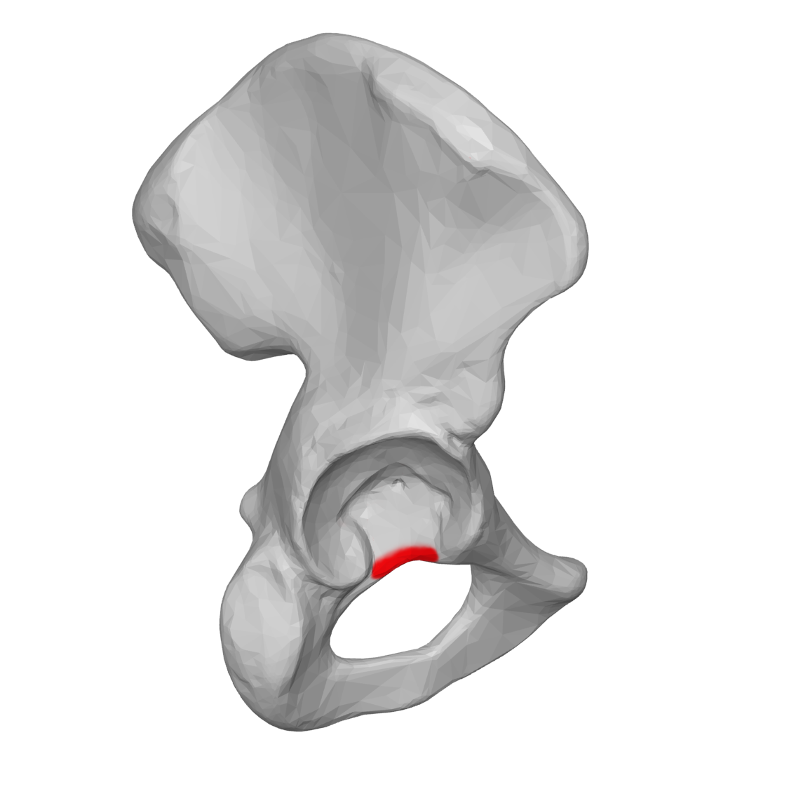
In the acetabulum, what part is covered in articular cartilage?
lunate surface
what part of the acetabulum is not covered in articular cartilage?
acetabular fossa
what sits on the acetabular fossa?
fat pad! - cushioning
what is located in the acetabulum that improves congruence & stability?
acetabular labrum
what tissue type is the acetabular labrum?
fibrocartilage!

what is the area between the anterior and posterior parts of the lunate surface called?
the acetabular notch
what ligament sits on the acetabular notch and connects anterior/posterior lunate surfaces
transverse ligament - since its blends in with labrum, it is considered a continuation of it
how much congruence is there between the femoral head and acetabulum
High congruence! - but still lots of mobility
what parts does the hip joint capsule have?
fibrous part
synovial part
what is the fibrous part of the hip joint capsule made of?
dense IRREGULAR ct
why does the synovial part of the hip joint capsule produce synovial fluid?
to nourish the articular cartilage
where does the synovial membrane attach to in the hip joint capsule?
edge of acetabulum
Why does the synovial membrane run underneath itself to reach edge of femoral head articular cartilage?
to allow for arteries (medial and lateral femoral circumflex) to sit on the surface of the bone and supply blood to neck and head of femur
what arteries are the primary supplies of blood to the femoral head?
medial and lateral femoral circumflex arteries!
where do the branches of the medial and lateral femoral circumflex arteries run?
deep to synovial membrane along femoral neck
what would happen to the to the femoral head if there was femoral neck fracture?
femoral head would DIE because the arteries would be severed- likely leading to a hip replacement!
What ligaments work to thicken and reinforce the hip joint capsule?
iliofemoral
ischiofemoral
pubofemoral
in the joint capsule, what kind of reinforcement does the iliofemoral ligament provide?
anterior reinforcement
AIIS & adjacent ilium to the intertrochanteric line
in the joint capsule, what kind of reinforcement does the pubofemoral ligament provide?
inferior reinforcement!
body, superior ramus of pubis to the intertrochanteric line
in the joint capsule, what kind of reinforcement does the ischiofemoral ligament provide?
posterior reinforcement!
body of ischium to greater trochanter, intertrochanteric line
what is a ligament doing when its resisting a motion?
stretching!
what action does the iliofemoral ligament resist?
hip extension
what action does the pubofemoral ligament resist?
hip abduction!
what action does the ischiofemoral ligament resist?
hip extension AND internal rotation!
where do all hip ligaments attach to?
the intertrochanteric line!
what reduces friction between muscles, bones, and other tissues?
bursa!
what is the superior boundary of the femoral triangle?
inguinal ligament
what is the lateral boundary of the femoral triangle?
sartorius
what is the medial boundary of the femoral triangle?
adductor longus
what is the floor of the of the femoral triangle?
pectineus and small parts of iliopsaos
What goes through the femoral triangle?
femoral nerve, artery, vein
what is the femoral artery a continuation of?
external iliac artery
what vein branches off in the femoral triangle?
great(er) saphenous vein
where is the adductor canal located?
deep to sartorious!
superficial to adductor longus and magnus!
what is the opening in the adductor magnus called?
adductor hiatus!
what is artery and vein located in the adductor canal?
femoral artery and vein!
how do the femoral artery and vein reach the posterior knee?
through the adductor hiatus!
what is the name change for the femoral artery and vein after it exits the adductor hiatus?
popliteal artery and vein
what nerves go through the adductor canal but NOT the adductor hiatus?
Nerve to vastus medialis
saphenous nerve
what does the saphenous nerve do?
provide cutaneous sensation to the medial aspect of the leg
what is the common attachment for all quadriceps femoris muscles?
tibial tuberosity!
what is the common attachment for the hamstrings?
ischial tuberosity
What is the common innervation for the hamstrings?
tibial branch of the sciatic nerve
what nerve of the posterior compartment goes through the greater sciatic foramen?
sciatic nerve!
What does the sciatic nerve split into in the popliteal fossa (posterior knee)?
tibial and common fibular branch
what compartment are the hamstrings located in?
posterior compartment!
where do the perforating arteries in the posterior compartment of the leg come from?
the deep femoral artery of the anterior compartment
what muscles are in the medial compartment of the LE?
gracilis
pectineus
adductor longus
adductor brevis
adductor magnus
obturator externus
what is located in the superficial layer of the medial compartment?
pectineus
adductor longus
what is located in the middle layer of the medial compartment?
adductor brevis
what is located in the deep layer of the medial compartment?
adductor magnus
obturator externus
what is located on the medial layer of the medial compartment?
gracilis!
Where does the obturator nerve trave?
along the medial side of the pelvic cavity! passes through the obturator foramen
What innervates basically all the muscles in the medial compartment?
obturator nerve
The hamstring part of the adductor magnus is innervated by the tibial branch of the sciatic nerve