lab 3: appendicular skeleton
1/166
There's no tags or description
Looks like no tags are added yet.
Name | Mastery | Learn | Test | Matching | Spaced | Call with Kai |
|---|
No analytics yet
Send a link to your students to track their progress
167 Terms
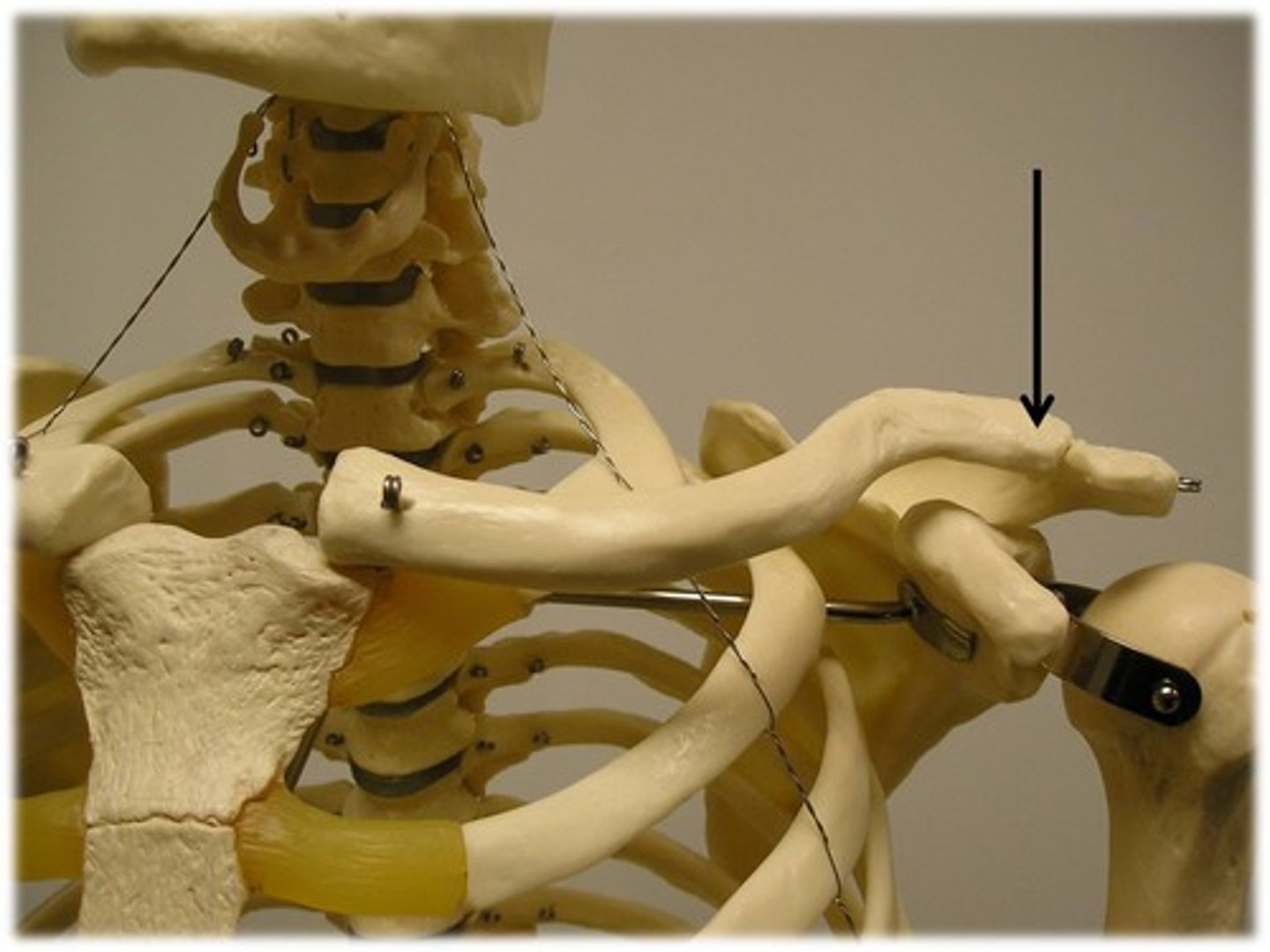
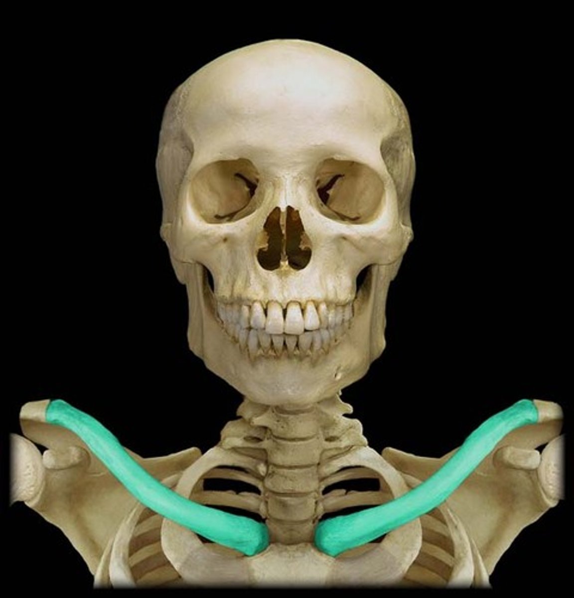
Clavicle
Bottom is rough, top is smooth. (Whole bone)
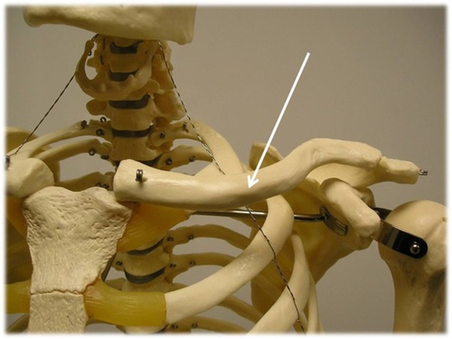
Sternal end
Medial end of clavicle; more flat; sits in sternum

Acromial end
Lateral end; paddle
Right vs. Left Clavicle
Sternal end is coming out to you first.
Acromial end's curvature is pointing towards the back.
Rough portion should be facing down, smooth portion up.
Coronoid (tip) facing back.
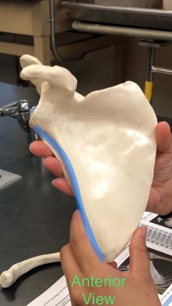
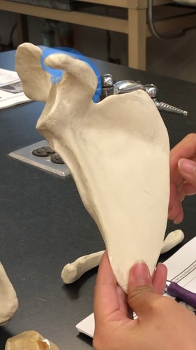
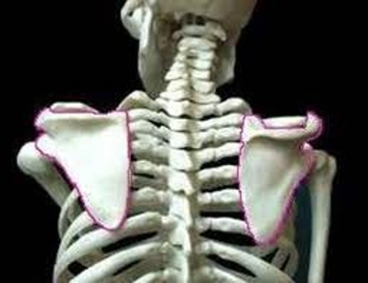
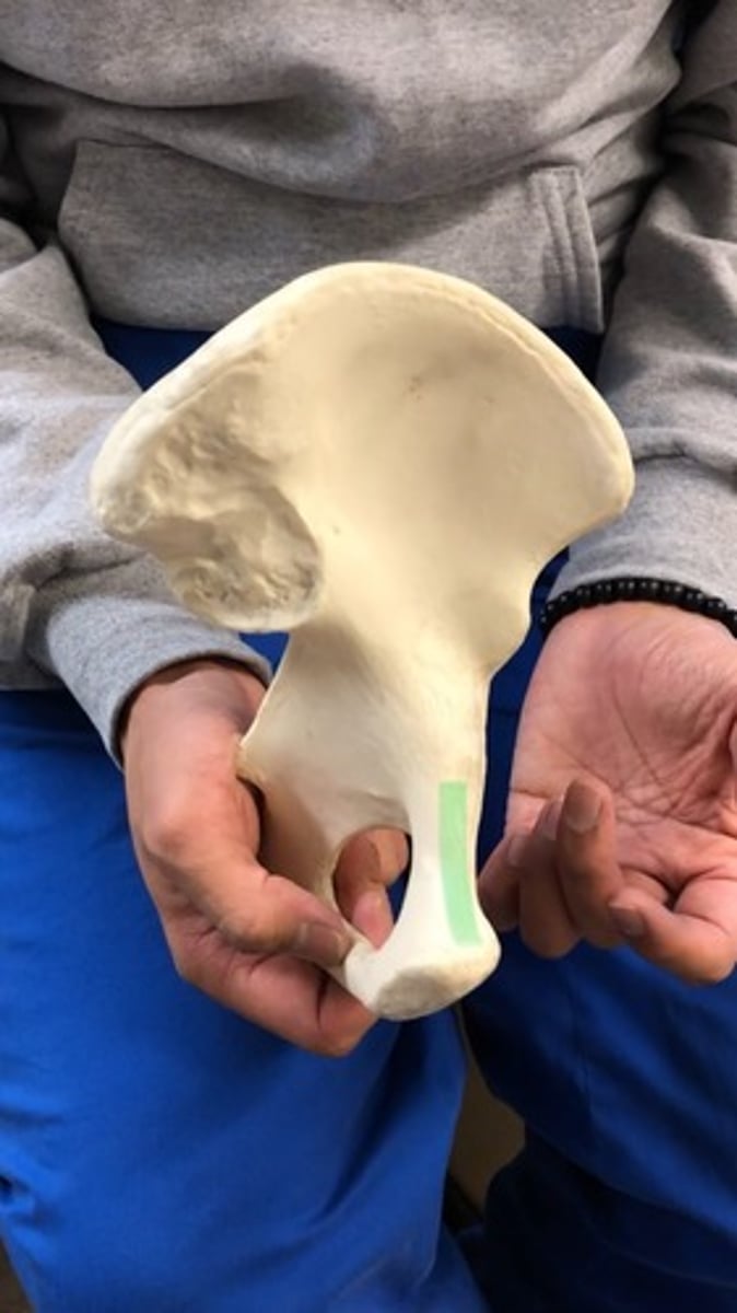
Scapulae
Name the entire structure:
Shoulder blades
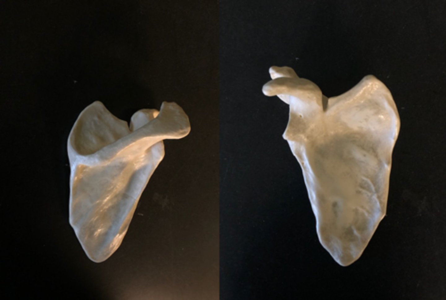
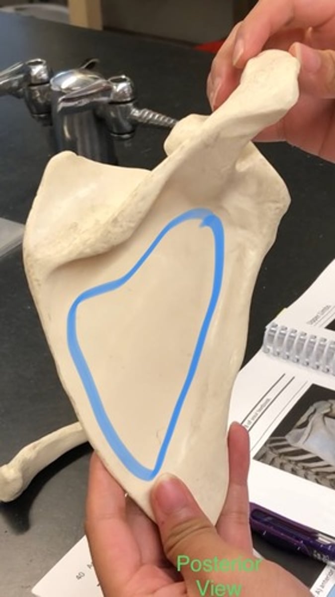
Body of scapula
Flat part below the scapular spine ENTIRE REGION
Superior border of scapula
top lining of scapula
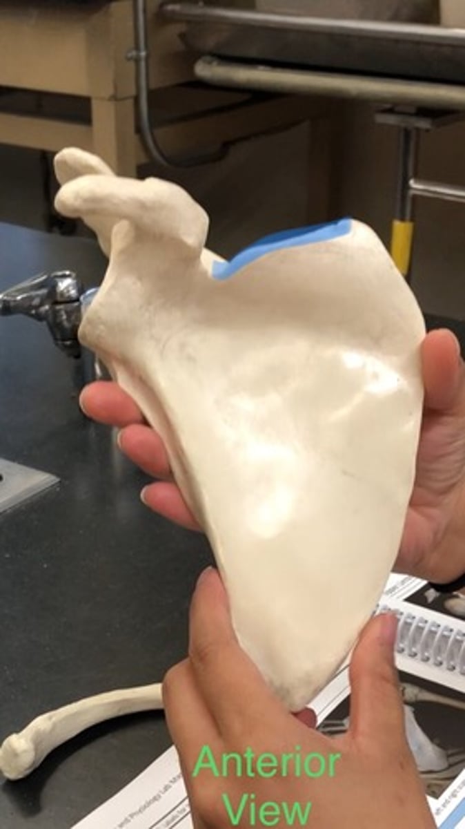
Medial border of scapula
Lining on side of scapula; medial on body; more curved
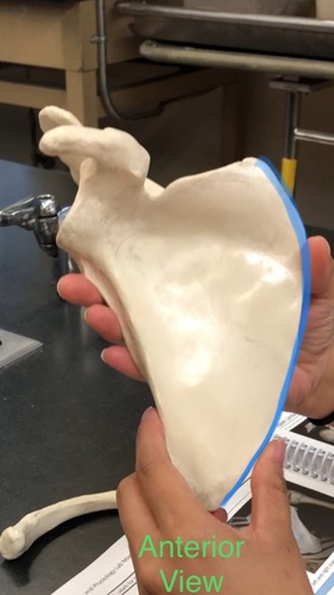
Lateral border of scapula
inside lining almost straight down; less curved
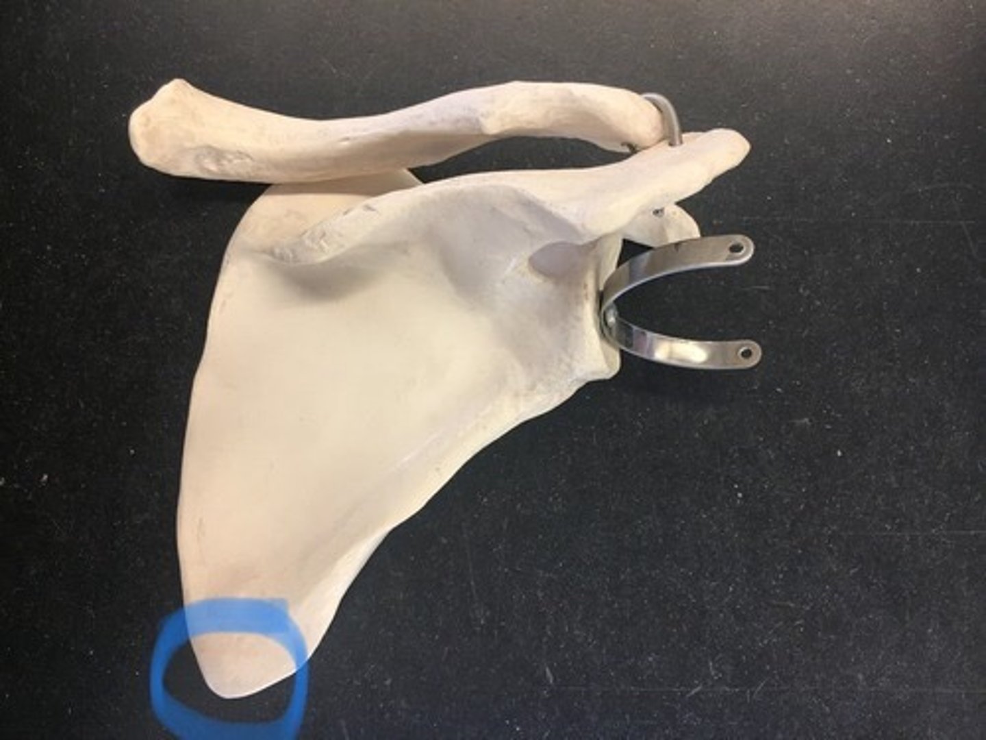
Inferior angle of scapula
bottom point on body of scapula; point where the medial and lateral borders meet
Glenoid cavity
Space where humerus goes in

Coracoid process of scapula
anterior to glenoid cavity; in the front
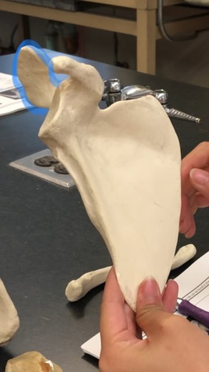
Acromion
behind coracoid process
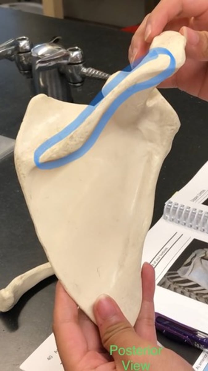
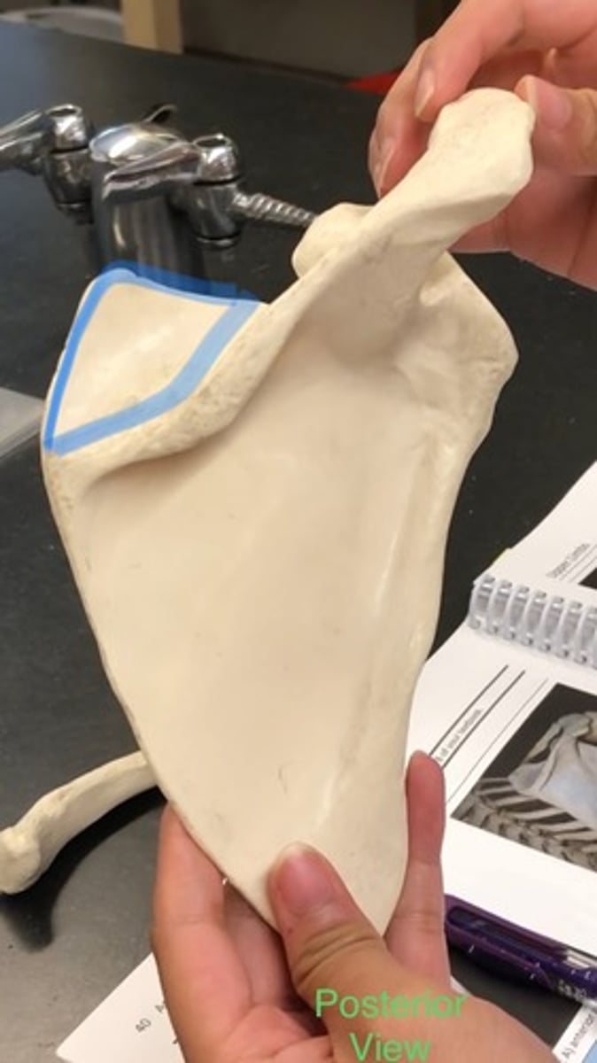
Scapular spine
posterior (back of); connected to acromion
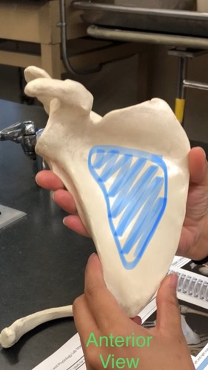
Subscapular fossa
Shallow depression in scapula, from superior border to inferior angle
Supraspinous fossa
above scapular spine (on back)
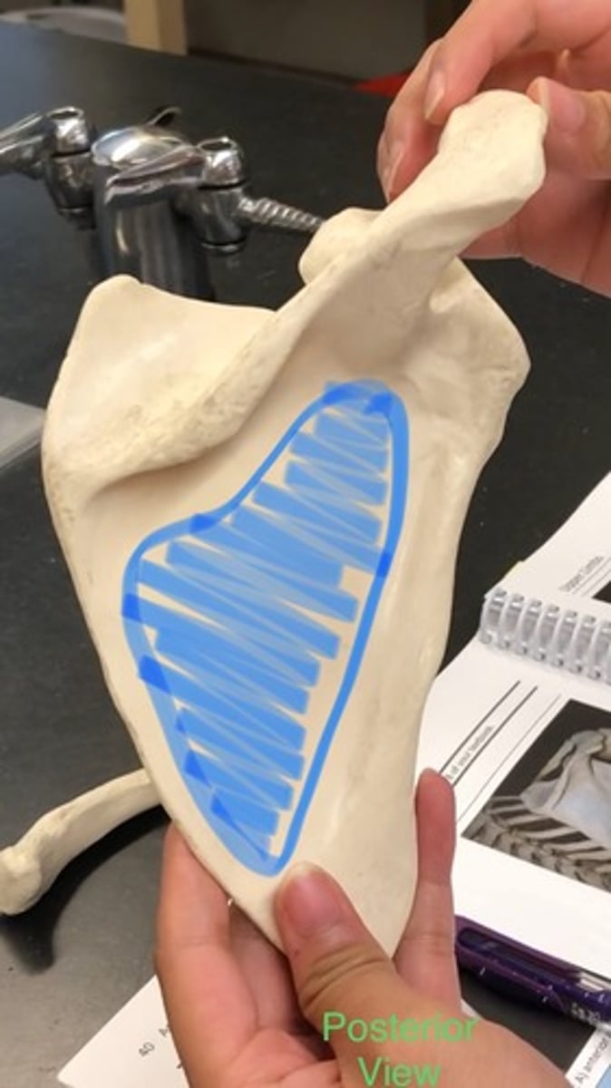
Infraspinous fossa
below scapular spine (on back)
Right vs. Left (Scapulae)
Glenoid cavity if lateral
Scapular spine is posterior; spine sticks out
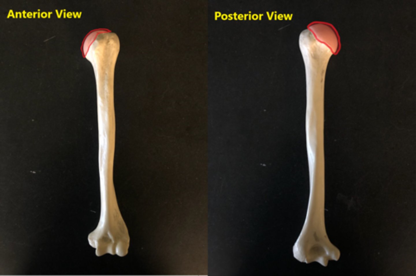
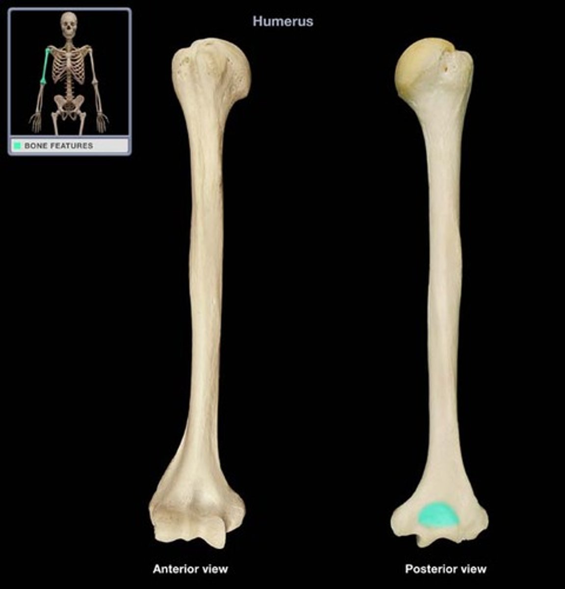
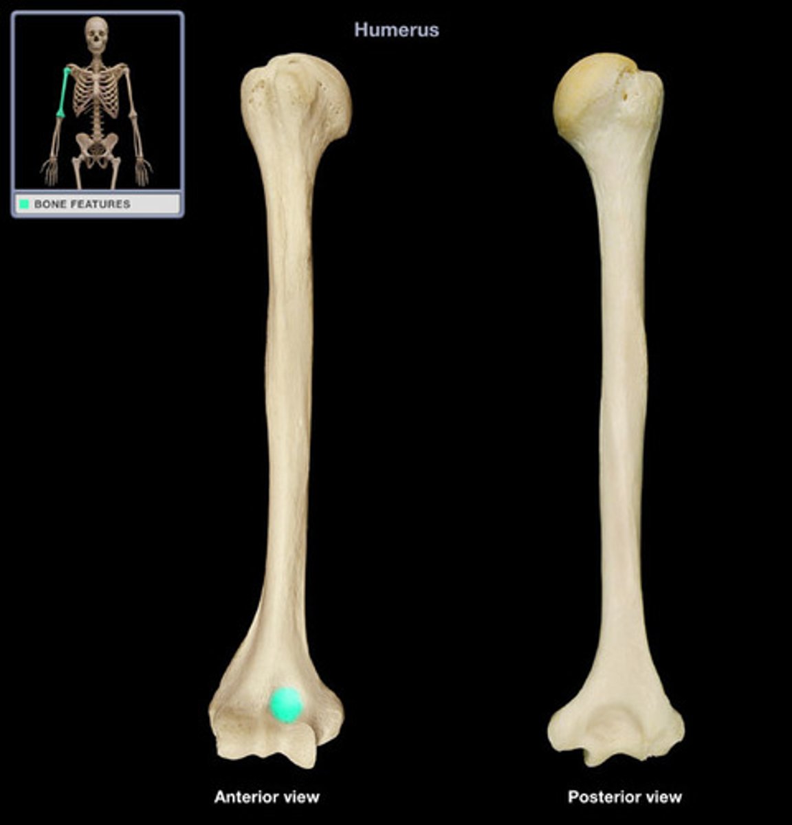
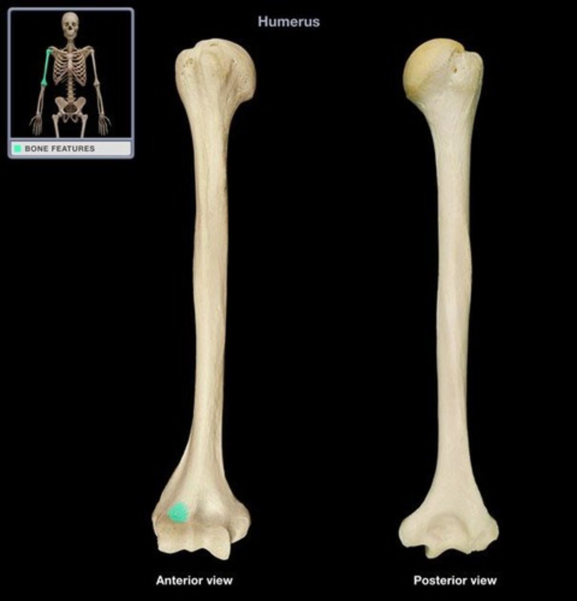
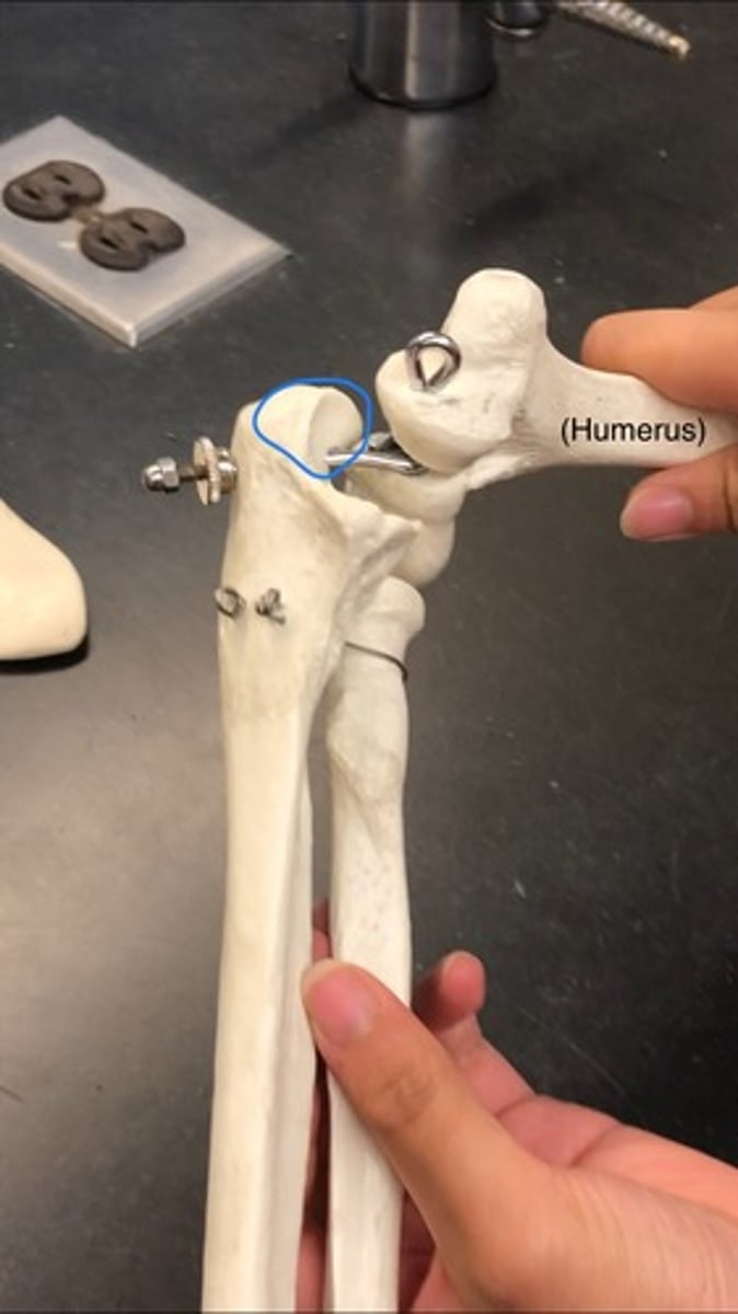
Humerus
Upper arm
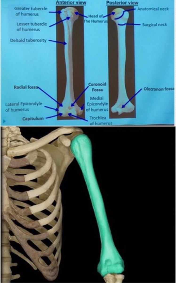
head of humerus
Medial, fits in scapula round portion (glenoid cavity)
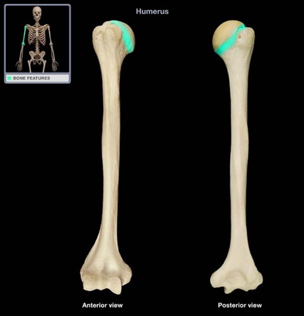
Greater tubercle of humerus
Top portion of hump, right next to head (laterally)
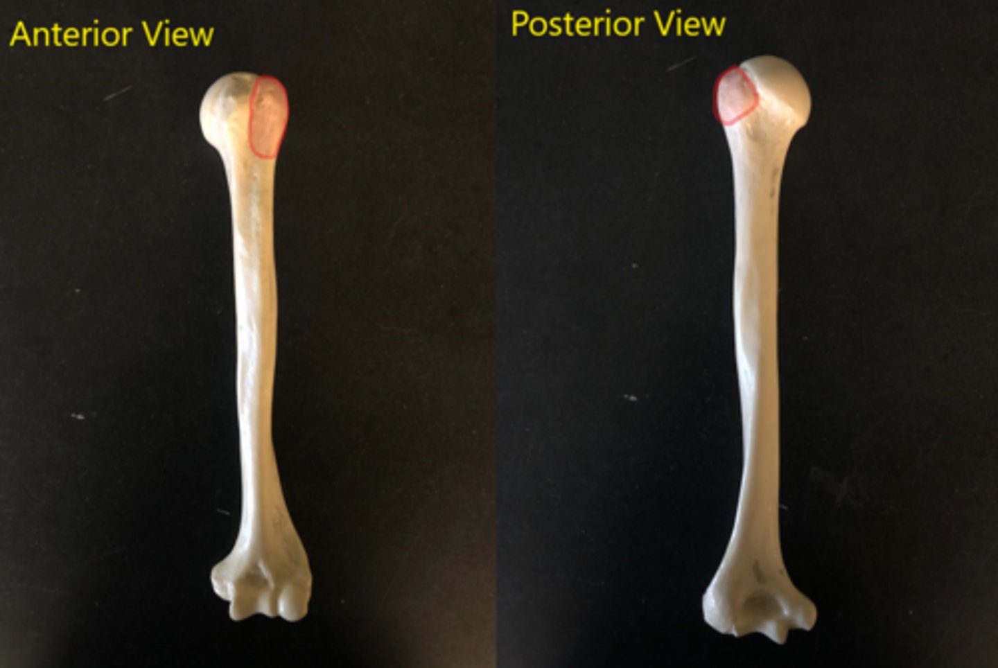
Lesser tubercle of humerus
faces forward (can only see from front); below the greater tubercle
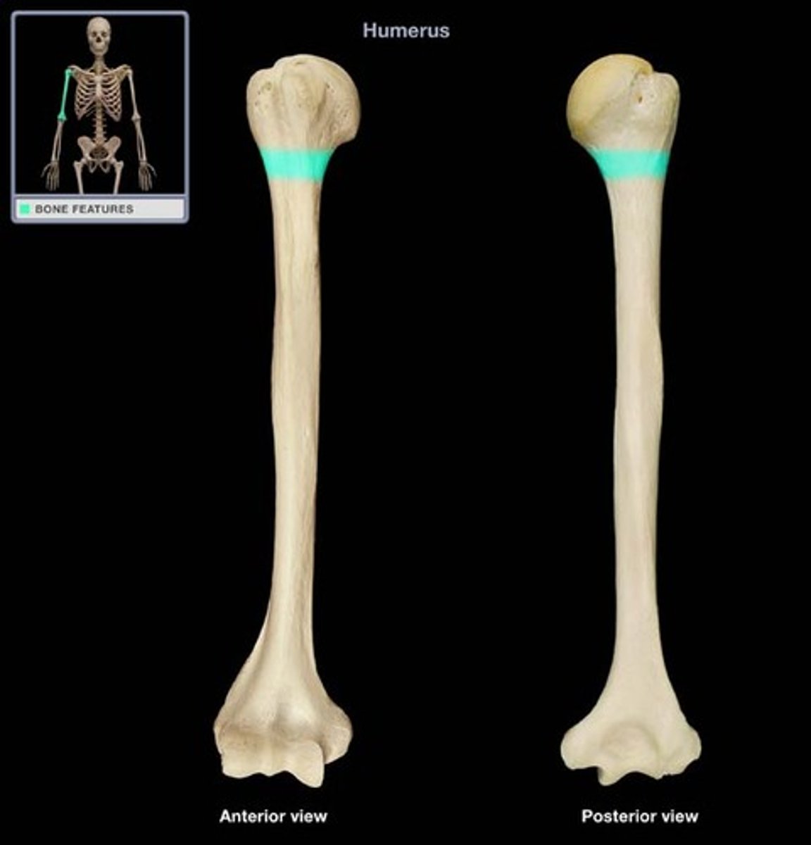
Anatomical neck
Separates head from tubercles
Surgical neck
Right below lesser tubercle; where everything starts to narrow
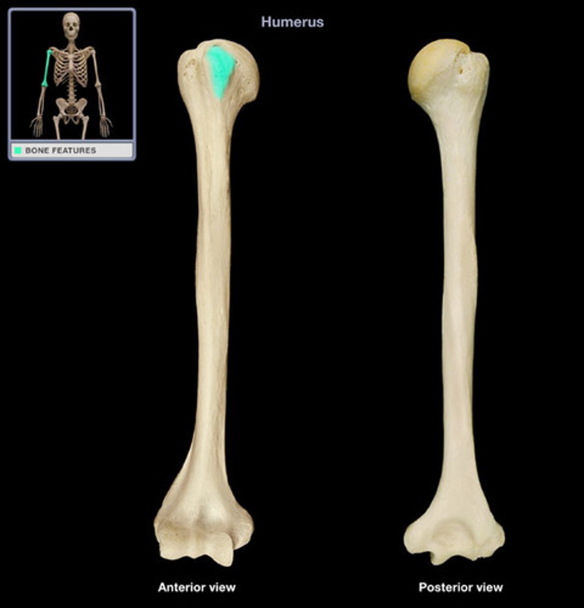
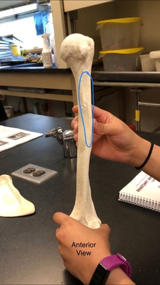
Deltoid tuberosity
Rough patch; think of deltoid muscle (this points to it)
Right vs. Left (Humerus)
Head of humerus is medial and proximal; olecranon fossa is posterior (big indent)
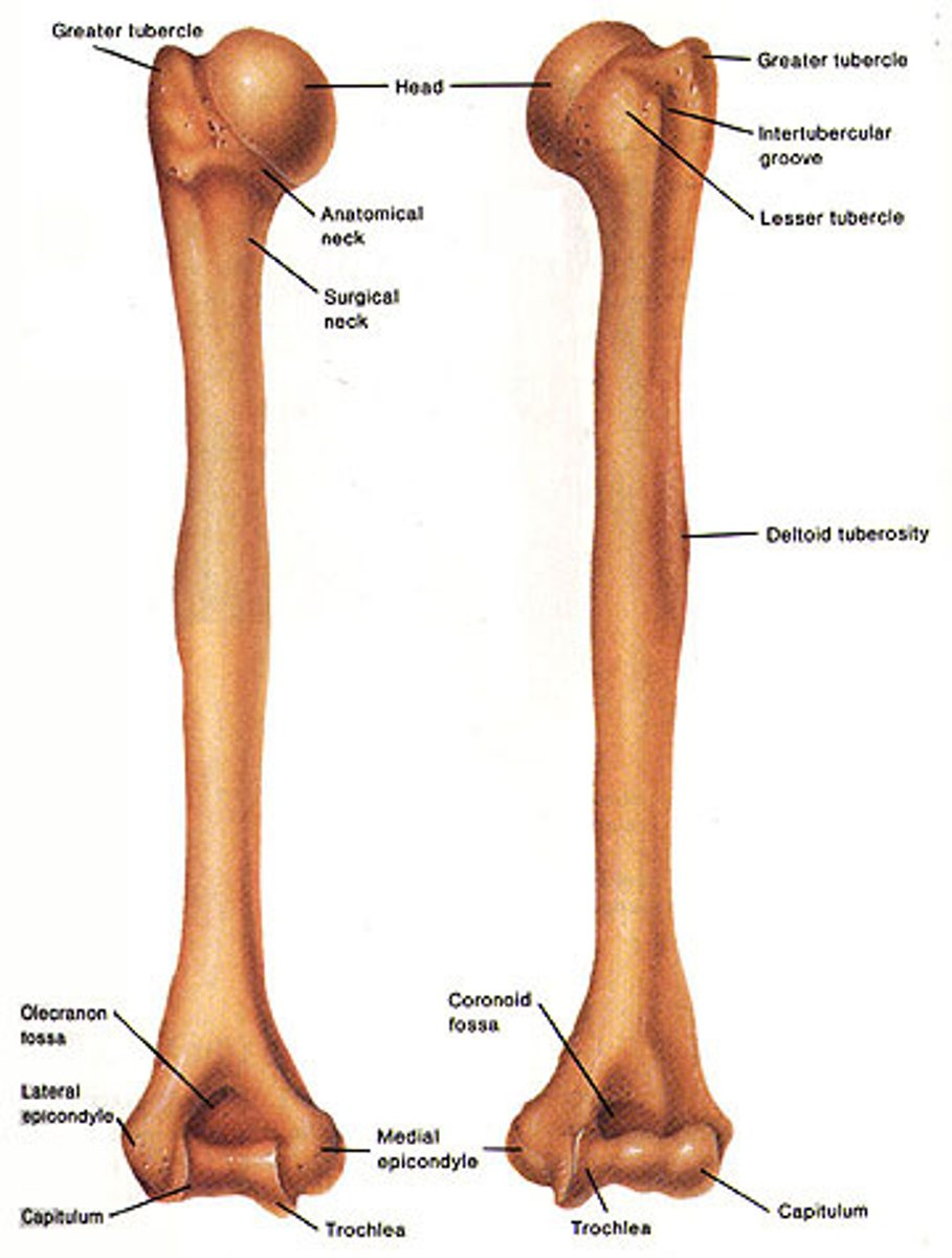
Medial epicondyle of humerus
parallel to the head of humerus; outermost
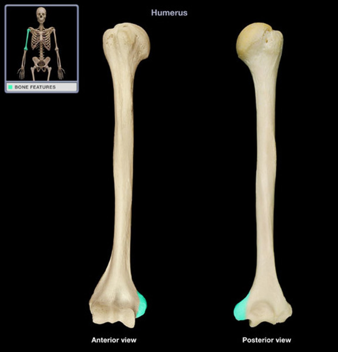
Lateral epicondyle of humerus
outermost projection on the lateral side
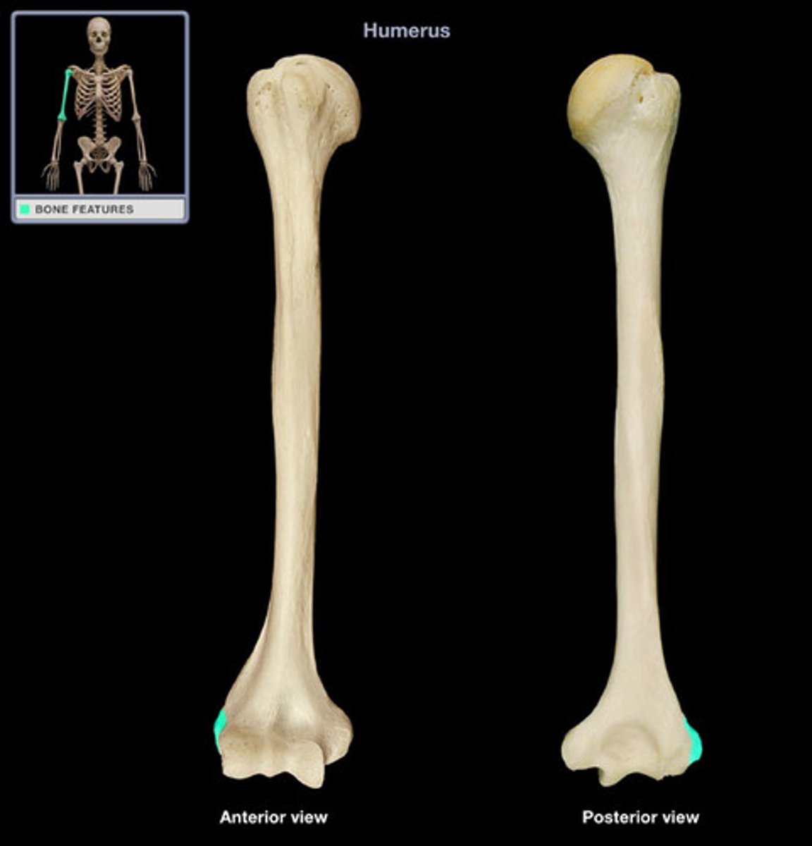
Capitulum
only on the front; inferior to the lateral epicondyle

Trochlea of humerus
"Hourglass"; bumps next to capitulum
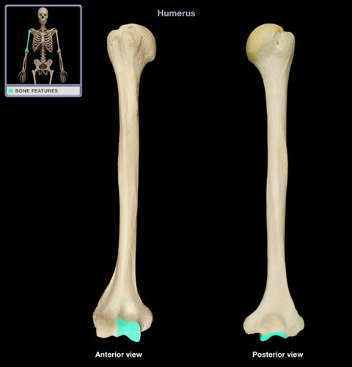
Olecranon fossa
posterior; large indent on bottom
Coronoid fossa
Biggest depression on anterior side of humerus. Right above trochlea of humerus ("Hour glass")
Radial fossa
Shallow depression right above capitulum; anterior
Antebrachium
Forearm
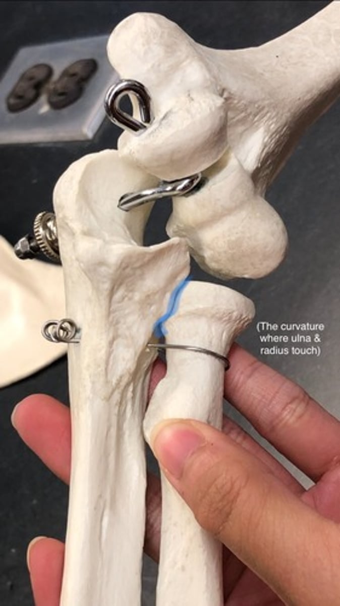
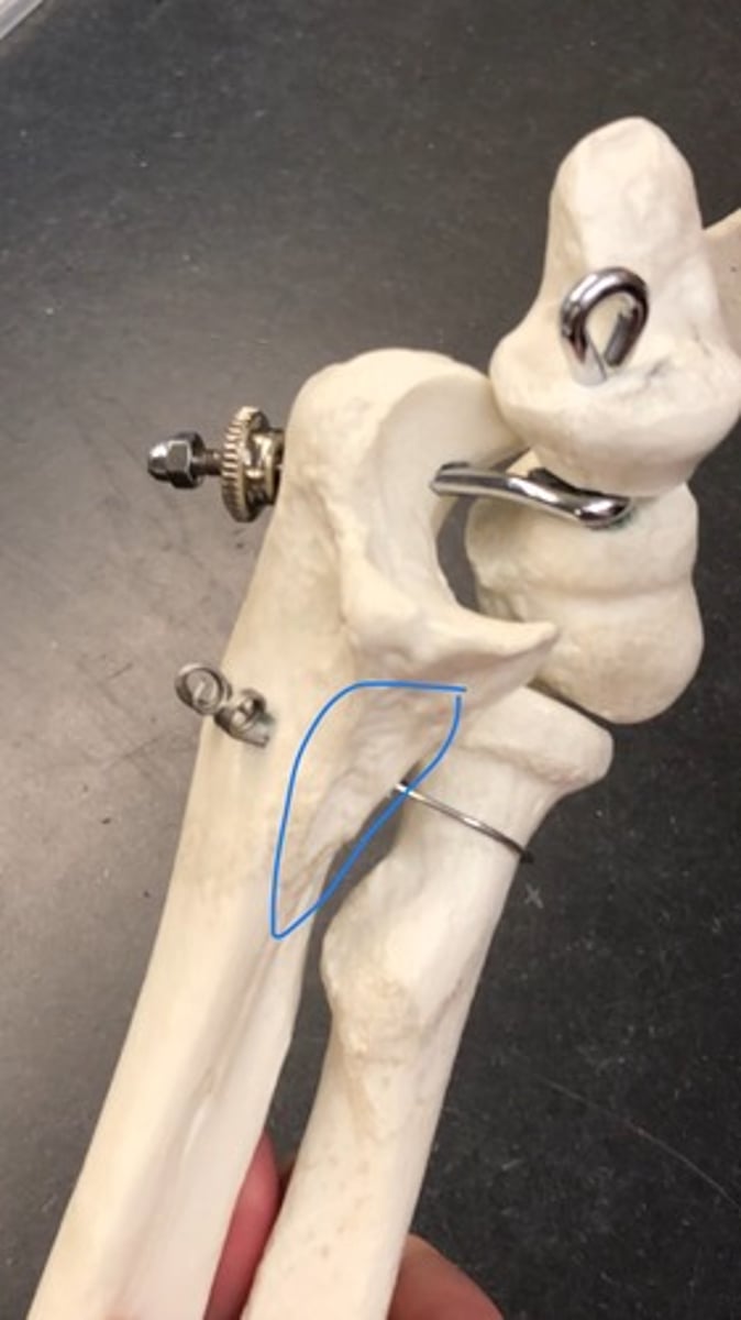
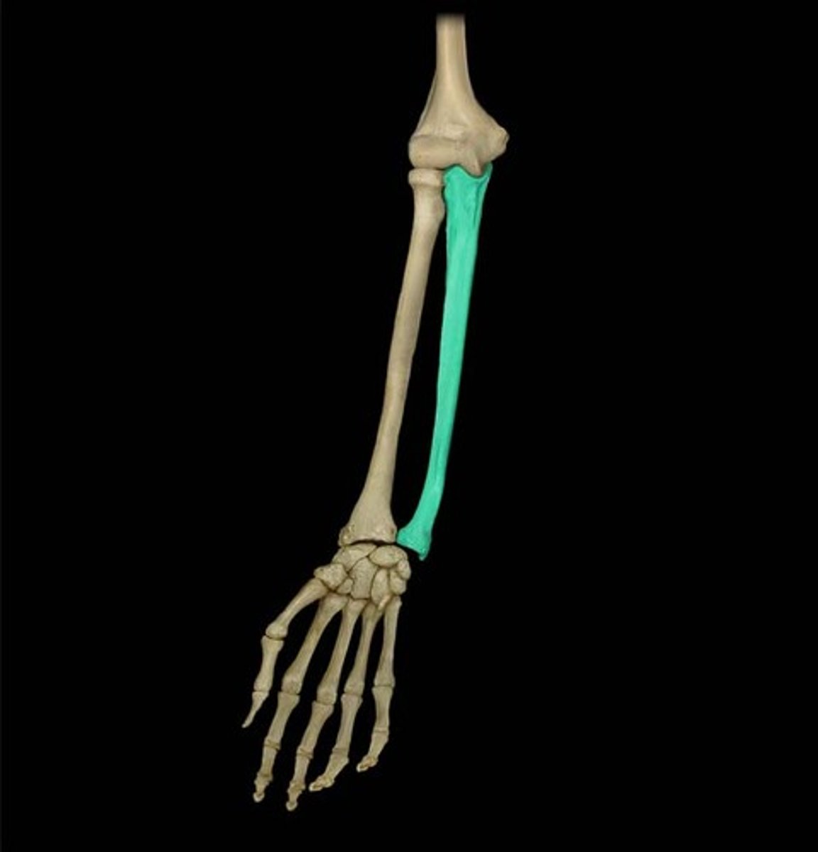
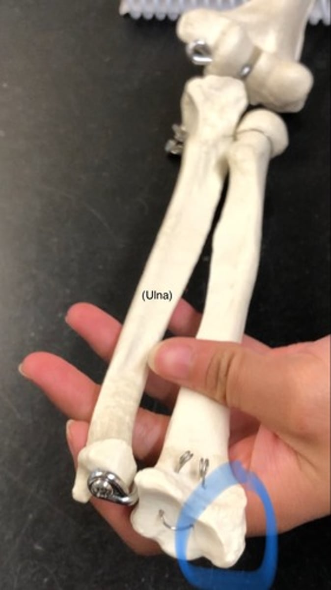
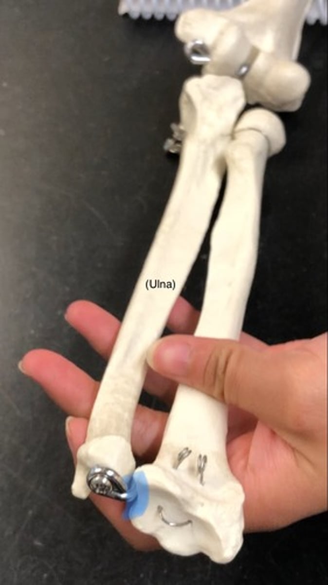
Ulna
On pinky side, most medial; "ice cream scoop"

Olecranon
"scooper"; posterior; fits into olecranon fossa
Trochlear notch
"where ice cream goes into"; depression below olecranon
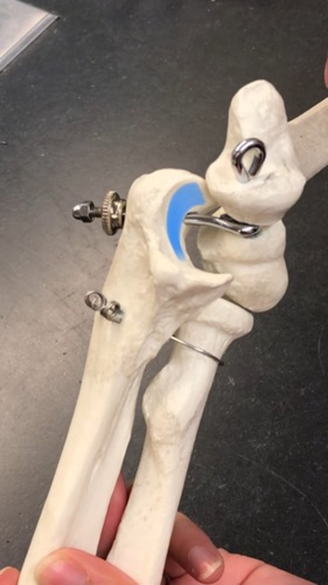
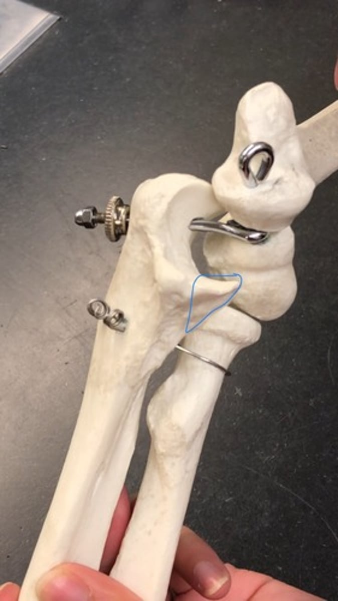
Coronoid process of ulna
"bottom of scooper"
Radial notch
lateral; side of coronoid process where radius fits in
Ulnar tuberosity
rough patches under coronoid process
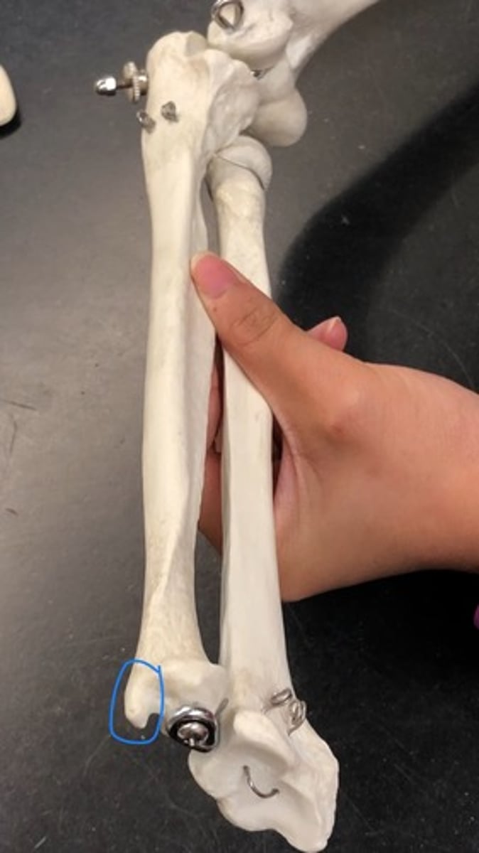
Head of ulna
circular knob on bottom

Styloid process of ulna
longest point on the bottom of bone
Right vs. Left (Ulna)
trochlear notch is anterior; radial notch is lateral
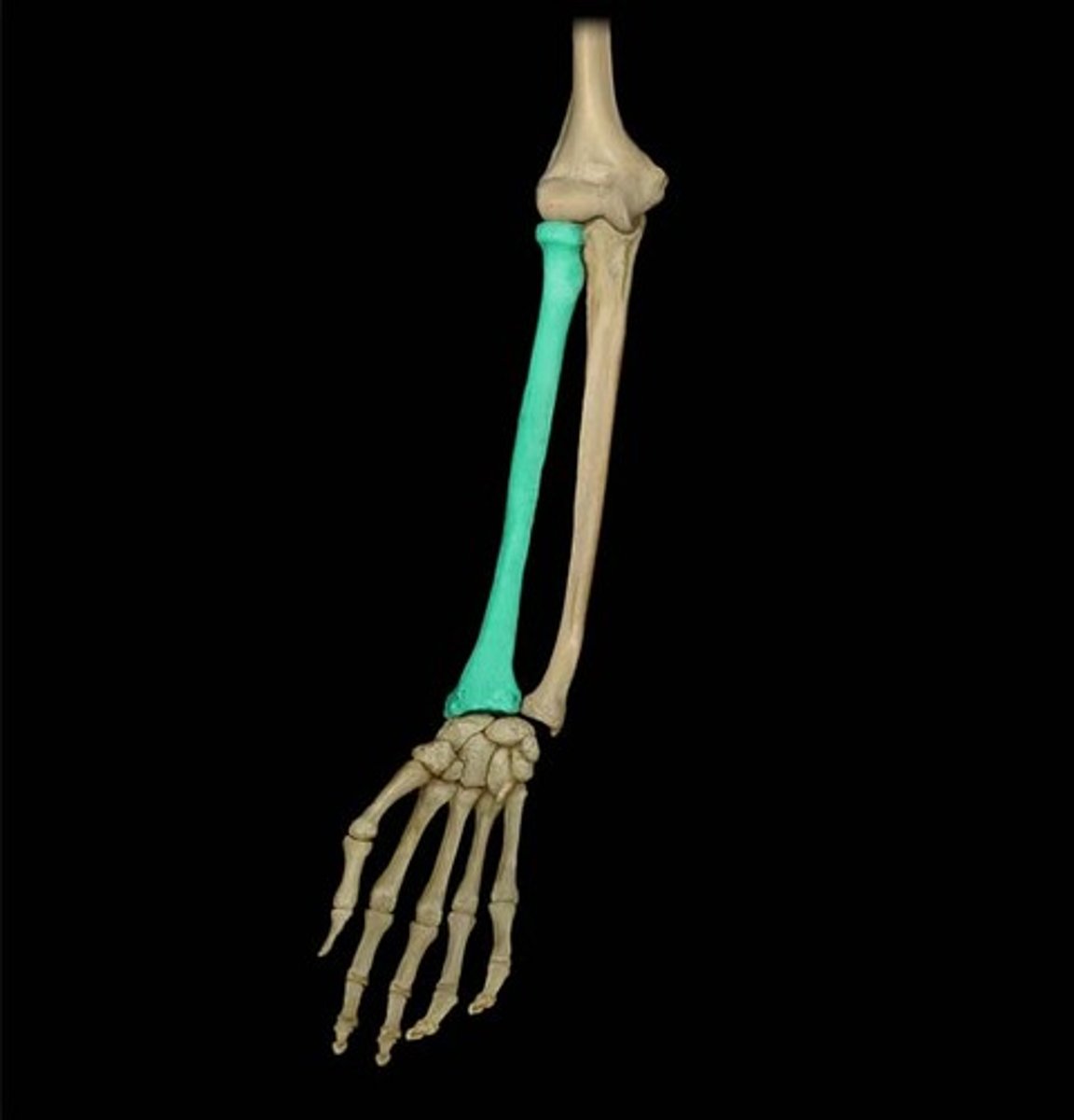
Radius
thumbs up = "rad"
Head of radius
knob on the top
Neck of radius
narrows after the head
Radial tuberosity
rough patch after the head
Styloid process of radius
lateral (pinky); bottom end of radius
Ulnar notch
head of the ulna fits in
Right vs. Left (Radius)
radial tuberosity = medial/anterior

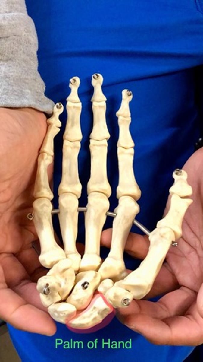
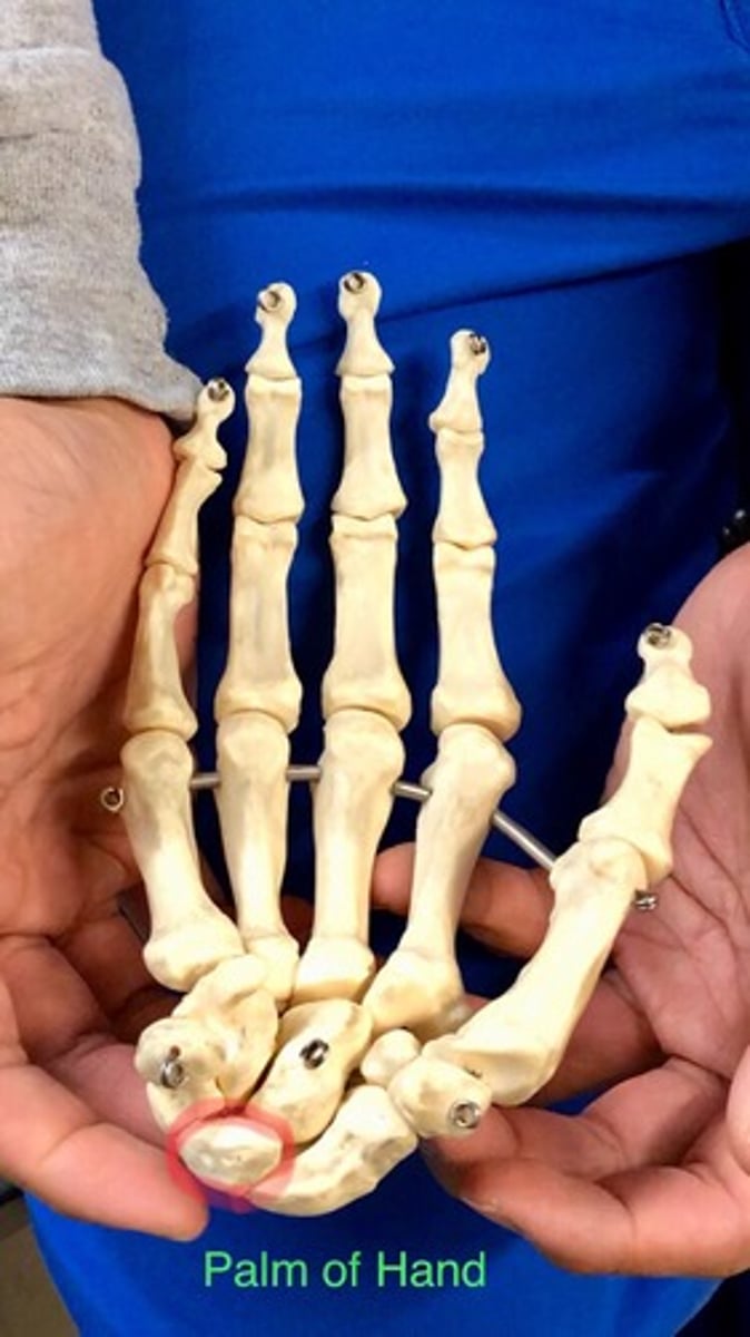
Carpal bones
8 total
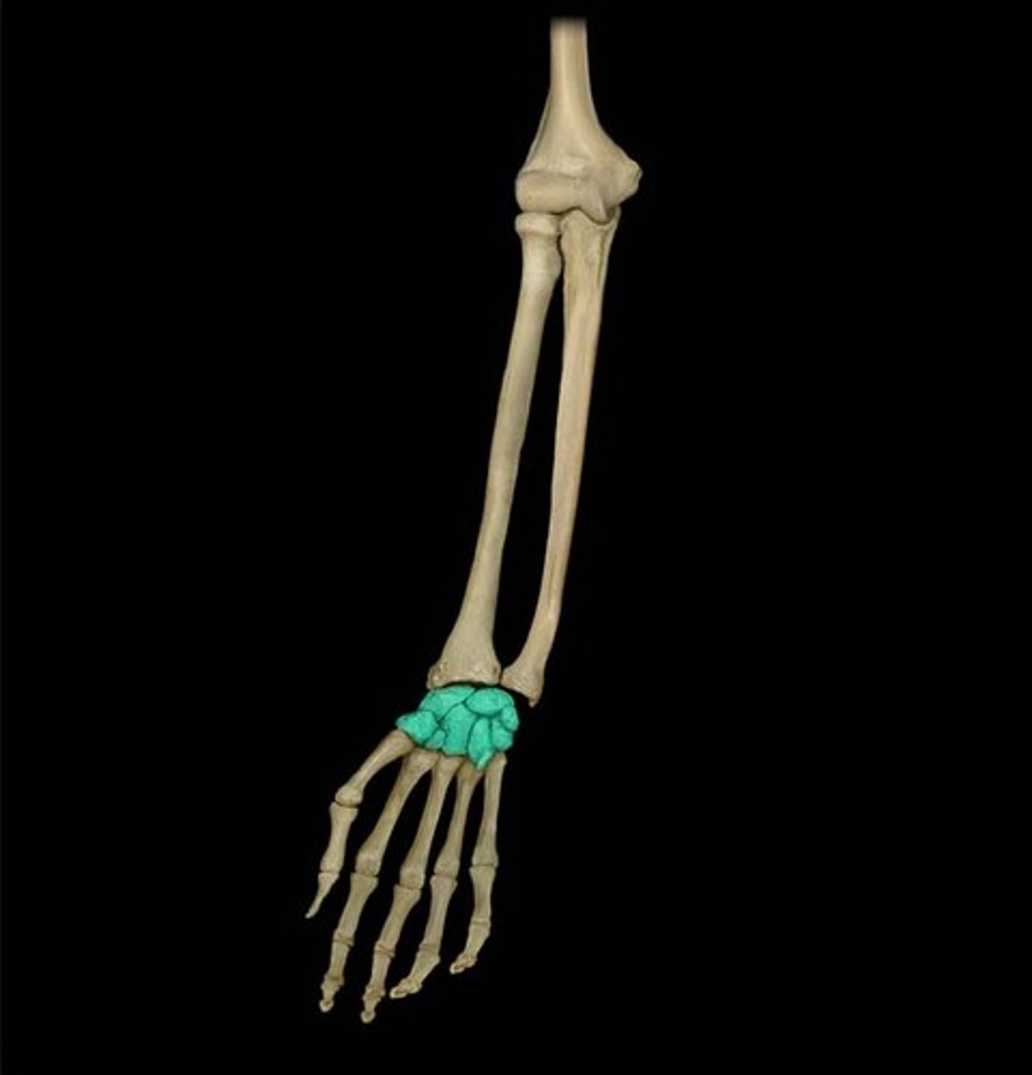
Scaphoid
"So"; longest one
Lunate
"Long"
Triquetrum
"To" ; small bone behind pisiform
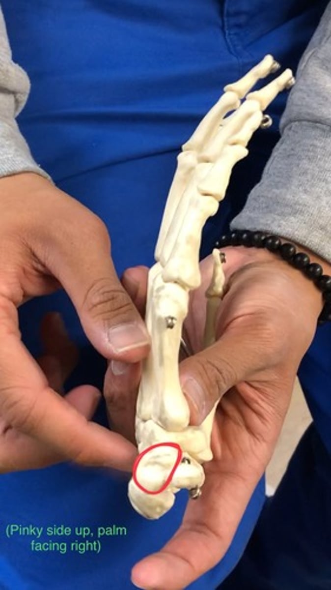
Pisiform
"Pinky"
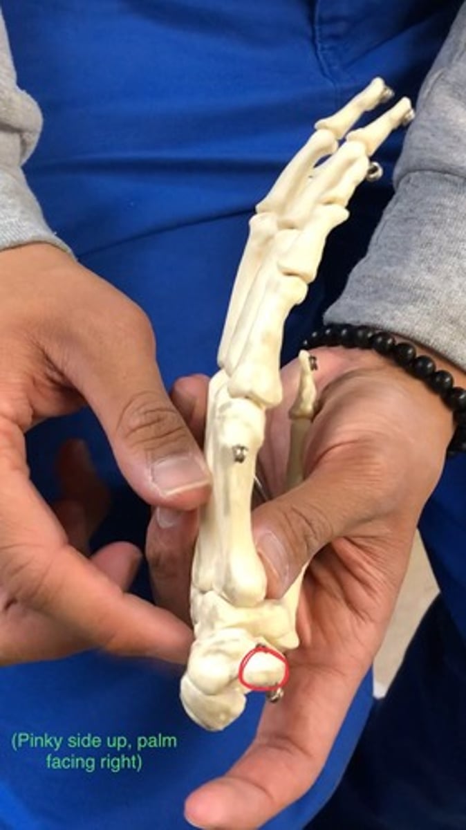
Hamate
"Here"; under ring finger
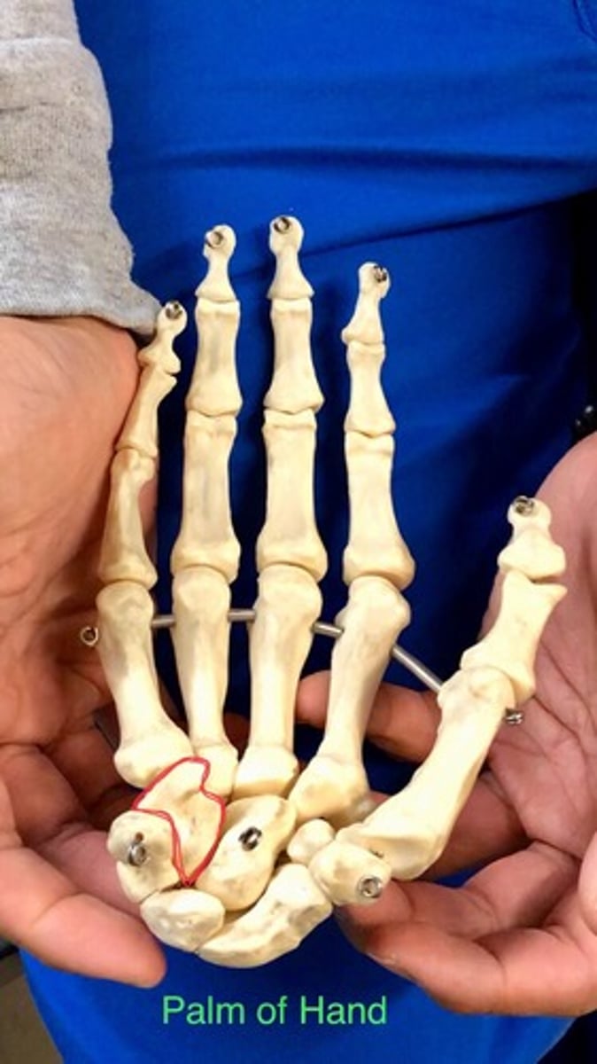
Capitate
"Comes"; underneath middle finger

Trapezoid
"The"; underneath index finger
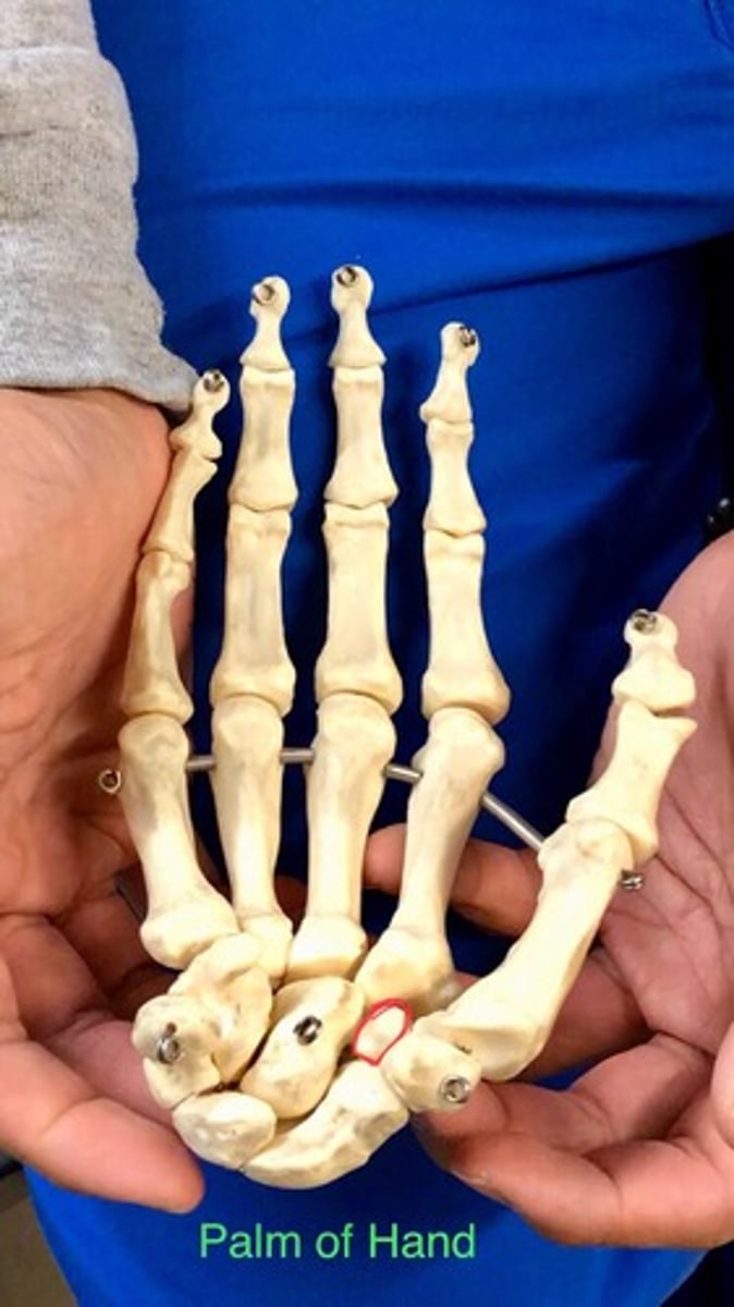
Trapezium
"Thumb"; right below the thumb (pollex)
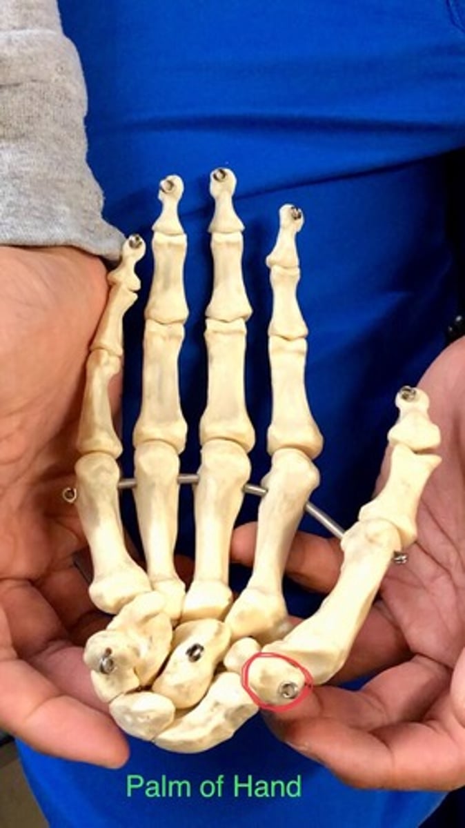
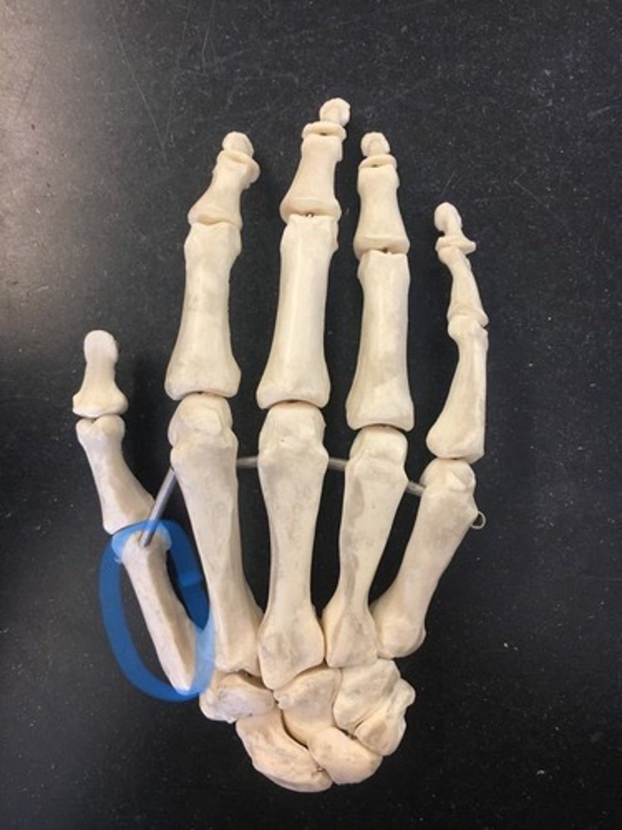
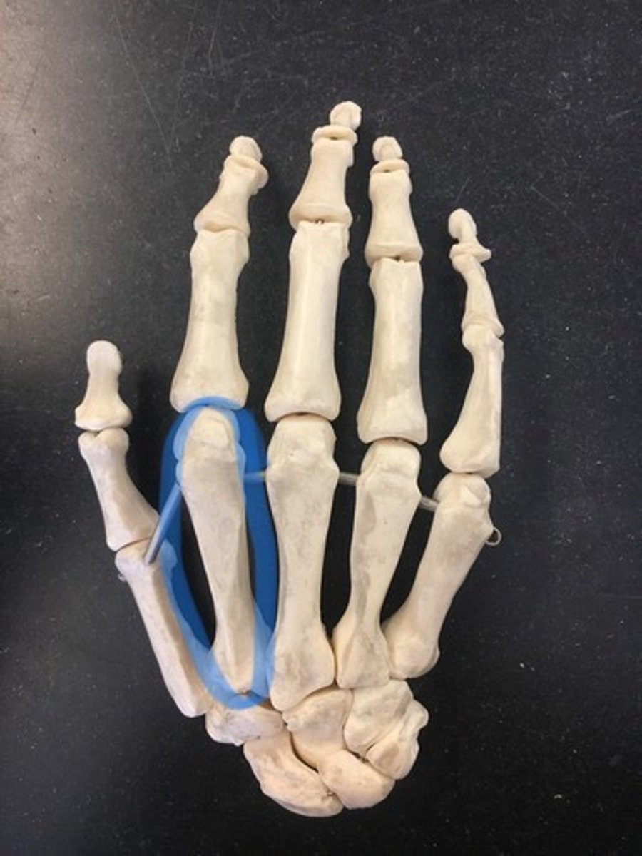
Metacarpal bones I
thumb (pollex)
Metacarpal bones II
index finger
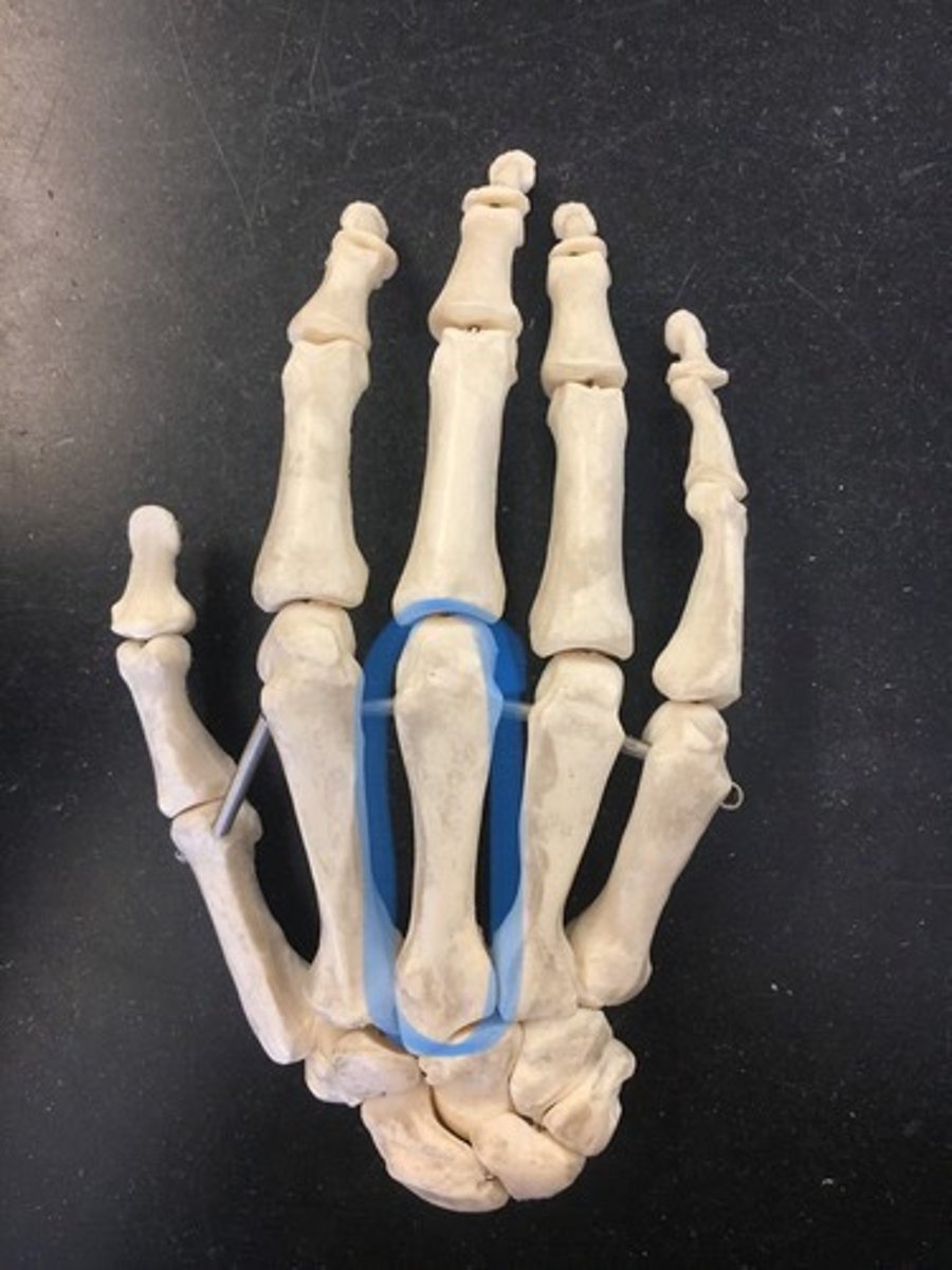
Metacarpal bones III
middle finger
Metacarpal bones IV
ring finger
Metacarpal bones V
pinky
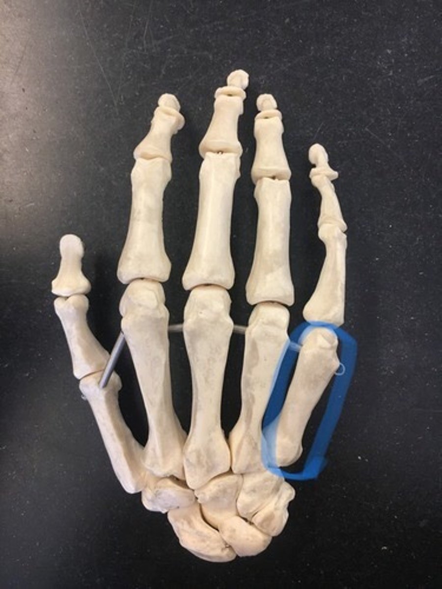
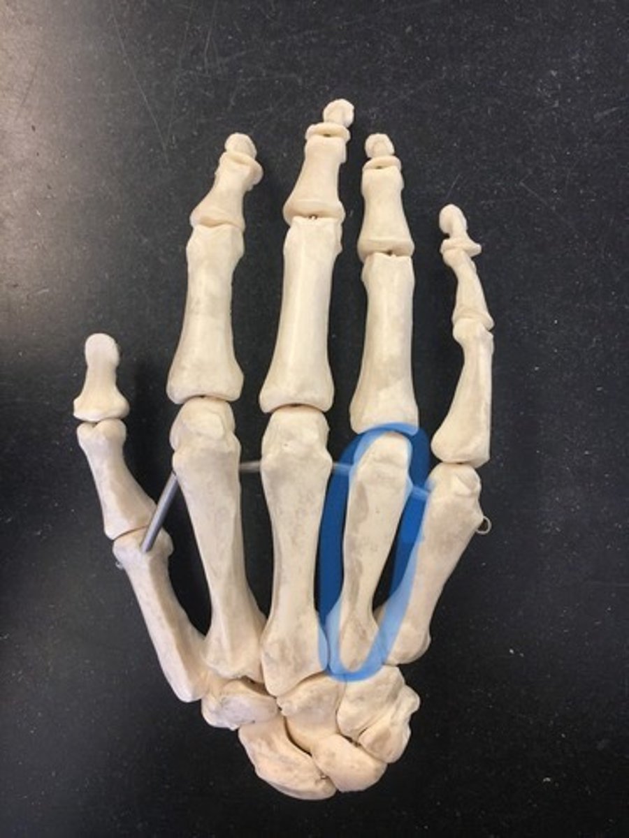
Phalanges (Thumb or pollex)
2 bones; proximal, distal
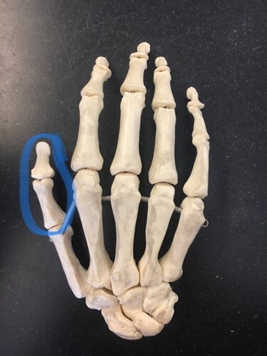
Phalanges (Index, middle, ring, pinky)
3 bones; proximal, middle, distal
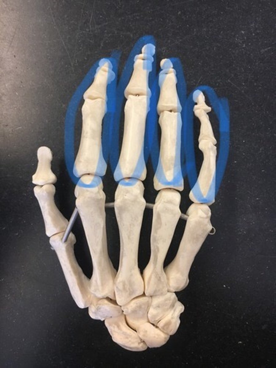
Metacarpal IV
(AND/OR)
Proximal phalanx of Metacarpal III
How to properly write the specific part of the metacarpals/phalanges
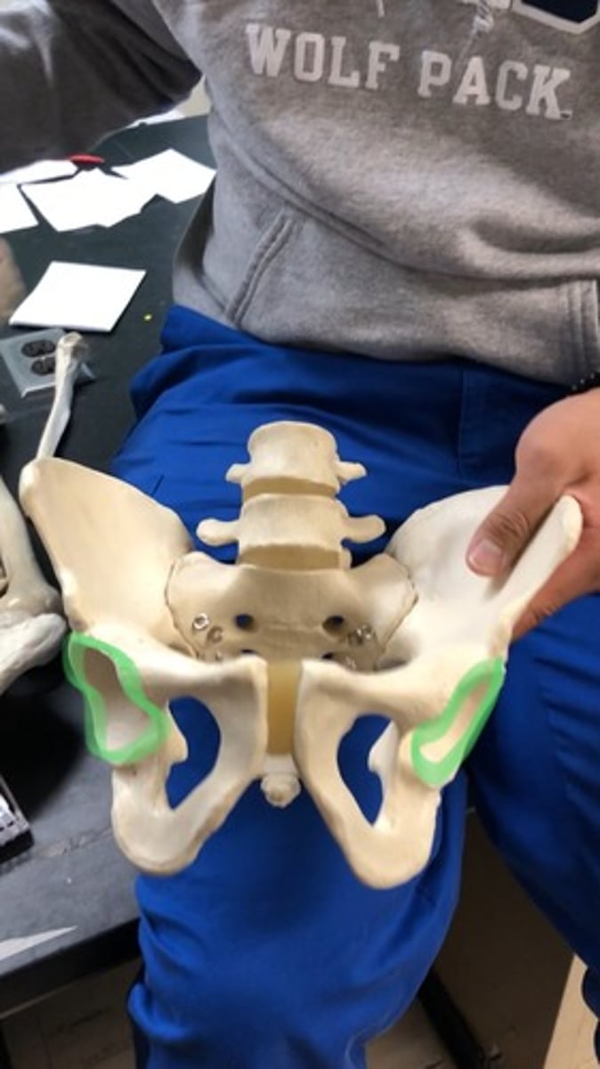
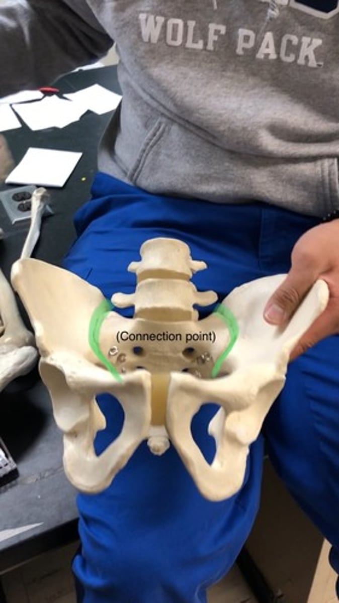
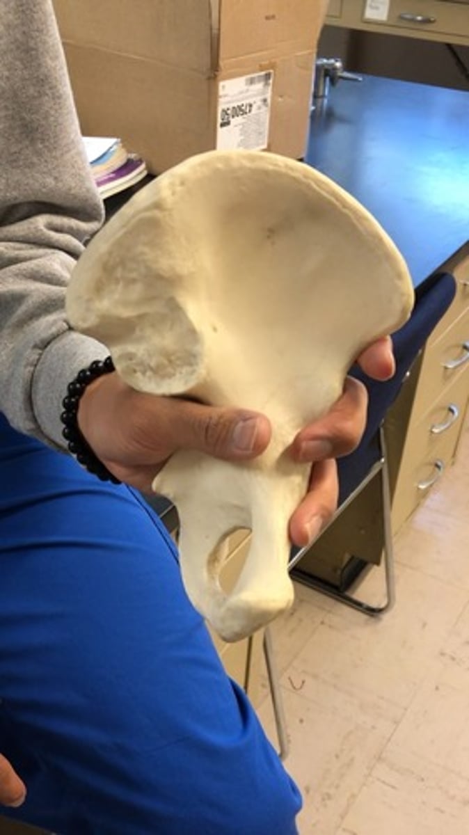
Ilium
top region
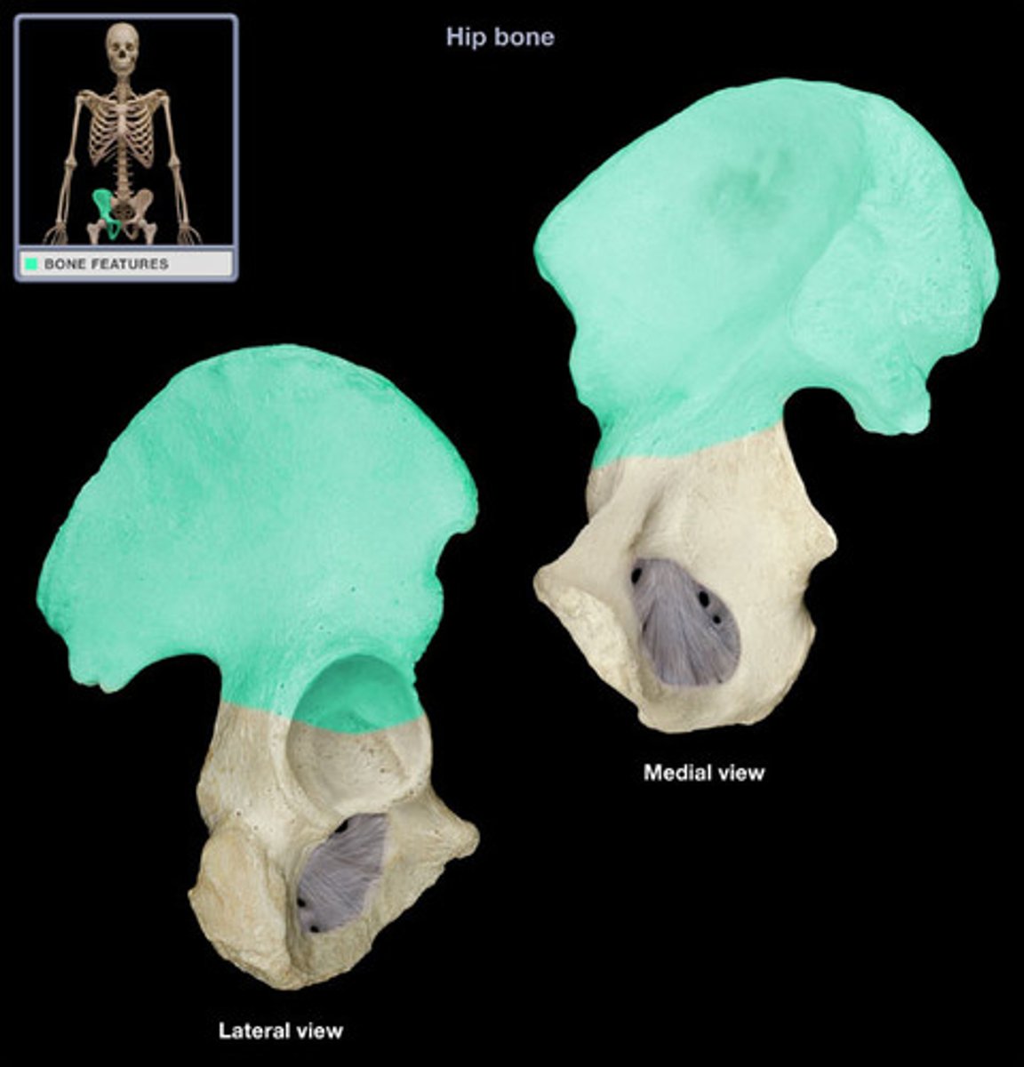
Iliac crest
ridge on top of illium
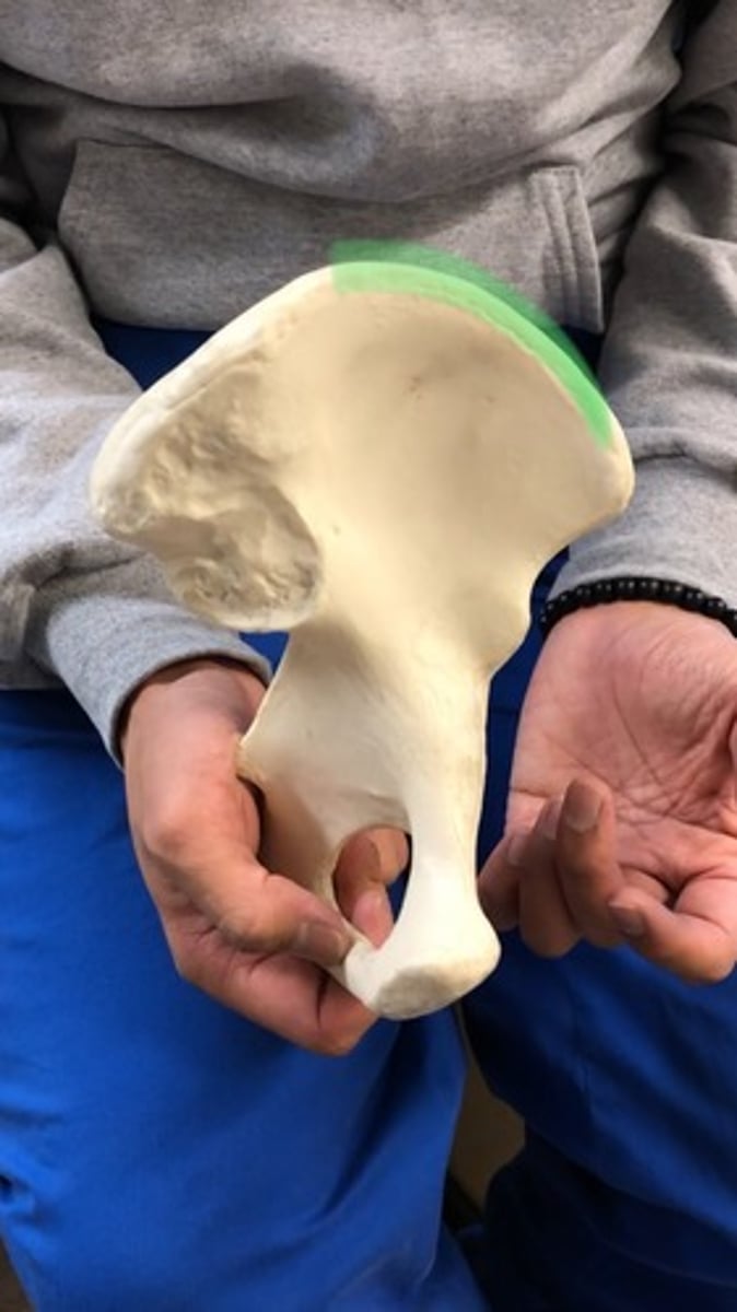
Anterior superior iliac spine
lateral; top point on lateral side

Anterior inferior iliac spine
second point below anterior superior
Posterior superior iliac spine
medial; above inferior

Posterior inferior iliac spine
medial; below superior

Greater sciatic notch
big notch below posterior inferior iliac spine (large indent)

Arcuate line
ridge below iliac fossa

Iliac fossa
smooth portion on top of surface

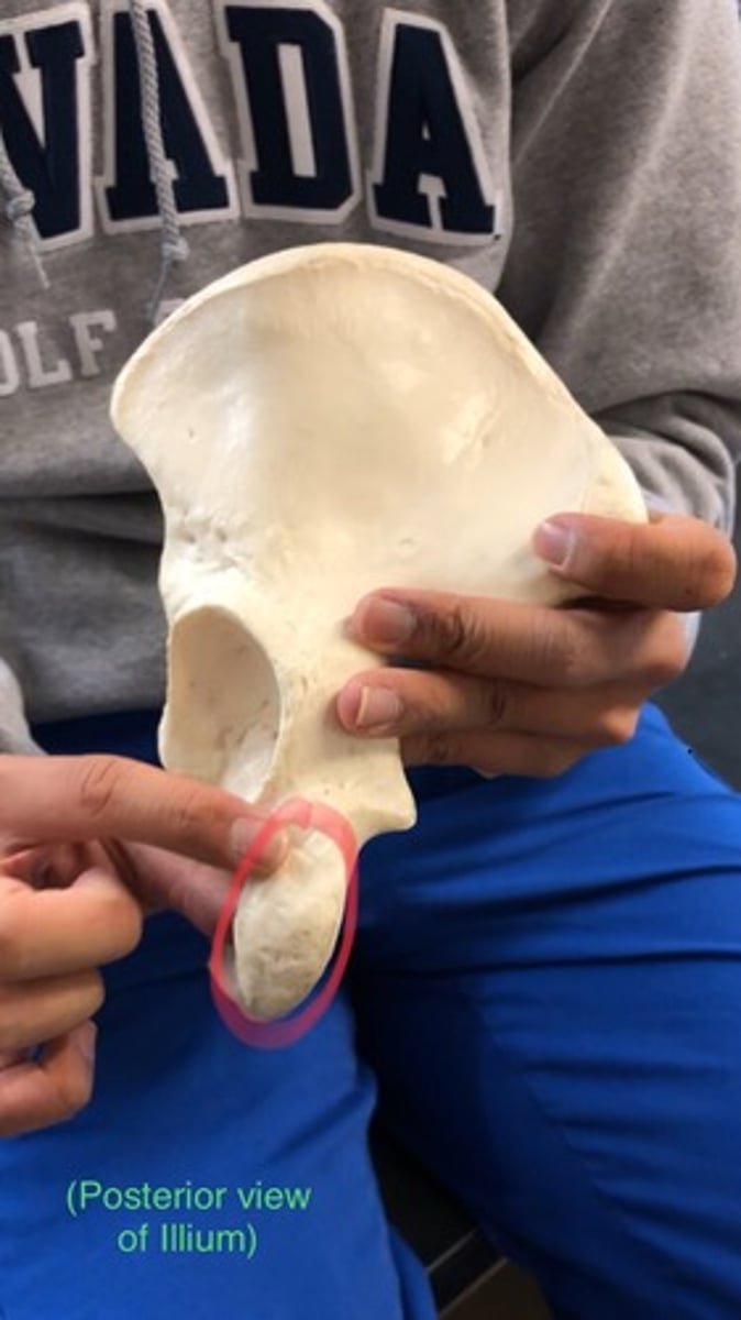
Auricular surface of ilium
rough patch where the sacrum fits into; lateral to iliac fossa

Ischium
what we're sitting on; right after notch
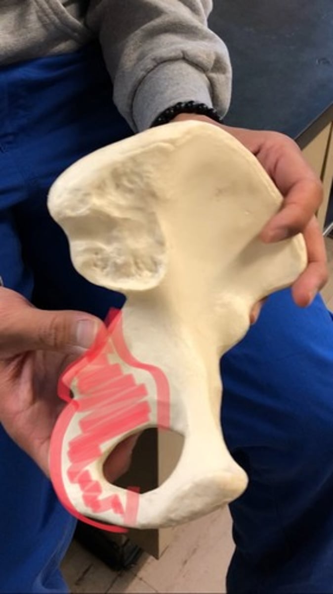
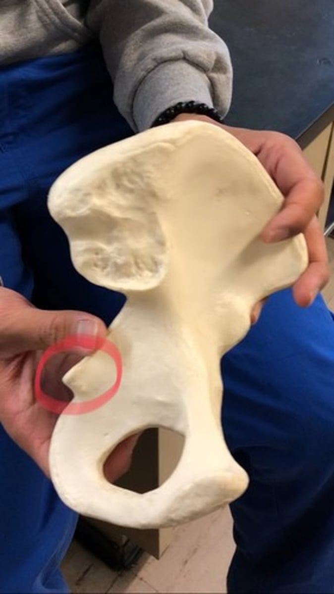
Ischial spine
point after greater sciatic notch
Lesser sciatic notch
depression/indent after ischial spine

Ischial tuberosity
rough patch; below the lesser sciatic notch (what you feel when you sit)
Ischial ramus
arch outside of notch; first half on the ischium
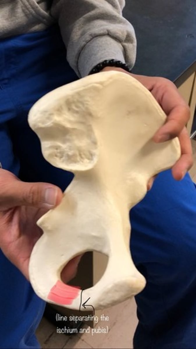
Pubis
region
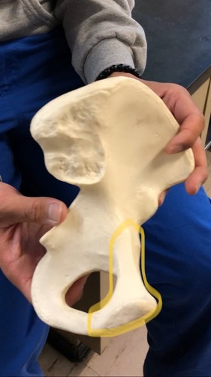
Superior ramus of pubis
top half

Inferior ramus of pubis
bottom half; after ischial ramus

Pectineal line
other half of ridge; goes up to the arcuate line in ilium
Pubic symphysis
where two coccyeal bones connect; padding where they connect

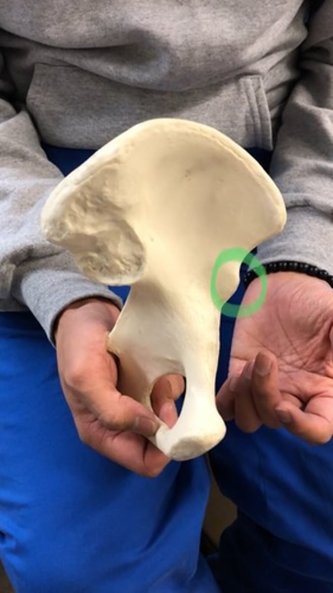
Acetabulum
where the femur fits into; hip socket
Obturator foramen
holes on the bottom (2) - in coccyeal bones
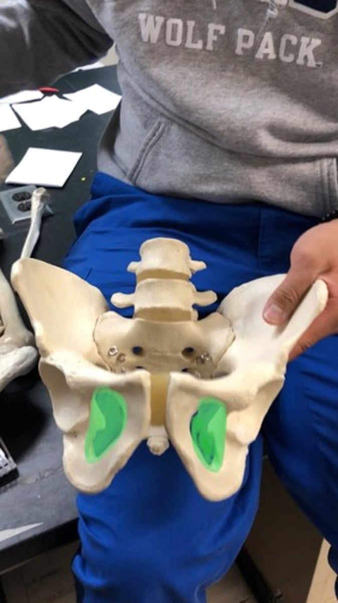
Sacroiliac joint
where the sacrum and illium come together; would only test together with sacrum
Right vs. Left (Pelvic Girdle)
Acetabulum = lateral
Iliac crest = posterior
Greater sciatic notch = posterior
(Hold like a telephone)
Male vs. Female (Pelvic Girdle)
Male: less than 90 degree angle under pubic symphysis
Female: greater than 90 degree angle under pubic symphysis. (use paper)
**Baby skull should be able to go through the female's pelvic girdle**

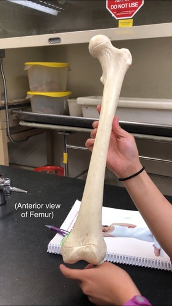
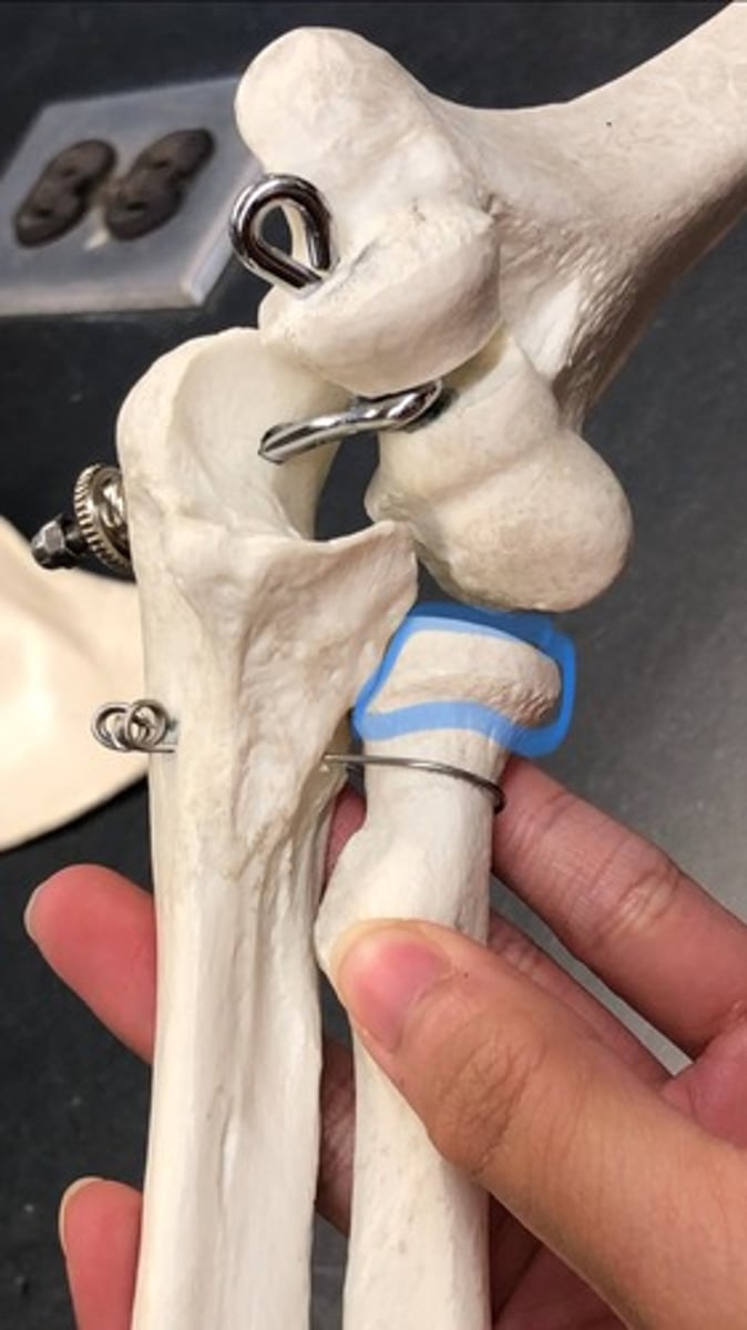
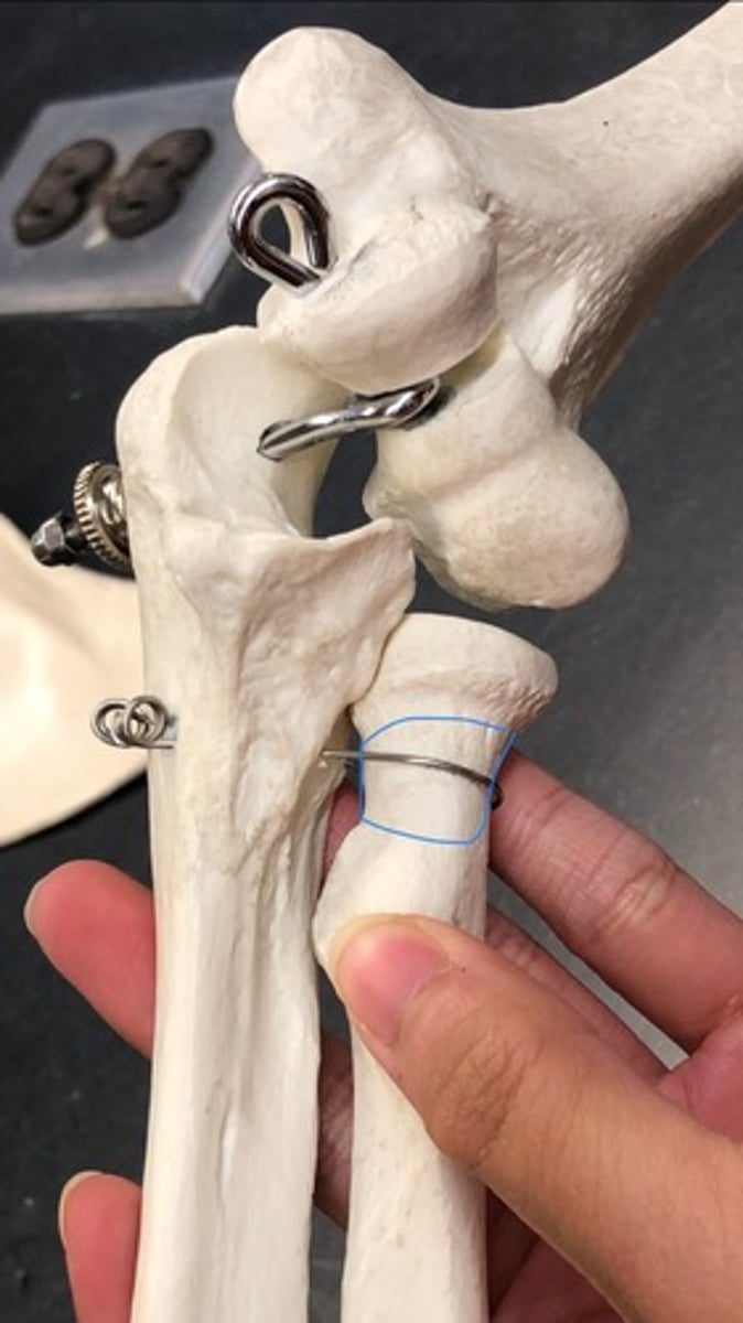
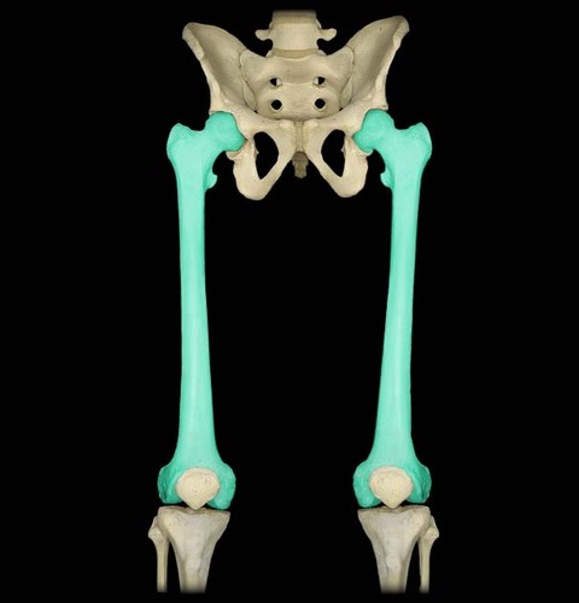
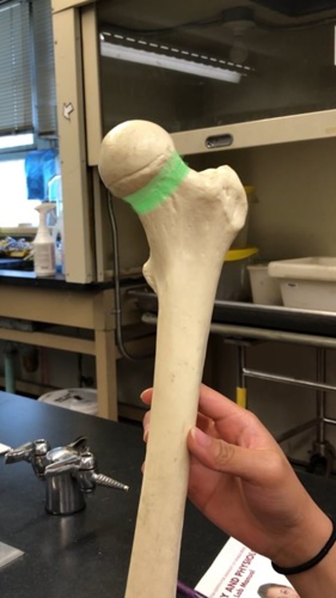
Femur
big caveman like bone
Femoral head
medial
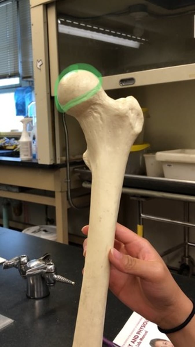
Fovea capitis
small indent in femoral head

Neck of femur
narrows after femoral head
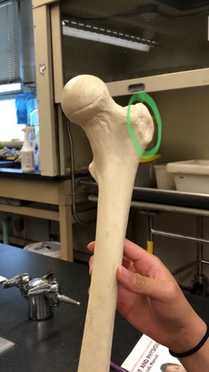
Greater trochanter of femur
lateral; side of head
Lesser trochanter of femur
medial (same side as femoral head); smaller bump

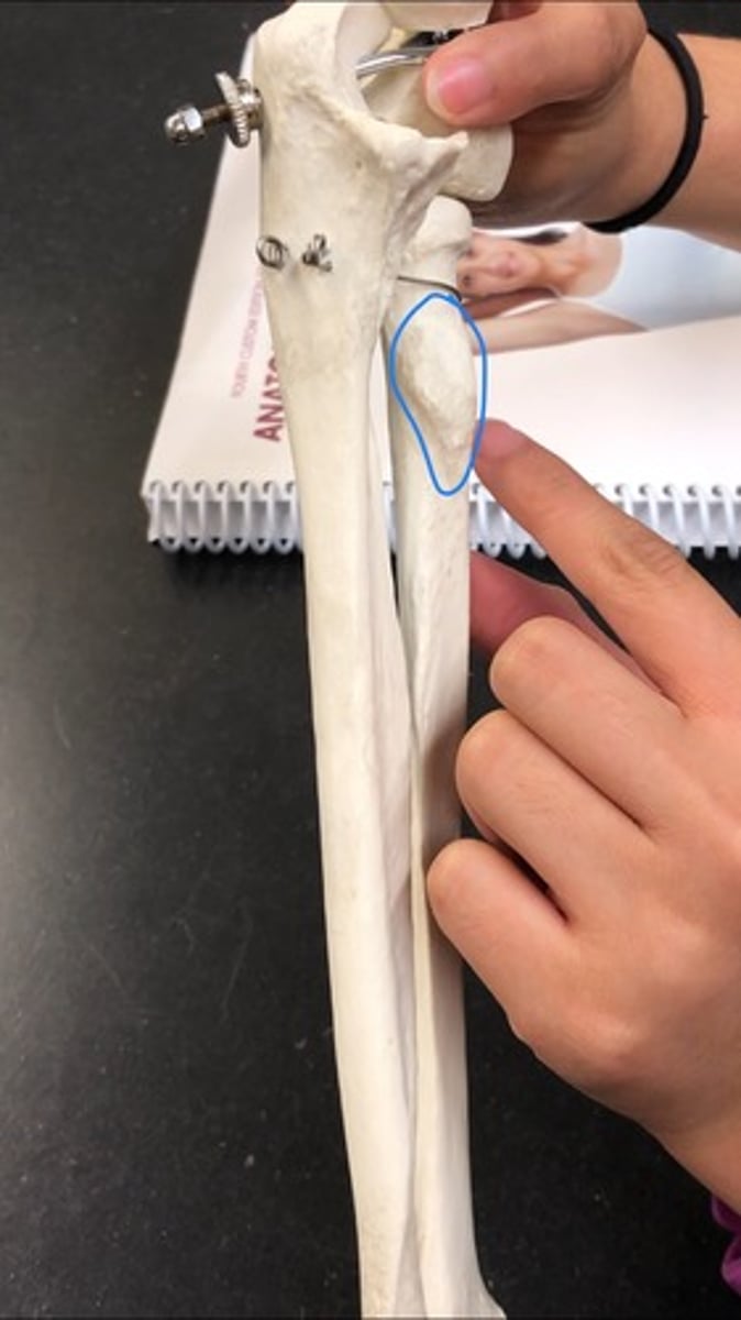
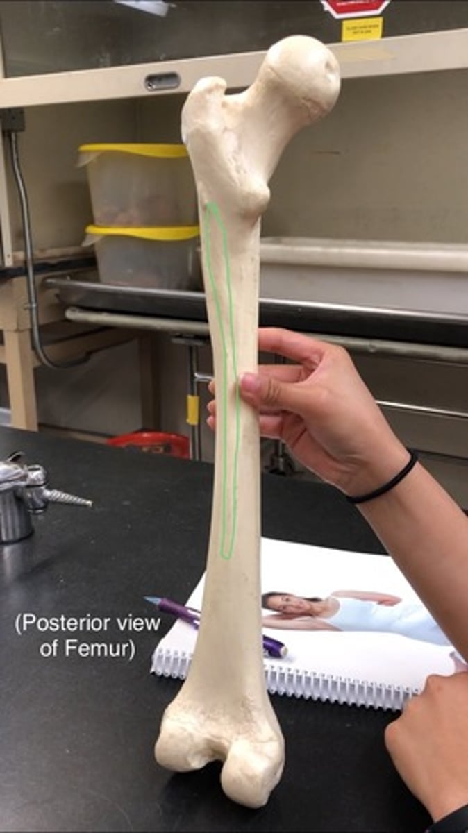
Linea aspera
posterior; ridge on posterior side of femur
Medial epicondyle of femur
most medial "outward" projection