Visual Field Analysis in Glaucoma
1/56
There's no tags or description
Looks like no tags are added yet.
Name | Mastery | Learn | Test | Matching | Spaced | Call with Kai |
|---|
No analytics yet
Send a link to your students to track their progress
57 Terms
SITA standard
HFV algorithm that is the gold standard in glaucoma testing. Reduces test time (50%) when compared to traditional full threshold testing, this improves test result reliability by reducing patient fatigue. Test is about 3-7 minutes per eye. Defects will appear shallower, but is still adequate for detecting them.
SITA fast
HFV algorithm that is similar to SITA standard but is even faster (60%), about 2-5 minutes per eye, being especially useful in patients having difficulty with SITA standard test length. May be more difficult for the patient due to stimuli tested closer to threshold more quickly. Has greater test variability and is not currently considered the test of choice for follow up care.
SITA faster
HFV algorithm that is similar to SITA fast but is even faster (30%, 50% faster than SITA standard), about 2 minutes per eye. It does this by not testing the blind spot location and therefore cannot detect false negatives. It only available in 24-2. Is not reliable in detecting early glaucoma defects.
30-2
HFV test parameter testing the central 30 degrees, at 76 locations, 6 degrees apart, at equidistance from the horizontal axis
24-2
HFV test parameter testing the central 24 degrees, at 54 locations, 6 degrees apart, at equidistance from the horizontal axis
24-2 C
HFV test parameter testing the SITA faster 24-2 with an additional 10 points incorporated
horizontal axis
In 30-1 and 24-1 testing grid points are located on the ____ and are therefore not used in glaucoma testing
10-2
HFV test parameter testing the central 10 degrees, at 68 locations, 2 degrees apart. Is useful in the detection of early glaucoma presenting with paracentral defects as well as for detecting changes in patients having significant VF loss due to advanced glaucoma.
12, 4, 68
30-2 and 24-2 testing tests ___ points within the central 10 degrees of the visual field with only ___ points in the macular region. 10-2 testing tests ___ point in the central 10 degrees of the visual field making 10-2 testing more sensitive to central and paracentral defects
12, 6
HVF testing in a glaucoma suspect or patient having early glaucoma should be performed at minimum every ___ months, but ideally every ___ months
6, 3
HVF testing in patient having late glaucoma should be performed at minimum every ___ months, but ideally every ___ months
moderate to advanced disease, 40
A detectable visual field loss is indicative of ____. This is due to the fact that ____% of retinal ganglion cells must be lost before being detectable as a visual field defect. Structures of the retina will degenerate prior to a visual field defect being detectable.
reduced mean deviation
Generalized constriction of the visual field seen in early non-specific changes in glaucoma will be presented as a ____ on HVF testing
0 to -5.99 dB
mild generalized visual field defect
-6 to -11.99 dB
moderate generalized visual field
> -12 dB
advanced generalized visual field defect
Siedel's sickle shaped scotoma
visual field defect which is an early significant sign of glaucoma. Is a paracentral scotoma that joins the blind spot.
Ring scotoma
visual field defect which is a late significant sign of glaucoma. Is two arcuate scotomas joining together.
anterior ischemic optic neuropathy
Altitudinal defects generally should not be associated with glaucoma and are suspicious of...
Arcuate, nasal step, paracentral
____ and ____ visual field defects are characteristic of glaucoma, whereas ___ defects are nonspecific
Hodapp Parrish Anderson
the most widely used classification system for visual field defects in glaucoma
Ptosis
Poor alignment
Trial lens artifacts
Patient fatigue
Small pupils
Co-morbidities
6 Factors Affecting VF Test Validity
20%
Fixation losses should be less than...
30%
False positive and negatives should be less than...
White scotoma
areas of higher than normal decibels on the grey scale of a HVF. Is characteristic of falsely elevated threshold measurements due to high false positive results.
gaze tracking, false negatives
Recent studies have shown that ____ may be better for determining reliability than ____ which may increase in progression of disease
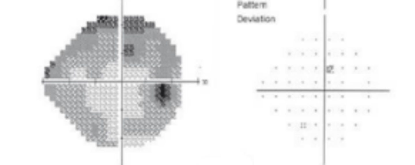
Total deviation plot
plots the difference (in dB) between test results and normative data from the patient's age group. Shows the probability of each point being normal.
Pattern deviation plot
plots the overall sensitivity changes in the hill of vision negating overall depression as caused by media opacities or refractive error. Reveals localized defects.
Mean deviation
represents the overall depression of the visual field. Should not exceed -2 dB.
Pattern standard deviation
represents the degree of departure from the normal hill of vision. This value may be high due to low reliability, VF defect, or both.
Visual field index
represents the percent of a normal age-adjusted visual field with greater weight given to points closer to fixation to adjust for ganglion cell density and visual function. Is less sensitive to cataract and media changes.
Glaucoma hemifield analysis
compares corresponding point values across the horizontal meridians. Fields are then classified as outside/within normal limits or borderline. Has good sensitivity and specificity for separating normal and glaucomatous fields.
2, 2
A good baseline visual field is at least ___ tests taken about ___ weeks apart. The results should be comparable, otherwise another test is indicated to establish a baseline
3
It may take some patients up to___ tests until they are able to properly perform a HVF test. Improvement of a defect in a glaucoma patient should indicate the need for establishment of a new baseline due to learning effect, glaucoma visual field defects will never actually improve
increase
Variability in tests results may (increase or decrease) in early glaucomatous damage
1.5-2.0
Fast progression of visual field defect is defined as a mean deviation >___ dB per year
Glaucoma progression analysis (GPA)
glaucoma progression analysis software that uses a point by point method to evaluate a visual field change seen from baseline. Requires 2-3 additional tests in order to confirm a real change.
Visual field index (VFI)
value that gives information on the rate of deterioration of the overall visual field based on pattern standard deviation (less sensitive to overall field depression i.e. cataract) and weighing heavily on central points. Is expressed as a percentage of normal visual field. I.e.) 100% means a totally normal VF
occult
initial stage of glaucoma where there is no detectable defect despite the fact that there is damage occurring
threshold
stage of glaucoma where there is a period of shallow defects that are often transient and barely detectable.
critical phase
stage of glaucoma where there are detectable visual field defects that are progressive and become dense
Octopus Perimeter
visual field test that performs a full threshold exam in 2:30 minutes. It has complete eye fixation control and does not present a stimulus if fixation is lost.
GTOP, G patter, dynamic
octopus test parameter used for glaucoma testing
unreliable
Because the Octopus perimeter will not display a stimulus if fixation is lost, it is especially useful in ____ patients. Additionally, the test is shorter than HVF. Is also useful if there is a suspected defect that was not detected on HVF
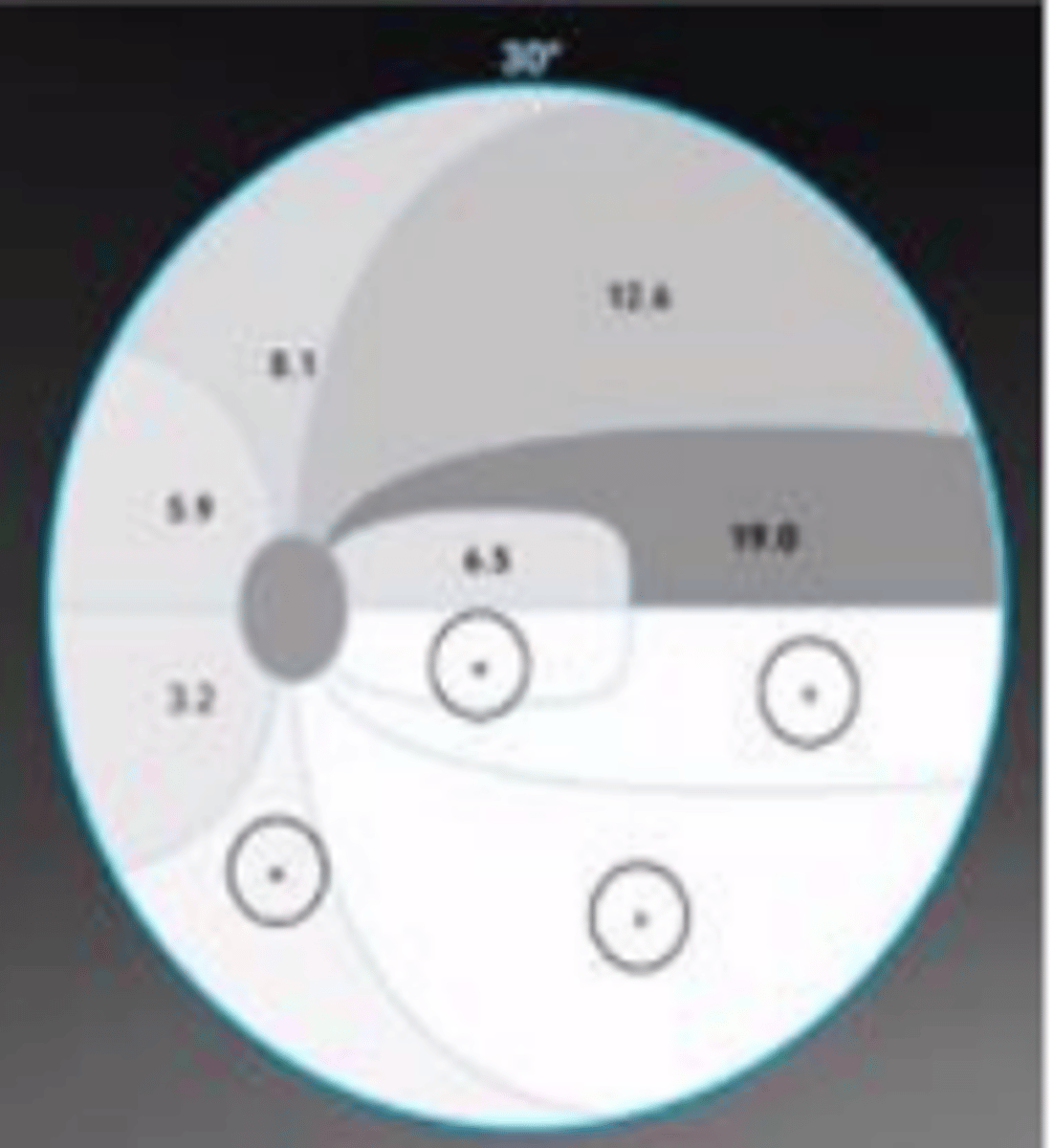
Cluster analysis
octopus analysis tool designed specifically for glaucoma being very sensitive to detection of subtle glaucomatous defects. A mean cluster defect is calculated for visual field locations corresponding to the same RNFL bundles.
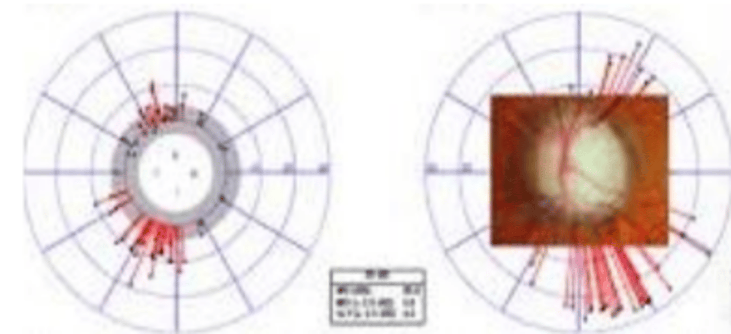
Polar analysis
octopus analysis tool that projects local visual field defects along the nerve fibers to the optic disc and displays them oriented as structural results.
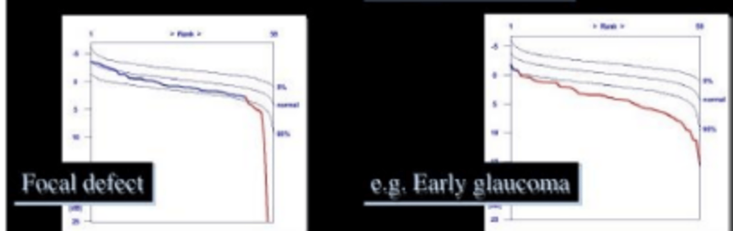
Defect curve
octopus analysis tool that is a graphical representation of visual field defects. Allows for distinguishment between local and diffuse defects.
Function specific perimetry
testing whose goal is to isolate sub-populations of ganglion cells by evaluating a specific visual function characteristically processed by that cell type. This results in less overlap of cell responses that could potentially mask ganglion cell loss. Therefore, this allows for easier detection of earlier glaucomatous damage.
Short wavelength automated perimetry (SWAP)
function specific perimetry designed to assess blue cones and K cells which encode blue yellow opponency via a blue stimulus presented against a yellow background. Although glaucoma does not preferentially target these cells, targeting these specific RGC (9%) reduced redundancy of testing for earlier detection of glaucomatous defects. This test is more affected by cataracts so it is important to analyze pattern deviation results.
3-5
SWAP testing detects defects ____ years earlier than standard automated perimetry testing
1-3
SWAP testing detects progression of visual field loss ____ years earlier than standard automated perimetry testing
deeper
In patients having visual field loss already detected by standard automated perimetry testing, defects appear ____ on SWAP testing
advanced
One major limitation of SWAP testing is that it may not be able to track ____ cases of glaucoma
Frequency doubling technology (FDT)
function specific perimetry designed to assess magnocellular RGCs (3-5%) via a rapidly flickering (25 Hz) low spatial frequency (0.25 cycle/degree) sine wave grating stimulus. The criteria for what results are considered abnormal are unclear, but it is important to be sure that the defect is repeatable.
12-24
FDT testing detects defects ____ months earlier than standard automated perimetry testing.
FDT matrix
FDT is less useful for glaucoma follow up due to large stimulus size and minimal number of targets presented. This updated version of FDT was developed to mimic the standard 24-2 and 30-2 testing patterns to address this problem.
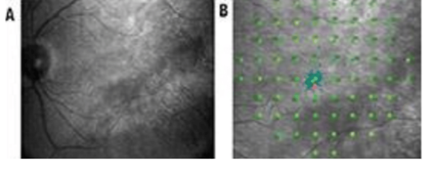
Microperimetry
test that uses real time tracking of retinal movements during VF testing to provide spatially registered measurements of retinal sensitivity projected onto a fundus image.