Spinal cord
1/12
There's no tags or description
Looks like no tags are added yet.
Name | Mastery | Learn | Test | Matching | Spaced | Call with Kai |
|---|
No analytics yet
Send a link to your students to track their progress
13 Terms
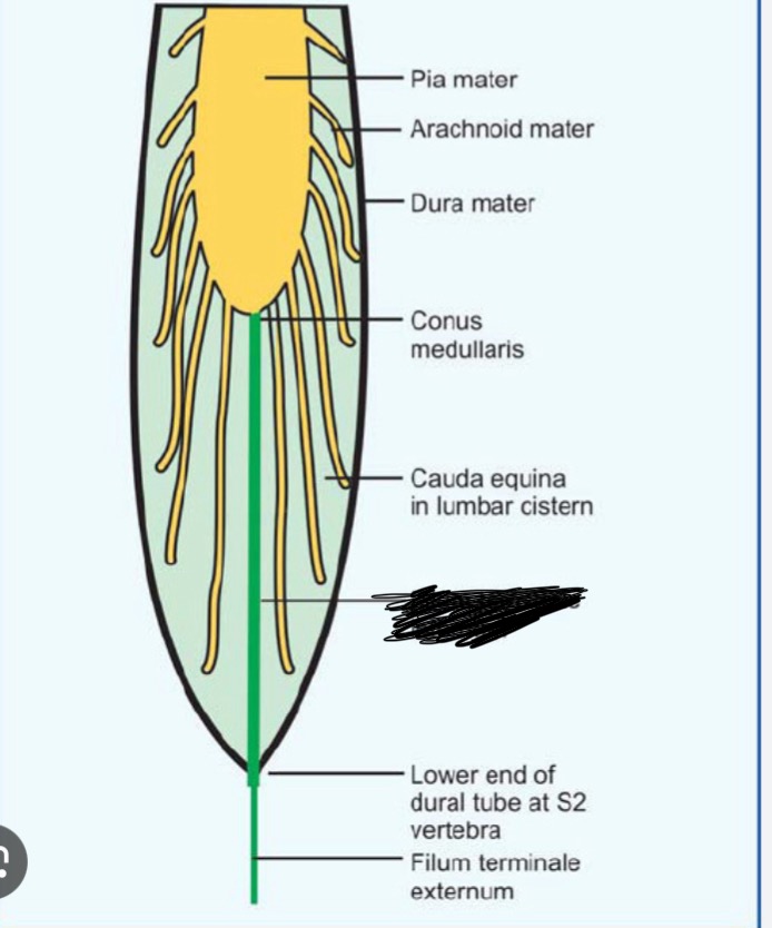
What is the conus medullaris?
Where the spinal cord ends (L1-L2)
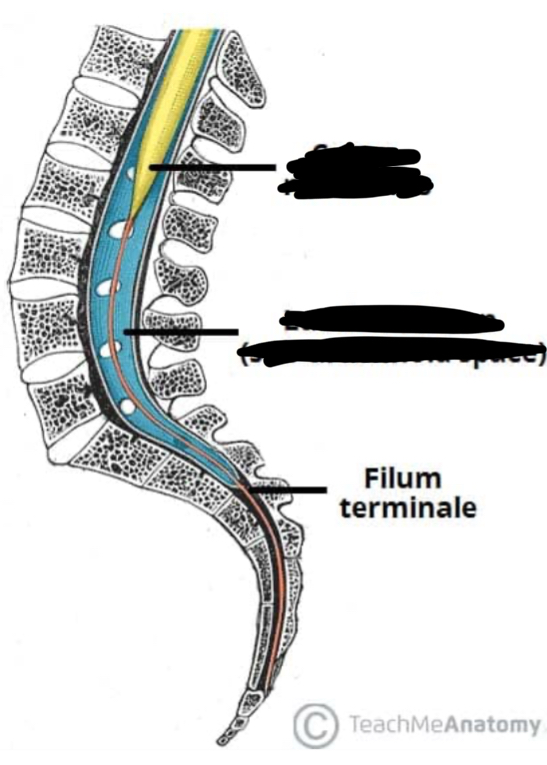
What is the yellow structure? Blue?
Yellow is conus medullaris, blue is lumbar cistern
What is the lumbar cistern? What is its clinical significance?
L2-S2, holds CSF and cauda equina. Clinically significant because this is the located an LP occurs.
What are the yellow squigly structures?
Fillmore terminala aka loose nerve at the end of the cauda equina
Where does the cervical enlargement occur?
C4-T1
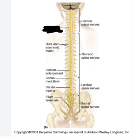
What does this figure show? What structure is associated with this?
This figure shows the dervish enlargement region. This is also where the brachial plexus is formed.
What is the brachial plexus and where is it formed?
A network of nerves from C5 to T1 that supplies the entire upper limb
Spinal nerve count mnemonic
8 Cats, 12 Tigers, 5 Lions, 5 Snakes, 1 Cobra
What is spina bifida?
A neural tube defect in which the vertebral arches fail to close completely during development, leaving part of the spinal, cord Unprotected.
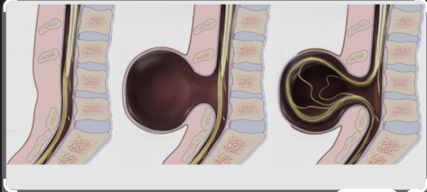
Label the spina bifida spectrum from left to right
Left: Spina Bifida Occulta, Middle: Spina Bifida Meningocele, Right: Myelomeningocele
What is the least severe form of spina bifida? How does it occur?
Occulta is the least severe. The vertebra is incomplete but the cord is fine. There is only a small tuft of hair or birthmark
What’s the slightly severe form of spina bifida?
Meningocele, sac is visible at birth. The cord is okay but the meninges herniate.
What is the most severe form of spina bifida?
Myelomeningcocele, usually lead to paralysis or hydrocephalus. Both the cord and meninges are herniated.