structure and function of the eye
1/22
There's no tags or description
Looks like no tags are added yet.
Name | Mastery | Learn | Test | Matching | Spaced | Call with Kai |
|---|
No analytics yet
Send a link to your students to track their progress
23 Terms
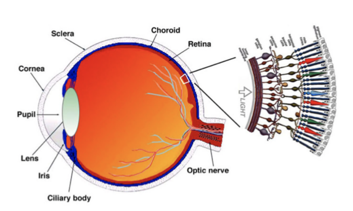
draw and label the eye ( include 9 labels )
briefly describe retinal development
retinal neurons are born in a sequential order then move into their proper positions and connect, forming synapses for vision.
(can also use model organisms to watch it happen) - dont memorise this bit
draw the retina
highlight each neuron type and support cell types
further label the layers and the direction of light
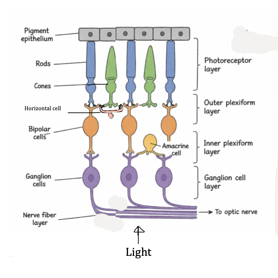
structure of photorecptor
what are the two types of photoreceptor
what do they do
what are each of their functions
rid and cone ( light sensitive cells of retina )
rod = low light ( dark ) ( so rods more snesitive )
cones = high light (bright) and colour
light enters through the inner segment of the cells
what type of synapse do retinal neurons use
ribbon synapse
= so neurotransmitters can be released easily ← dont memorise
what is the role of the disks in photoreceptors
active photoreceptors burn up disks throughout the day and they regenerate in the evening
bipolar cells
what does it mean by they can be on or off
what does it connect to
connect retina inner and outer layers
transmit signals from photoreceptor to retinal ganglion cells
classed as ON or OFF
ON = depolarised ( activated) with light
OFF = depolarise in dark
retinal ganglion cells ( 3 little points)
what is it located to etc
output neurons of retina
synapses with bipolar and amacrine cells
axons form optic nerve to brain
amacrine and horizontal cells
what do they do
excitatory or inhibitoy
what do they synapse with
modulate and integrate visual info
inhibitory neurons
horizontal cells between photoreceptor and bipolar cells ( outer retina)
amacrine cells between bipolar and ganglion cells ( inner retina)

why do we have muller glia ?
what demand is it for filling
lots of energy needed for visual processing → lots of input and waste
→ glial cells helps clean up waste by recycling neurotransmitters
what is the role of muller glia ( 4 points)
span entire retina (photoreceptors → ganglion cells)
part of tripartite synapses
convert glutamate → glutamine (detox + recycling)
return glutamine to presynaptic neurons
where is the region for high acuity vision
which photoreceptor do we associate with this area
fovea
( cones are here mostly rods in periphery)
means cones = high acuity vision
after activation do rods or cones hyperpolarise faster
cones
what is a receptive field
small area of retina affects one neuron’s firing
Example:
One Retinal ganglion cells might respond to light falling on 5–10 nearby photoreceptors.
Because many cells are close together, their receptive fields overlap
^^^ context dont memorise
what happens when receptive field overlap? ( 3 key things)
convergent excitation
Signals from multiple photoreceptors combine → signal stronger
surround inhibition
receptive field = centre + surround
centre excites
surround inhibits
lateral inhibtion
strongly activated photoreceptors inhibit nearby ones
this exaggerates differences between light and dark
why does it look like there's a white line between different shades of grey
slight overlap in lateral inhibition at those exact points so can't tell exactly the end point of each shade
^^ dont reallyyy need to memorise
all photoreceptors …. in responser to light
hyperpolarise
more about cones
how many types and what are they
what does this mean cones can do
3 types of cones , each more receptive to a specific wavelength of light
L ( long wavelength cones), M ( medium wavelength cones), S (short wavelength cones)
means they can tell colours
why do the l/m/s photoreceptors respond maximally to different wavelengths of light
they all have the same chromophore but opsins have slightly different amino acid sequences
what is the chromophore in photoreceptors
11 cis retinal ( -> all trans retinal)
( basically changes cis to trans when light is absorbed in eye) ←- dont need to memorise
explain the main cascade that leads to hyperpolarisation in photoreceptors.
good luck babe
to memorise:
Light activates rhodopsin → activates transducin → activates PDE → cGMP decreases → CNG Na⁺/Ca²⁺ channels close → reduced Na⁺ influx → hyperpolarisation
concept behind it explained:
Photoreceptors naturally depolarised because CNG (cyclic nucleotide-gated) channel open allows Na+ and Ca2+
This is kept open by cGMP
Now light comes in…
activates rhodopsin ( because goes from cis-trans) -> activates transductin
-> this decreases cGMP so now it cant keep CNG Na+ and Ca2+ channels open :(
The reduction of Na+ and Ca+ make photoreceptor more negative = HYPERPOLARIZE
explain the mini cascade that keeps the CNG channel open
Ca2+ inhibits GCAP which activates GC which activates cGMP
which types of bipolar cell so rods and cones synapse with
rods - on only
cones - both