Axial Skeleton (Lab 3)
1/33
There's no tags or description
Looks like no tags are added yet.
Name | Mastery | Learn | Test | Matching | Spaced | Call with Kai |
|---|
No analytics yet
Send a link to your students to track their progress
34 Terms
Describe the composition and role of the axial skeleton
It is composed of the skull, vertebrae, ribs, and sternum.
It protects the central nervous system within the cranial and vertebral cavities, protects the heart and lungs within the thoracic cage, and facilitates ventilation of the lungs.
Describe the curvature of the vertebral column from a posterior perspective
The cervical and lumbar curvatures are concave, while the thoracic and sacral curvatures are convex
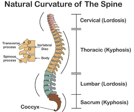
How many vertebrae make up the vertebral column
About 33 vertebrae

#1: Name and function
Body
The weight-bearing region of the vertebra. Vertebral bodies become more substantial in the lower regions of the vertebral column and bear more of the body’s weight

#2: Name and function
Vertebral foramen
Location of the spinal cord

#5 and #7: Names and function
#5 is the spinous process and #7 is the transverse process
Both are sites of muscle attachments
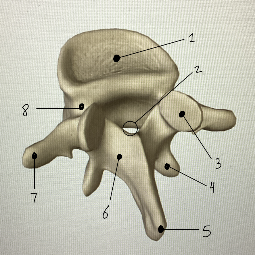
#3 and #4: Names and function
#3 is the superior articular process, and #4 is the inferior articular process
Both form joints with those of adjacent vertebrae. They are responsible for restricting the movements of the vertebral column

#8: Name and function
Pedicle
A stout connection between the body and the rest of the vertebra

#6: Name and function
Lamina
A flat region that connects the spinous process to the rest of the vertebra
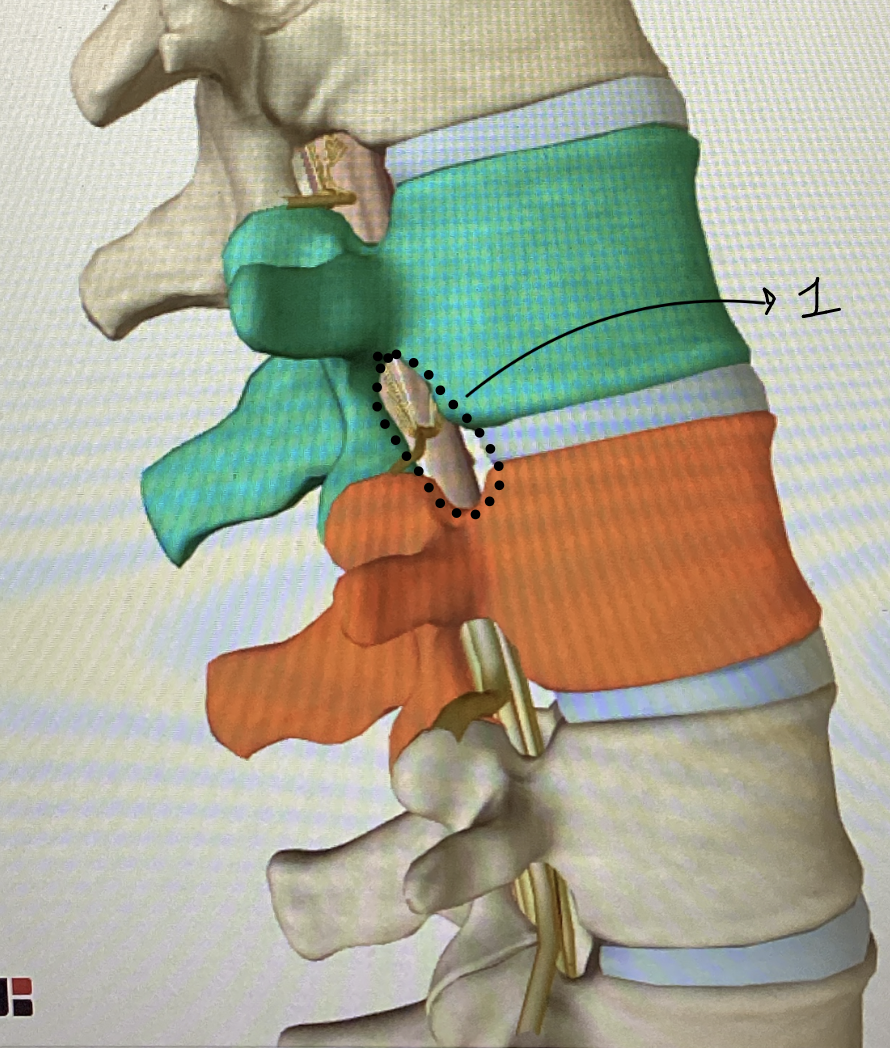
#1: Name and function
Intervertebral foramina
Above and below the pedicle. Notches allow for the passage of spinal nerves. Two adjacent vertebrae for one intervertebral foramen.
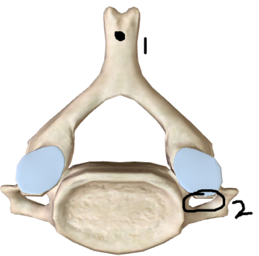
What type of vertebrae is this? Identify each point
This is a typical cervical vertebra (C3-C7). Features unique to these vertebrae are the bifid (forked) spinous process (#1) and the transverse foramen, which allow the passage of the vertebral artery to the brain (#2)
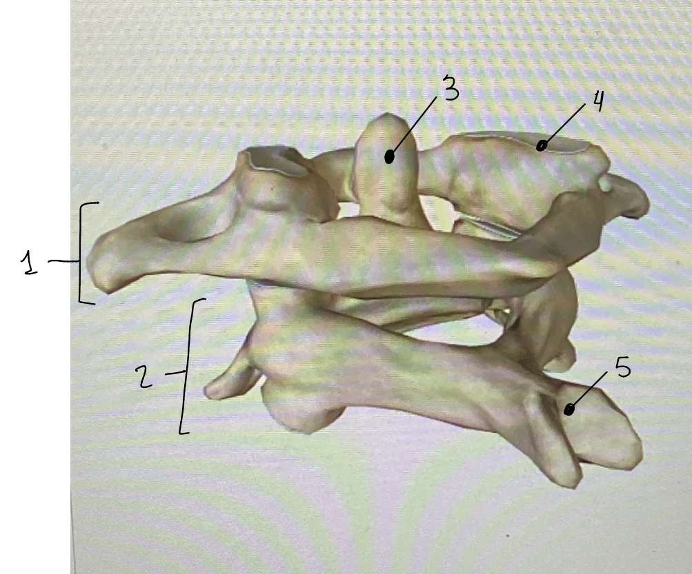
Identify all
#1: C1 (Atlas)
#2: C2 (Axis)
#3: Dens - A projection of the axis that prevents the atlas from slipping posteriorly
#4: Atlanto-occipital joint - Joint formed between the occipital bone of the skull and the superior articular process of C1. Allows you to nod your head “yes.”
#5: Bifid (forked) spinous process - typical of most cervical vertebrae
Which of the cervical vertebrae is unique?
C7 is unique because of its long spinous process that can be easily palpated. It is often called vertebra prominens.
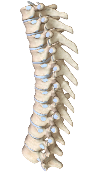
What type of vertebrae are these? How do you know?
These are thoracic vertebrae
The long, downward-sloping spinous processes indicate that these are thoracic vertebrae
***The most distinguishing characteristic of the 12 thoracic vertebrae is their articulation with the ribs
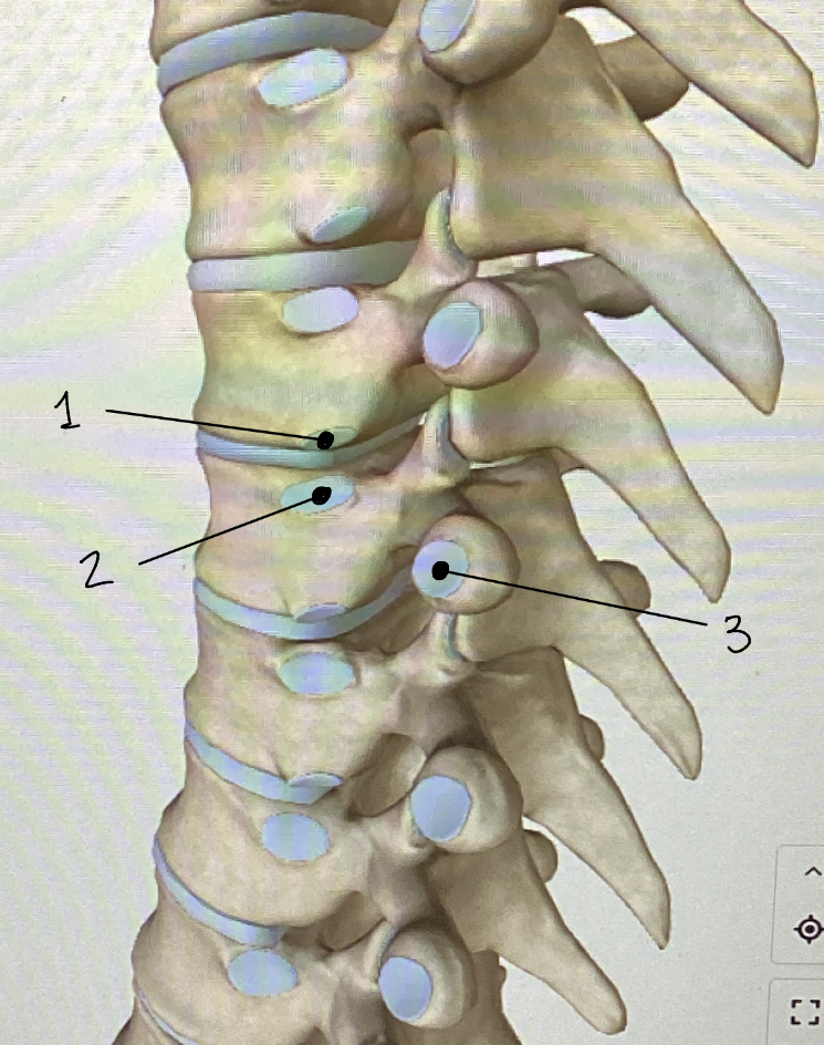
What type of vertebrae are these? Identify each point
These are thoracic vertebrae
#1: Inferior costal facet - point of articulation with the ribs
#2: Superior costal facet - point of articulation with the ribs
#3: Transverse costal facet - indicate where the transverse processes form a joint with the rib
***There are 6 costal facets on each thoracic vertebra. 2 of each of the above
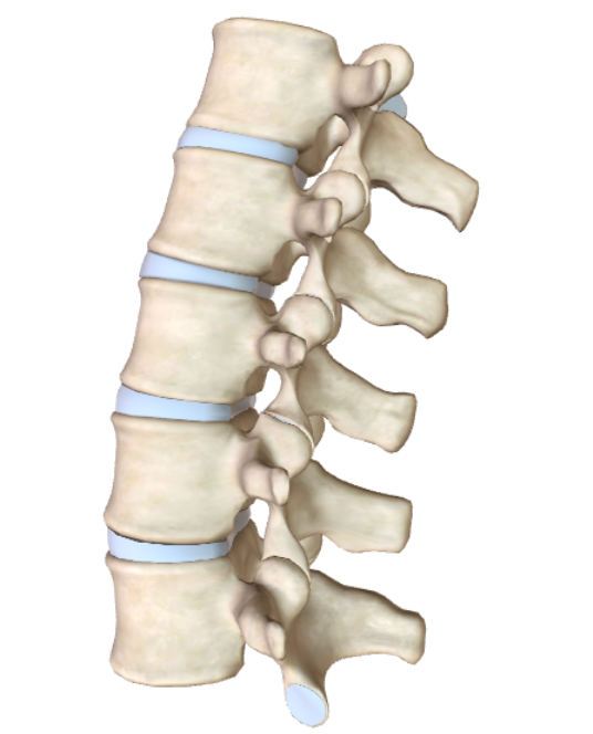
What type of vertebrae are these? Describe them.
Lumbar vertebrae
They have the largest vertebral bodies (because they hold the most body weight). They have no articulation with the ribs. They have strong, stout transverse and spinous processes.
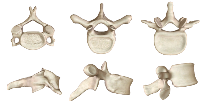
Identify each vertebrae
Left: Cervical
Middle: Thoracic
Right: Lumbar

#1: Name and function
Superior articular process
Articulates with the inferior articular process of L5

#2: Name and function
Median sacral crest
Is formed by the fusion of spinous processes
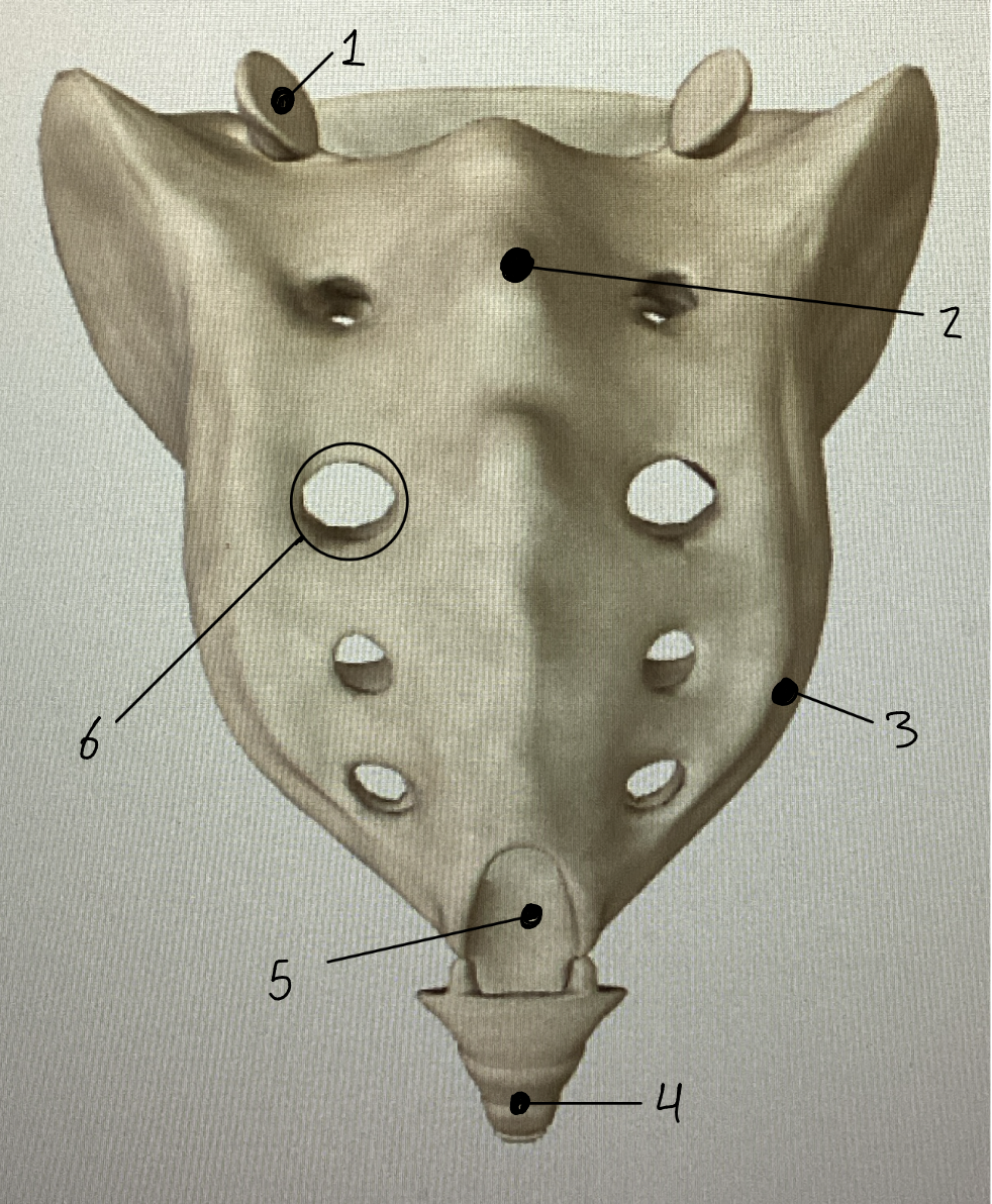
#3: Name and function
Ala (singular), Alae (plural)
The “wings” of the sacrum are the lateral margins, formed from fused transverse processes

#4: Name and function
Coccyx
The “tailbone” is formed by the fusion of 3-4 coccygeal vertebrae

#5: Name and function
Sacral canal
The sacral canal is the continuation of the vertebral canal. It houses the spinal nerve roots of the cauda equina.

#6: Name and function
Posterior sacral foramen
The dorsal rami of the spinal nerves leave the sacral canal through 4 pairs of posterior sacral foramina
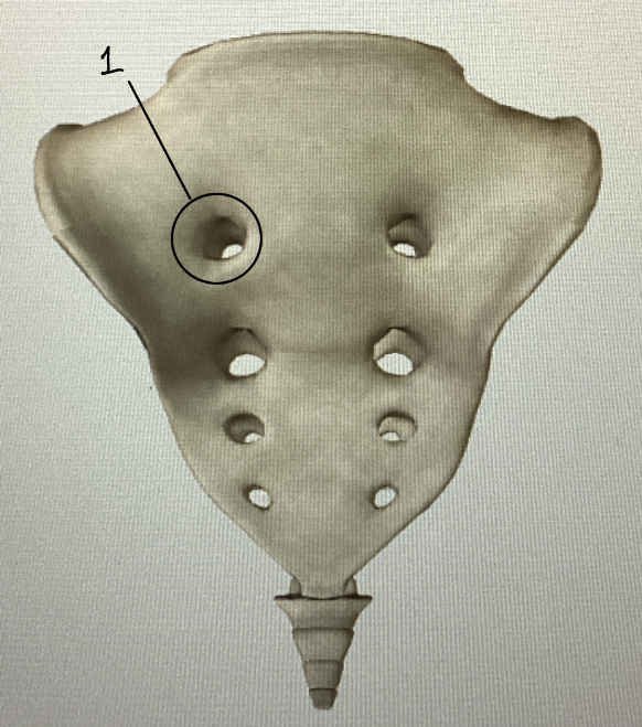
#1: Name and function
Anterior sacral foramen
The ventral rami of spinal nerves leave the sacral canal through 4 pairs of anterior sacral foramina
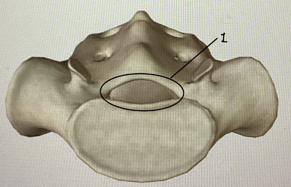
#1: Name and function
Sacral canal
The sacral canal is the continuation of the vertebral canal, and it leads into the sacral canal. It houses the spinal nerve roots of the cauda equina
Role of the sternum?
The sternum is commonly called the breastbone. It is the central bone in the anterior chest wall that anchors the ribs. It is composed of the manubrium, body of the sternum, and the xiphoid process. The sternal angle is the line of fusion between the body and the manubrium.
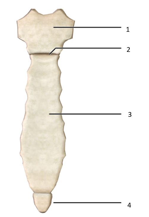
What bone is this? ID each part
This is the sternum
#1: Manubrium
#2: Sternal angle
#3: Body of sternum
#4: Xiphoid process
True vs false ribs
True ribs: Ribs 1-7 articulate directly (via their costal cartilage) with the sternum
False ribs: Ribs 8-12 do not directly attach to the sternum. Some connect to other costal cartilages, while others do not connect to the sternum at all (these are called floating ribs (ribs 11 and 12)).
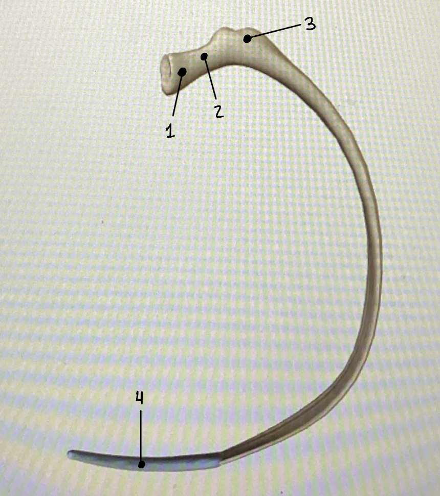
What bone is this? ID each part
This is a rib
#1: Head of rib - articulates with the costal facet of a thoracic vertebral body
#2: Neck of rib
#3: Tubercle of rib - articulates with the costal facet of a thoracic vertebra’s transverse process
#4: Costal cartilage - The ribs terminate at the costal cartilages, which span the gap to the sternum
Name these joints (and function)
#1: Atlanto-occipital joint - A condyloid joint. Movement between the occipital bone of the skull and the superior articular process of C1 is in the sagittal plane (shaking head “yes”)
#2: Atlanto-axial joint - This is a two-part joint. The dens and atlas form a pivot joint, and the articular processes form a plane joint. Movements are rotational. (shaking head “no”)
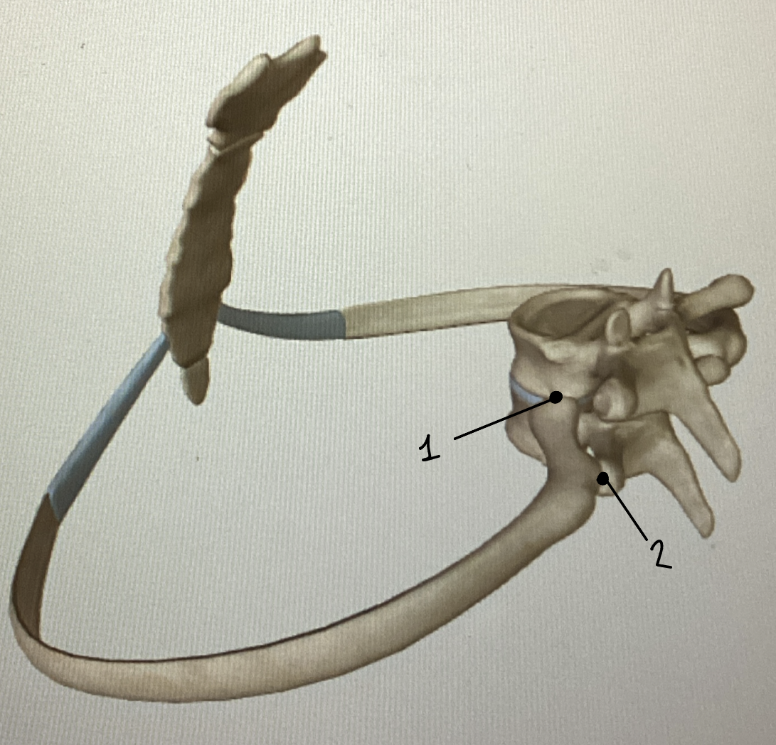
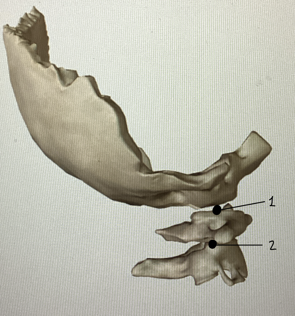
Name these joints (and functions)
#1: Costovertebral joint - The joint formed between the head of the rib and the superior and inferior costal facets
#2: Costotransverse joint - The joint formed between the tubercle of the rib and the transverse costal facet
***Both are plane joints that permit the ribs to be elevated and depressed
Name these joints (and functions)
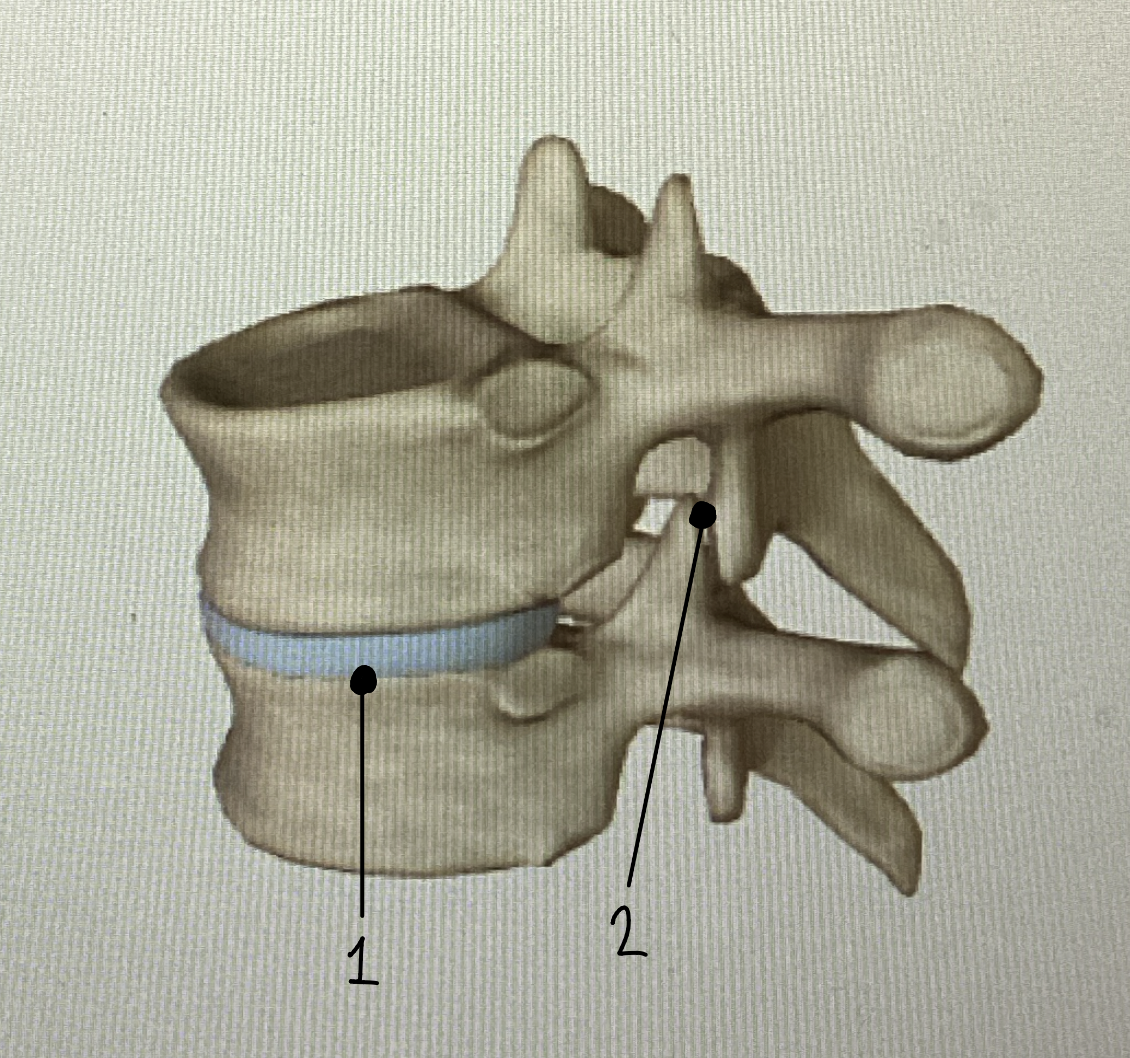
#1: Intervertebral joint (cartilagenous) - The intervertebral disc is a cartilage pad that forms a cartilaginous joint. These joints are found between the bodies of all non-fused vertebrae
#2: Intervertebral joint (synovial) - Formed between the superior and inferior articular processes, these intervertebral joints permit gliding motions.
***These are plane joints
Name this joint (and function)
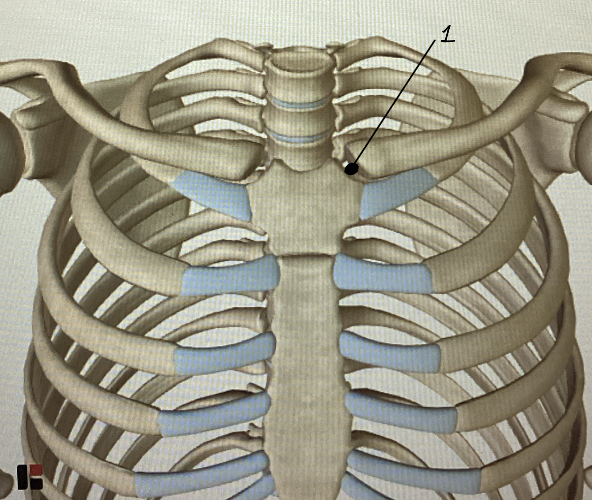
Sternoclavicular joint: This is where the sternum meets the appendicular skeleton at the clavicle. It is a saddle joint. It permits the lateral end of the clavicle to move up and down (aka shrug your shoulders)
Name this joint (and function)
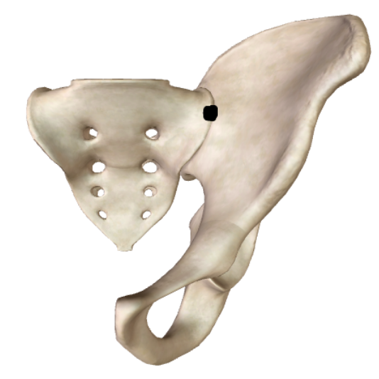
Sacroiliac joint: Formed between the alae of the sacrum and the ilium of the pelvis. Allows for the efficient transfer of weight from the body to the legs.