respiratory system :Larynx, Trachea and Lungs
1/49
There's no tags or description
Looks like no tags are added yet.
Name | Mastery | Learn | Test | Matching | Spaced | Call with Kai |
|---|
No analytics yet
Send a link to your students to track their progress
50 Terms
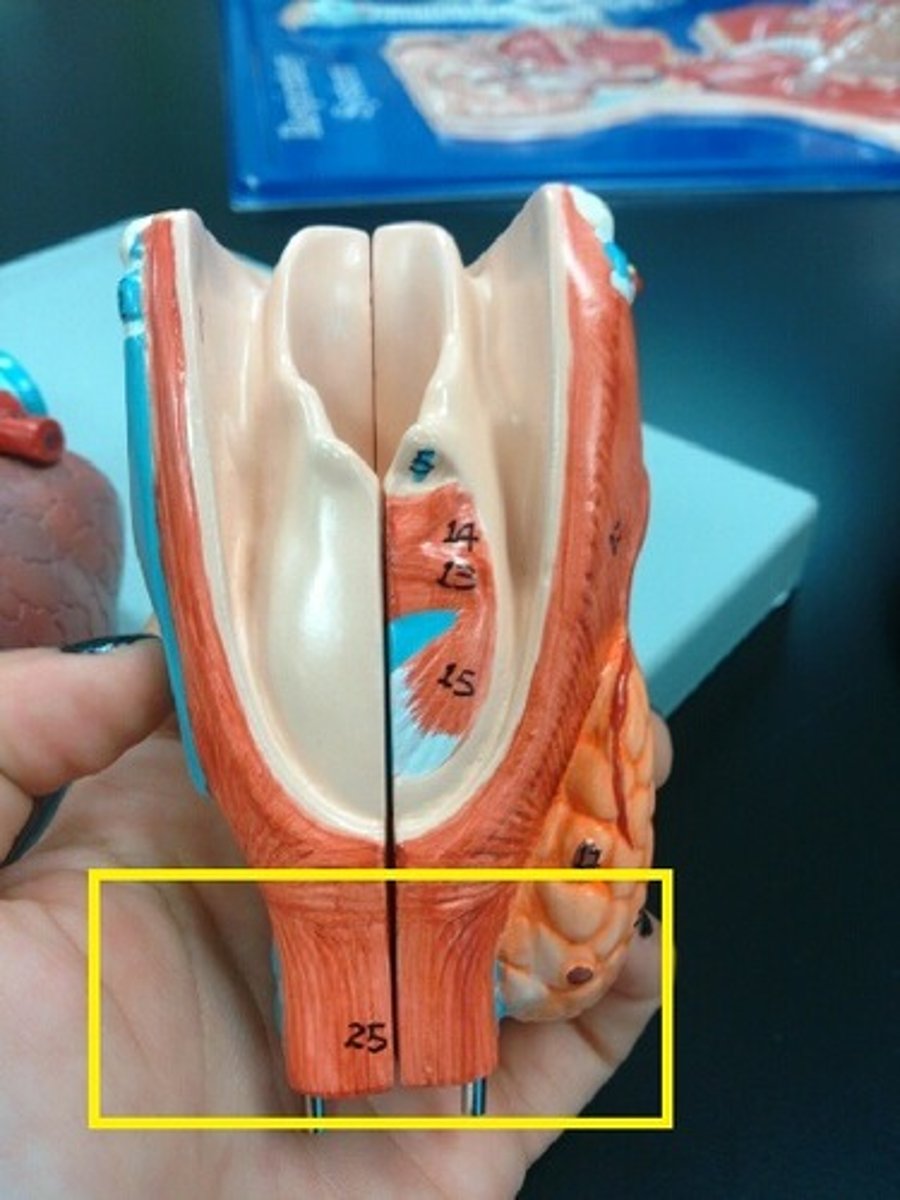
The Larynx
voice box; passageway for air moving from pharynx to trachea; contains vocal cords
opposite the 3rd, 4th, 5th and 6th cervical vertebrae
ts lower end is continuous with ithe trachea at the level of the sixth cervical vertebra.
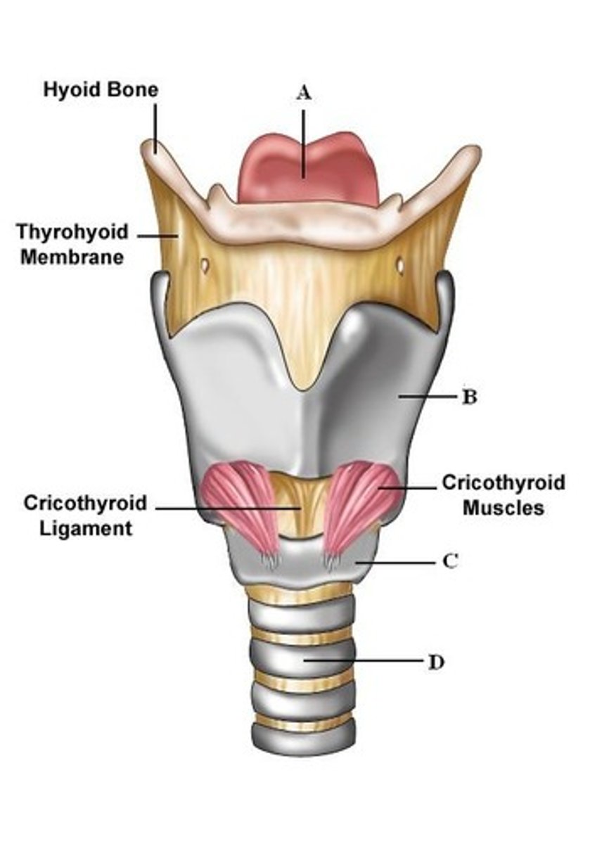
Larynx boundaries
epiglottis , cricoid cartilage.
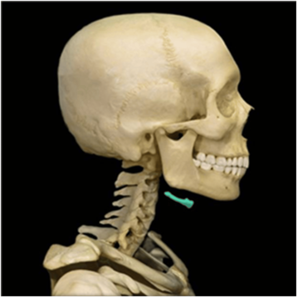
laryngeal inlet., hyloid bone,
hyloid bone,
attachs to the temporal bones and supports the base layer of the tounge
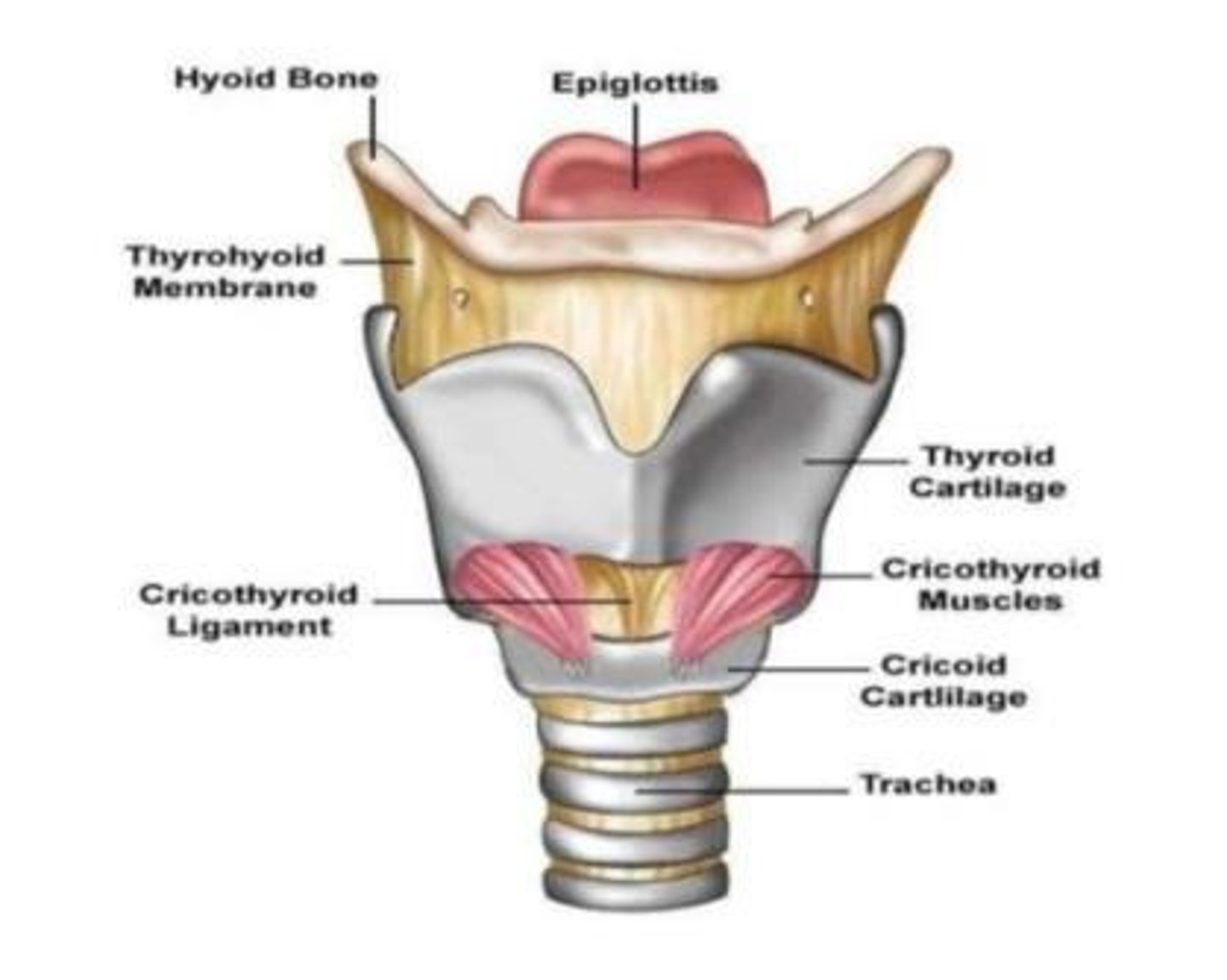
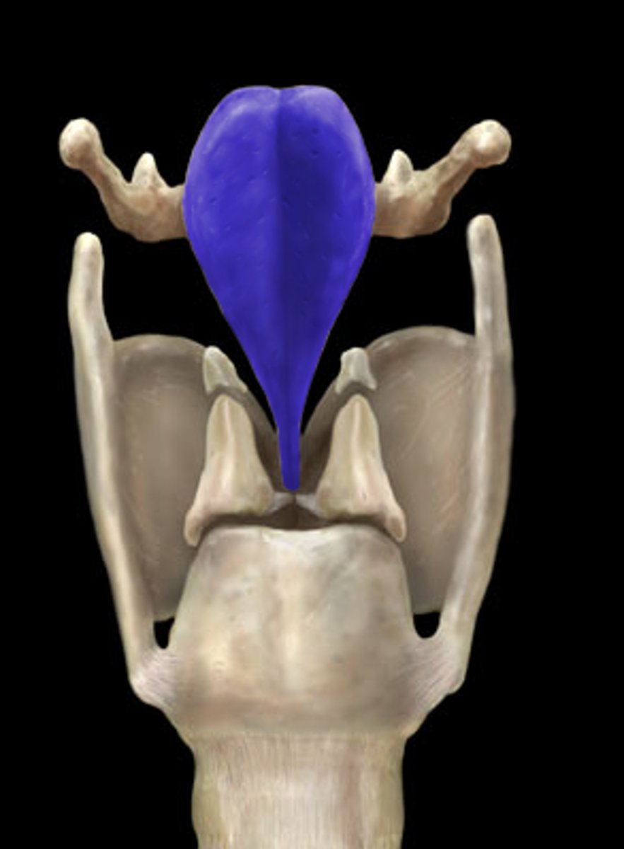
epiglottis
A flap of tissue that seals off the windpipe and prevents food from entering.
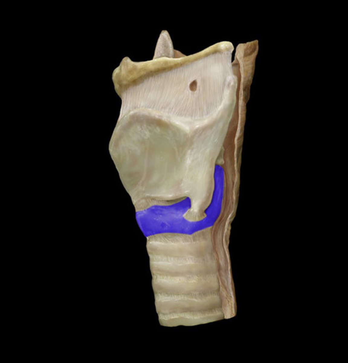
cricoid cartilage.
the ring-shaped structure that forms the lower portion of the larynx
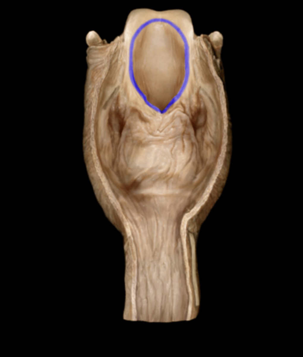
laryngeal inlet.
opening that connects the pharynx and larynx
laryngeal cartilages types
Single:
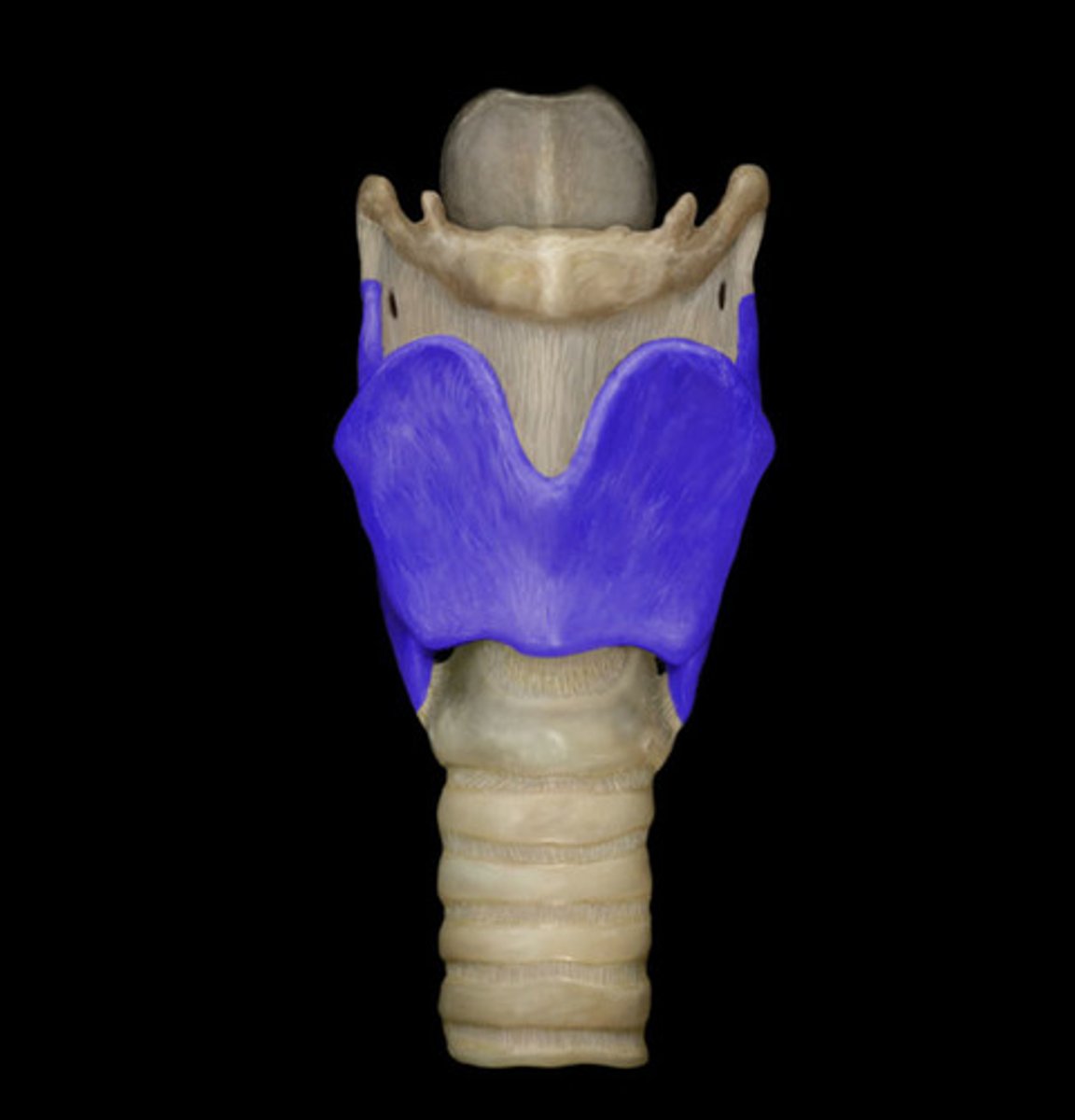
Thyroid cartilage.
Cricoid cartilage.
Epiglottis cartilage.
Paired:
Arytenoid cartilages.
Corniculate cartilages.
Cuneiform cartilages.

Thyroid cartilage.
A firm prominence of cartilage that forms the upper part of the larynx; the Adam's apple.
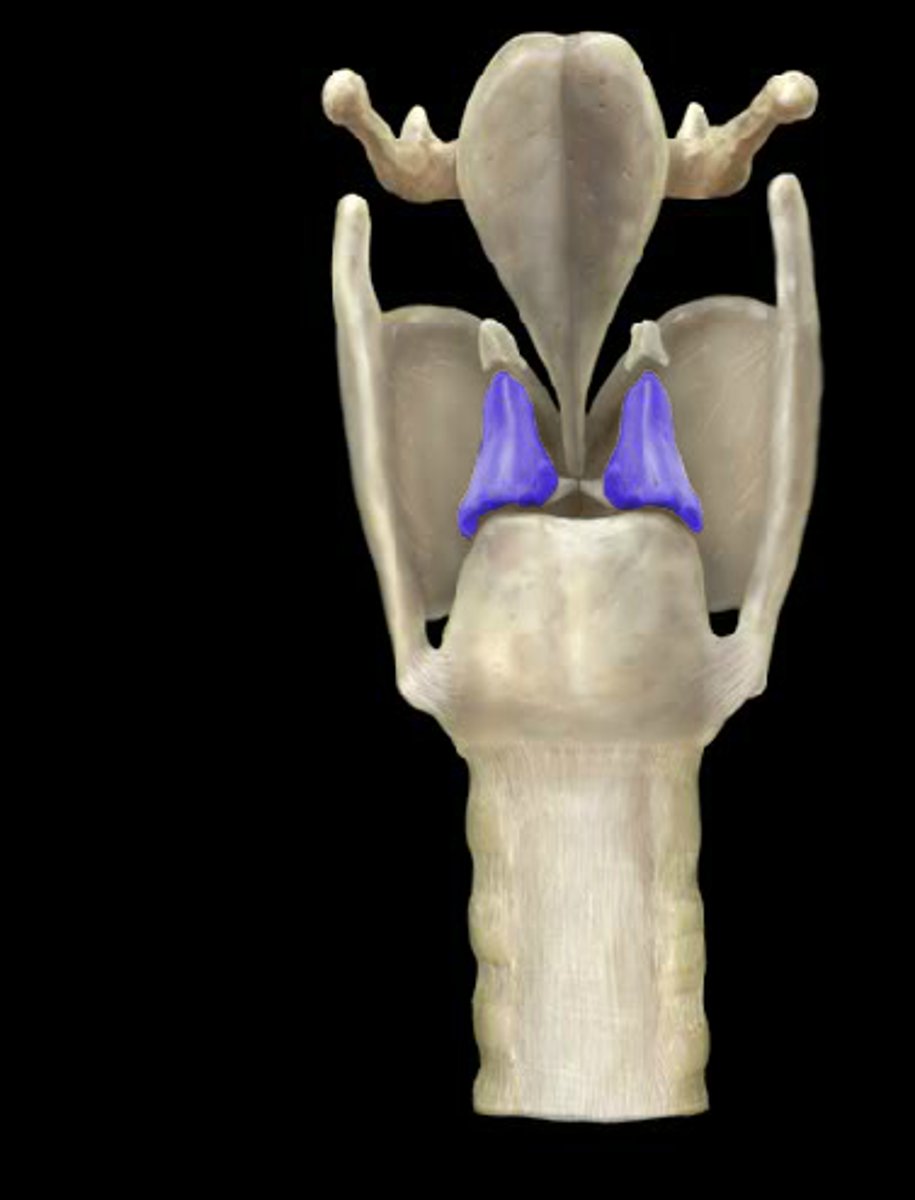
Arytenoid cartilages.
Pyramid-like cartilaginous structures that form the posterior attachment of the vocal cords.
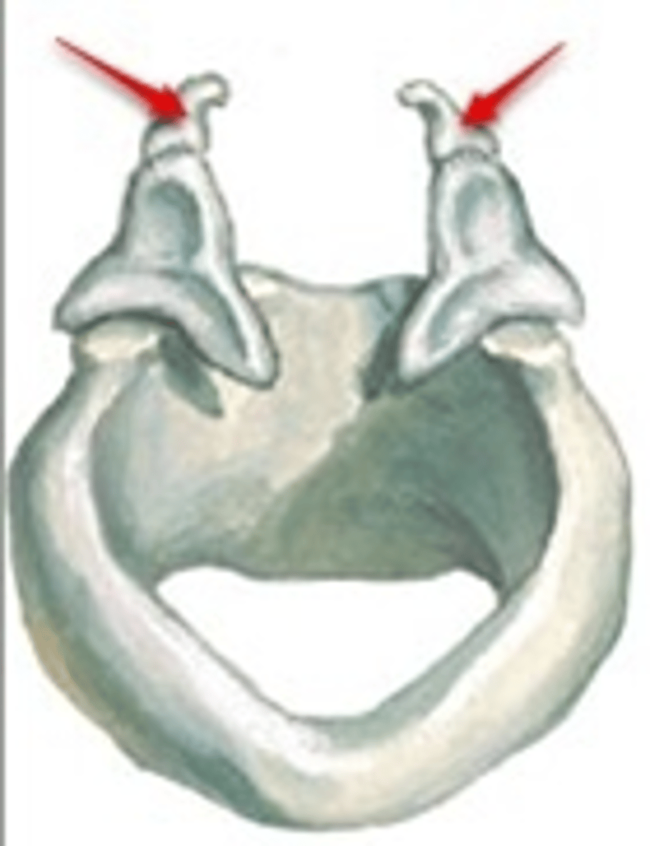
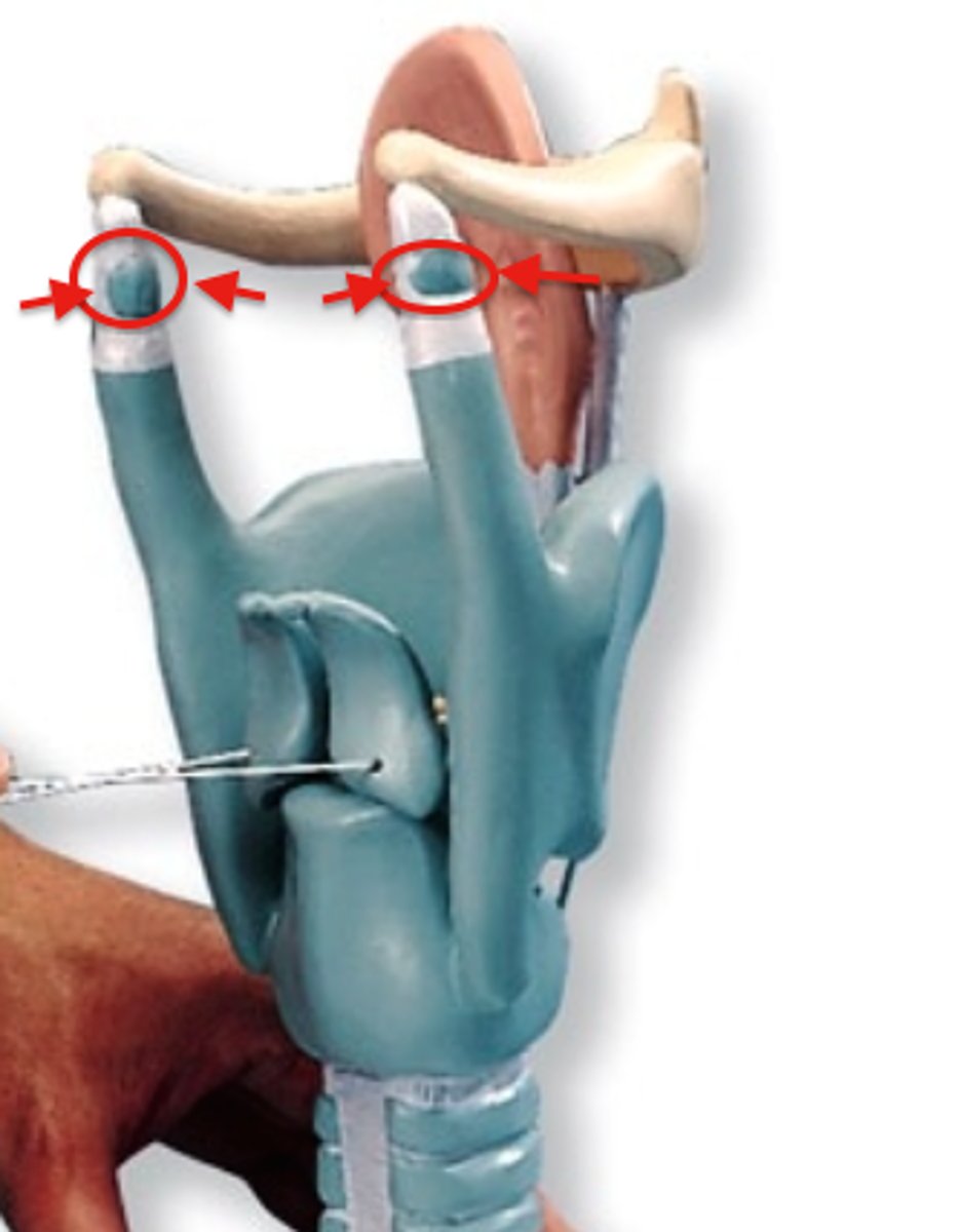
Corniculate cartilages.
attached to arytenoid cartilages like a pair of little horns
Cuneiform Cartilages
support soft tissue between arytenoids and epiglottis
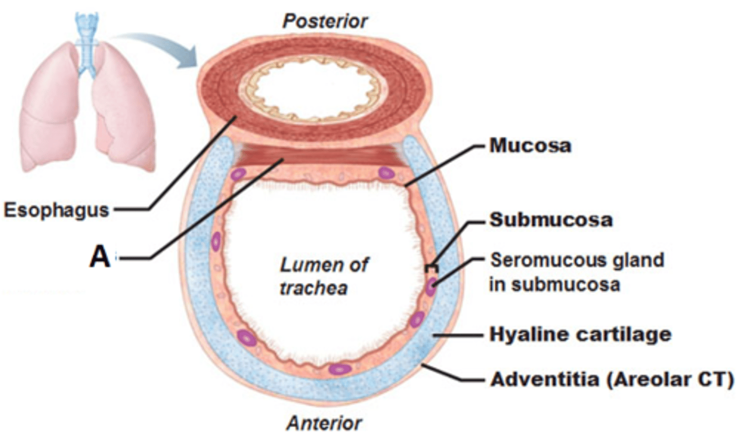
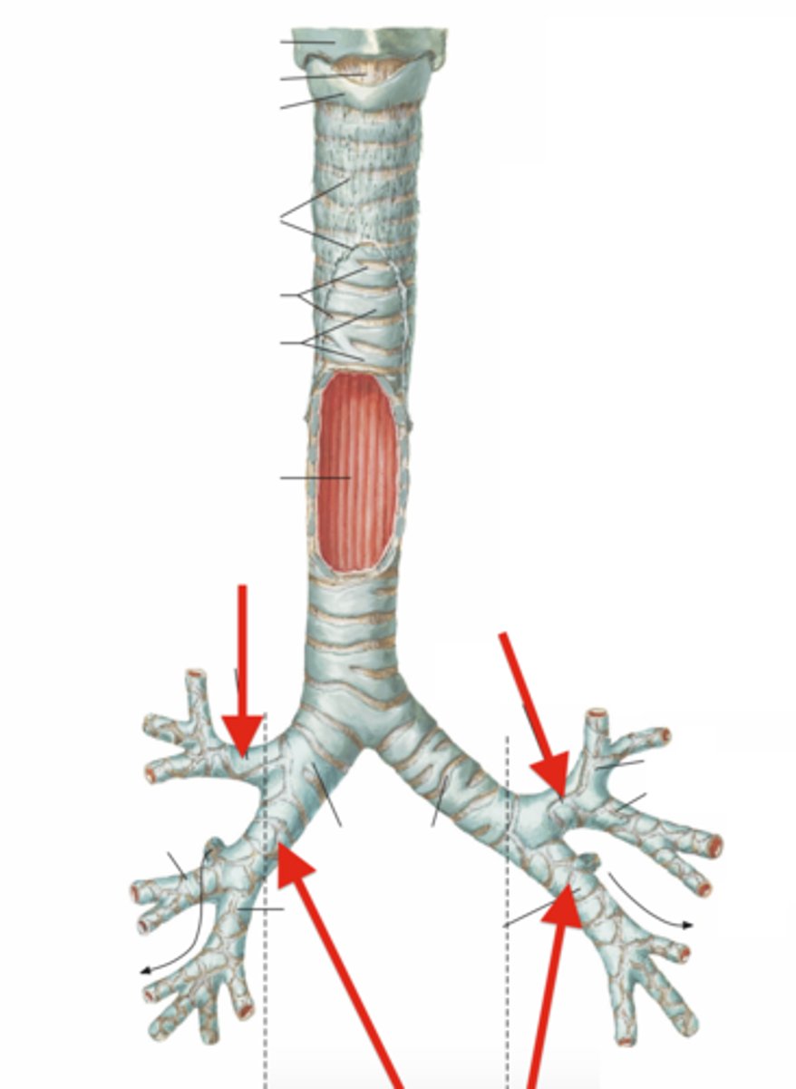
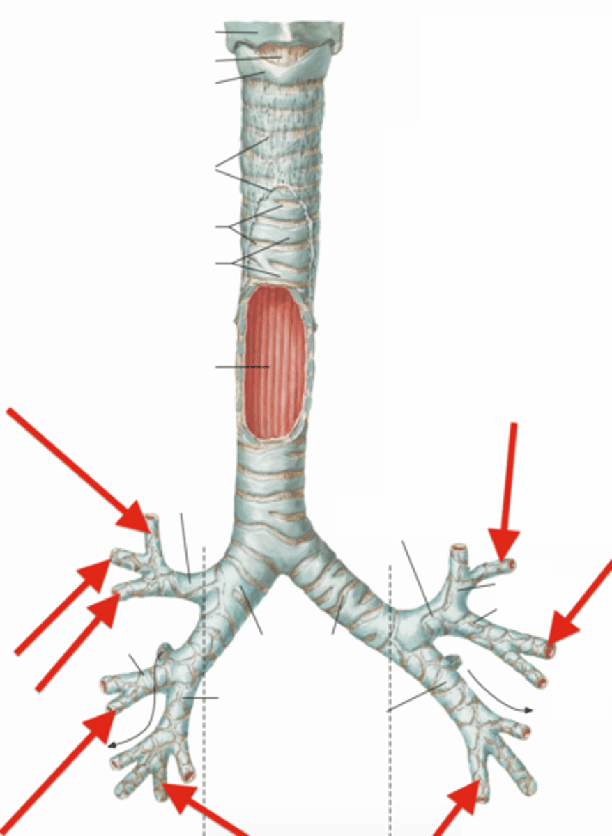
Trachea:
Allows air to pass to and from lungs. It is called also the windpipe.
C-shaped hyaline cartilage (non-collapsible).
It is lined with pseudo-stratified columnar ciliated epithe
Both ends of the cartilage is connected with smooth muscle which is called trachealis.
trachealis muscle
Consists of smooth muscle fibers that connect posterior parts of cartilage rings
Contracts during coughing to expel mucus
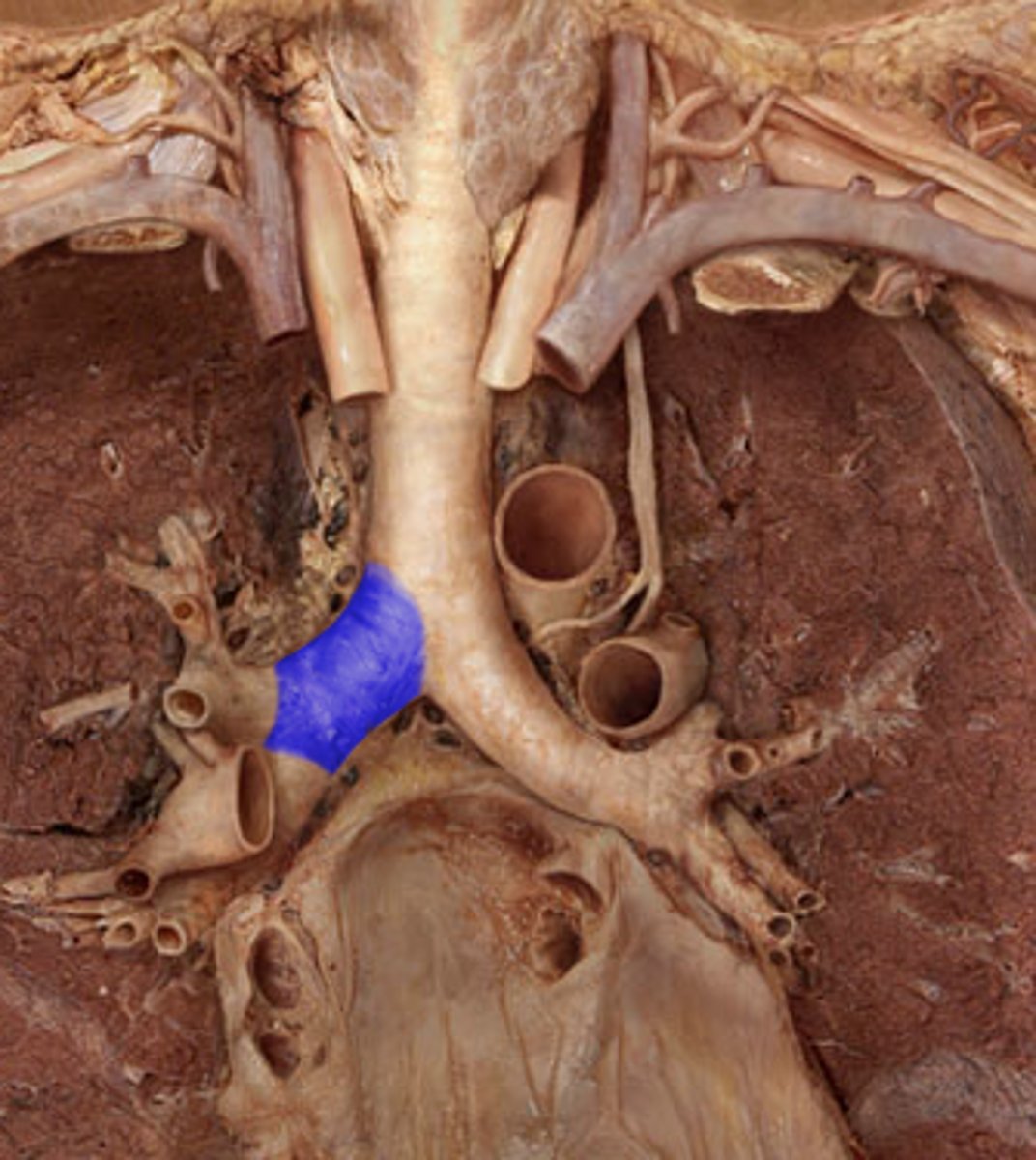
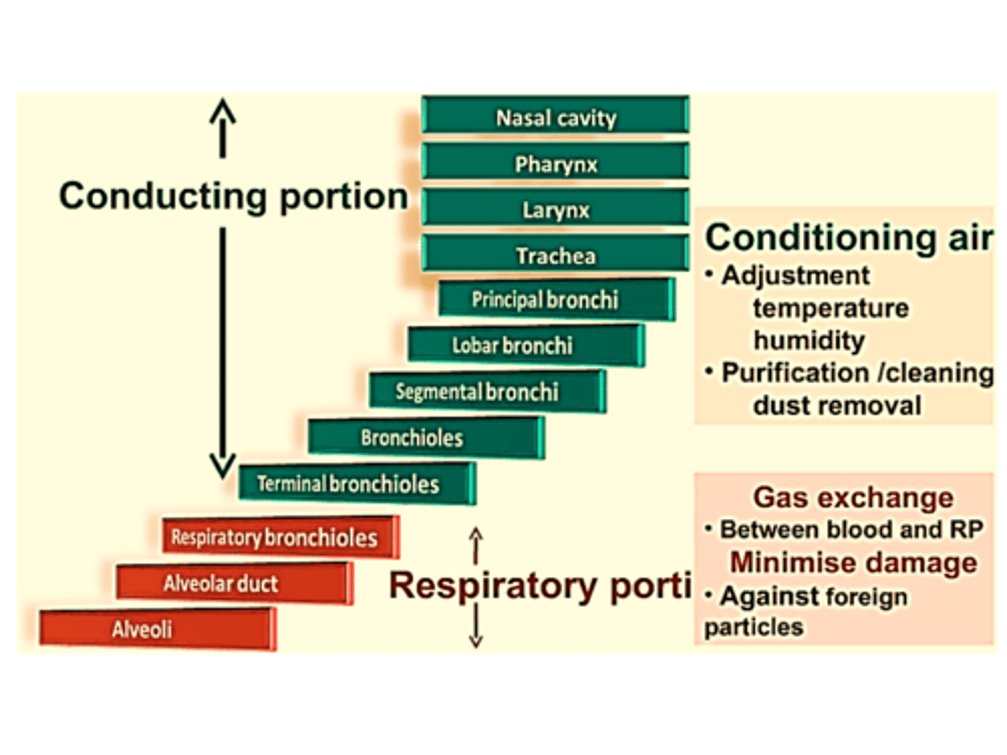
Bronchial tree:
branched airways that lead from the trachea to the microscopic air sacs called alveoli.
bronchi which are extra-pulmonary. (anything situated, occurring, or originating outside the lungs)
primary bronchus will divide into lobar (secondary) bronchi,
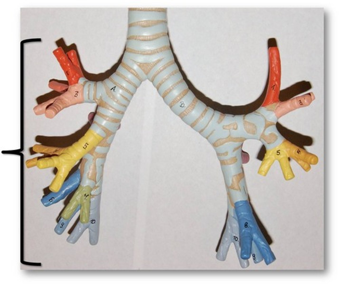
primary bronchus
a pair of branches of the trachea that lead to the right and left lung; consist of incomplete rings of cartilage and are lined by pseudostratified ciliated columnar epithelium
lobar (secondary) bronchi
branch from main bronchi; 2 on left, 3 on right
will divided into multiple segmental (tertiary) bronchi.
segmental (tertiary) bronchi.
supported by crescent-shaped cartilage plates
10 on right, 8 on left
Air passage ways connecting trachea with alveoli.
Warms and moistens incoming air.
Lungs
two spongy organs, located in the thoracic cavity enclosed by the diaphragm and rib cage, responsible for respiration
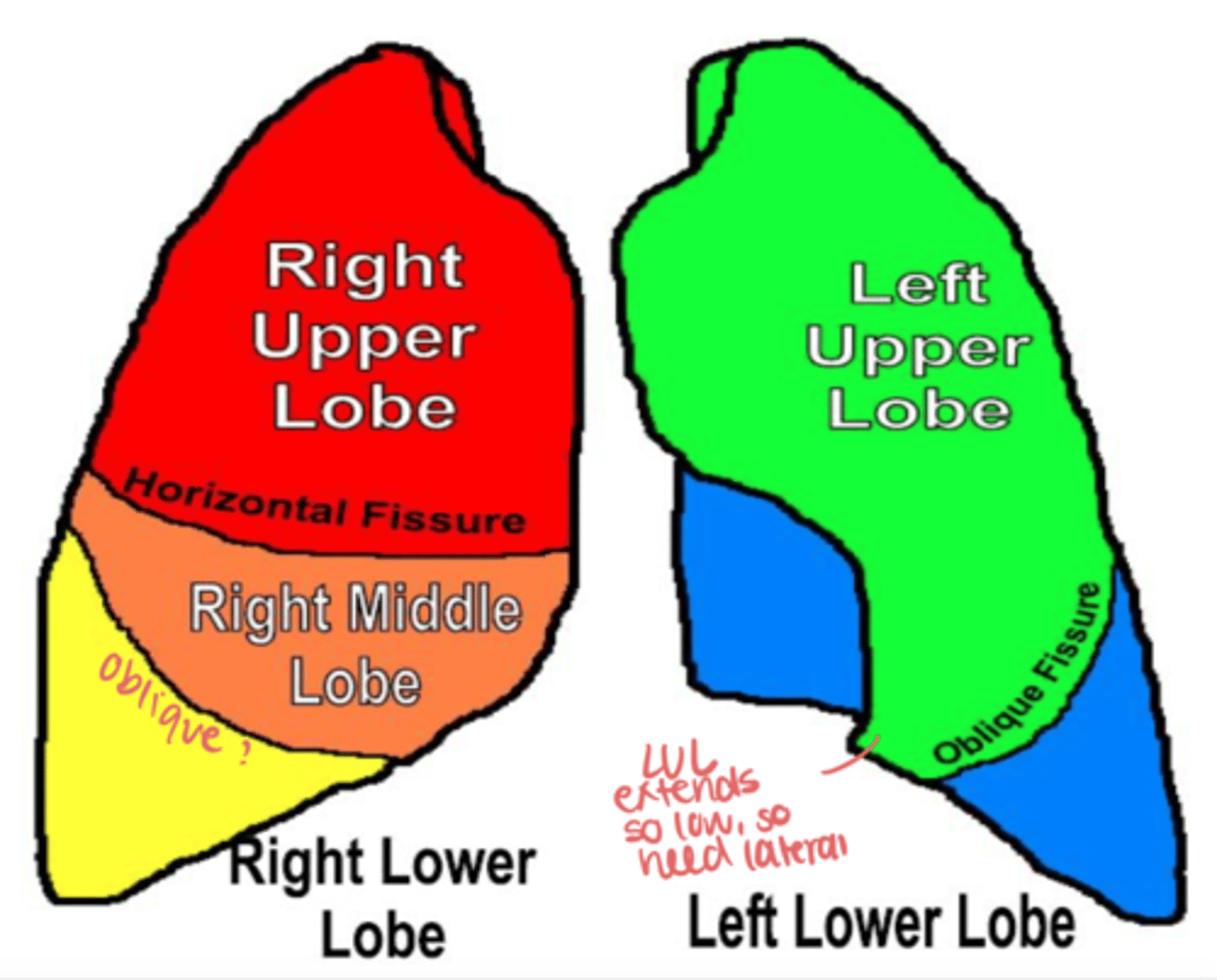
right lung
3 lobes (superior, middle, inferior) 600 gm, is broader and shorter, pushed up by the liver.
has two main fissures—the oblique (major) fissure and the horizontal (minor) fissure.
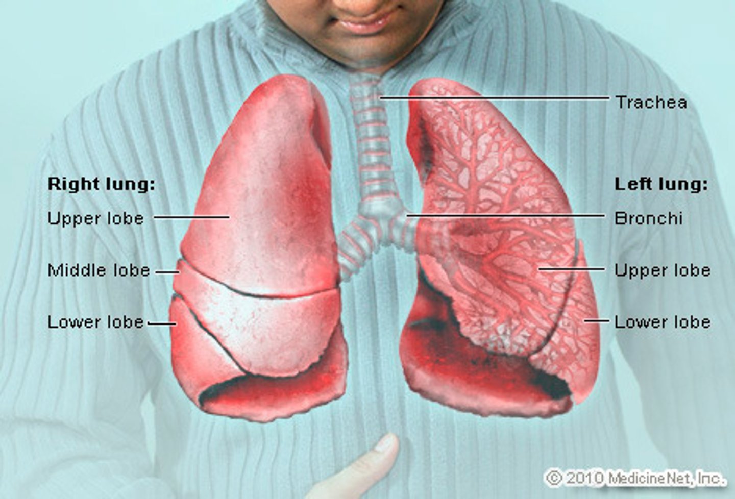
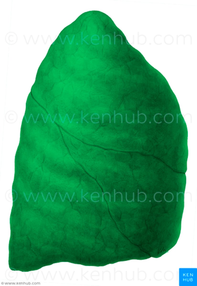
left lung
2 lobes: superior and inferior. 550 gm, is longer and narrower
has one main fissure, the oblique (major) fissure.
a prominent cardiac notch, and the lingula

cardiac notch
a concave space on the left lung in which the heart lies
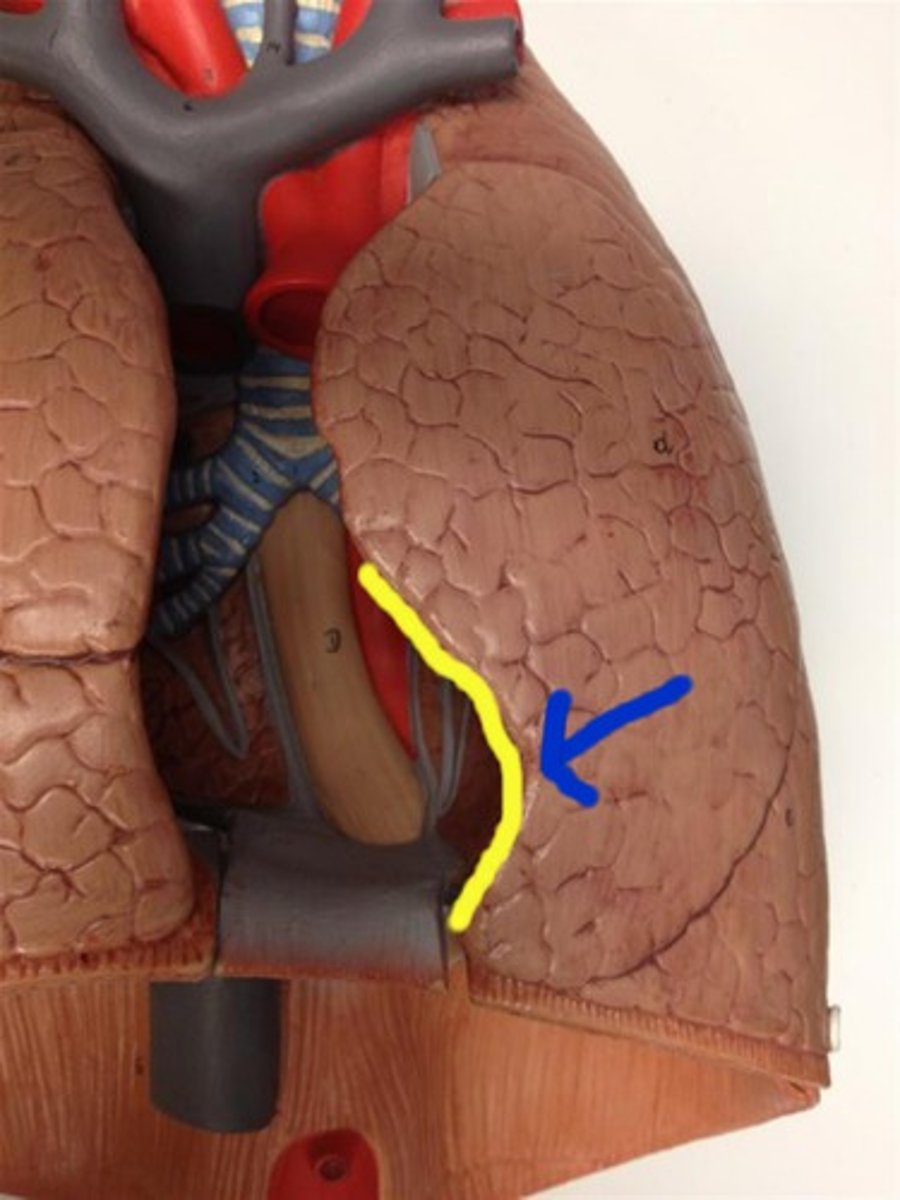
Lingula
The region of the left lung that corresponds with the right middle lobe.
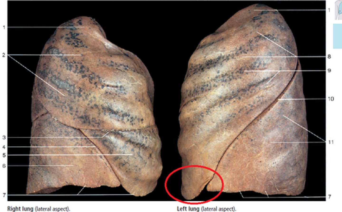
oblique (major) fissure
Divides upper and lower lung portions.

horizontal (minor) fissure
Divides right upper and middle lobes.
Borders of lungs
anterior, posterior, inferior
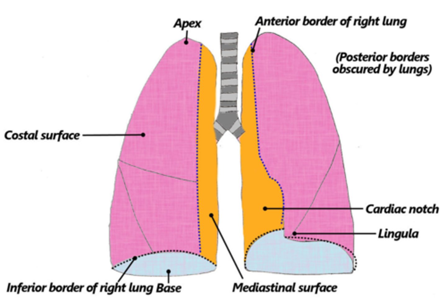
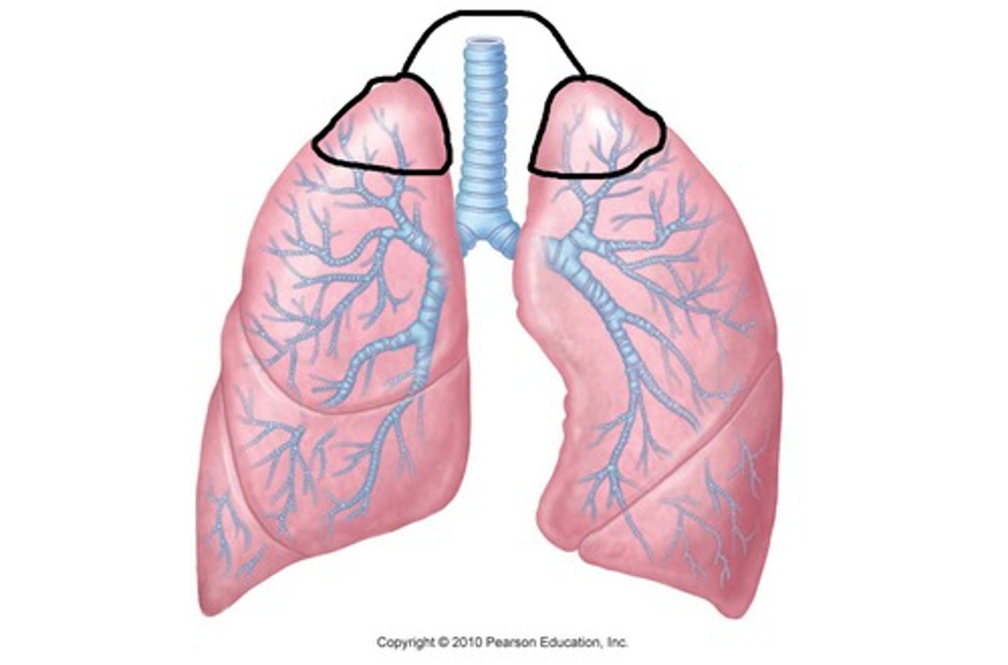
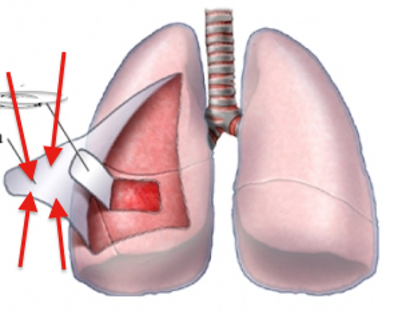
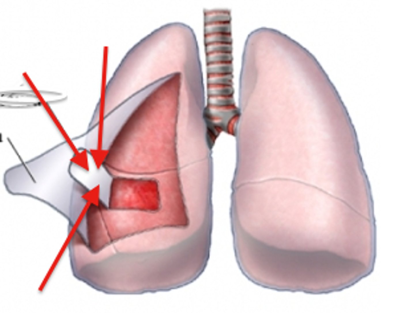
apex of the lung
tip or uppermost portion of the lung.
Area above the impression of Ist rib
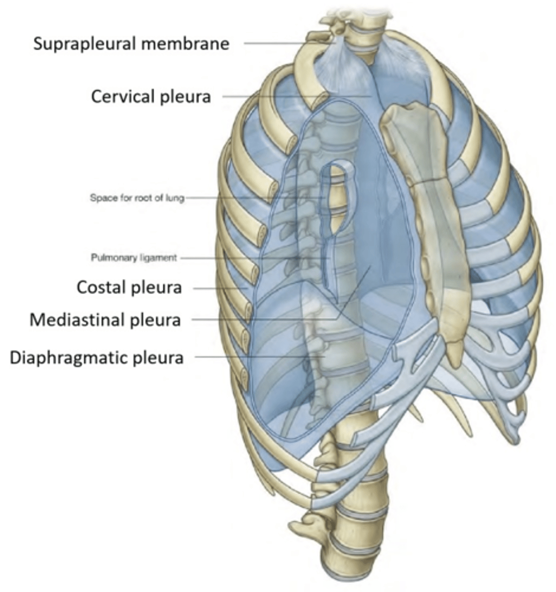
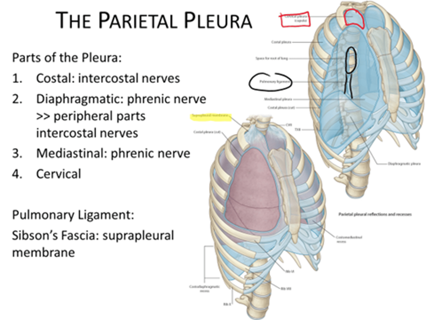
It is covered by the cervical pleura above which lies the suprapleural membrane.
cervical pleura
-extends through the superior thoracic aperture into the root of the neck
-forms a cup-shaped pleural dome over the apex of the lung
suprapleural membrane.
The thickening of the fascia over the apex of the lung
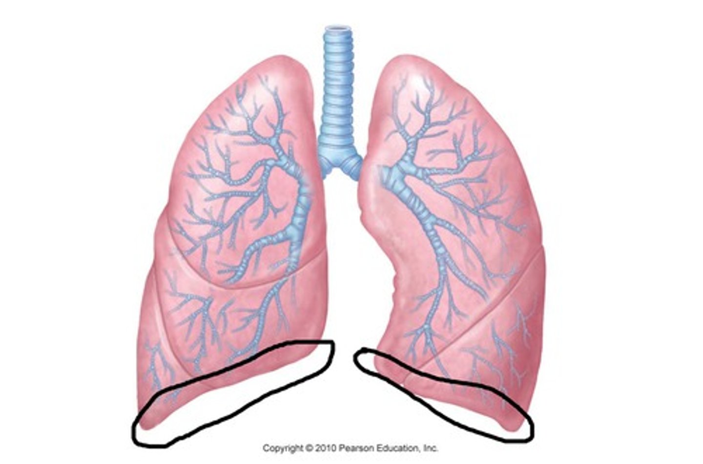
base of the lung
lowest part of the lung, resting on the diaphragm
Right side- right lobe of liver
Left side- left lobe of liver, fundus of stomach and spleen.
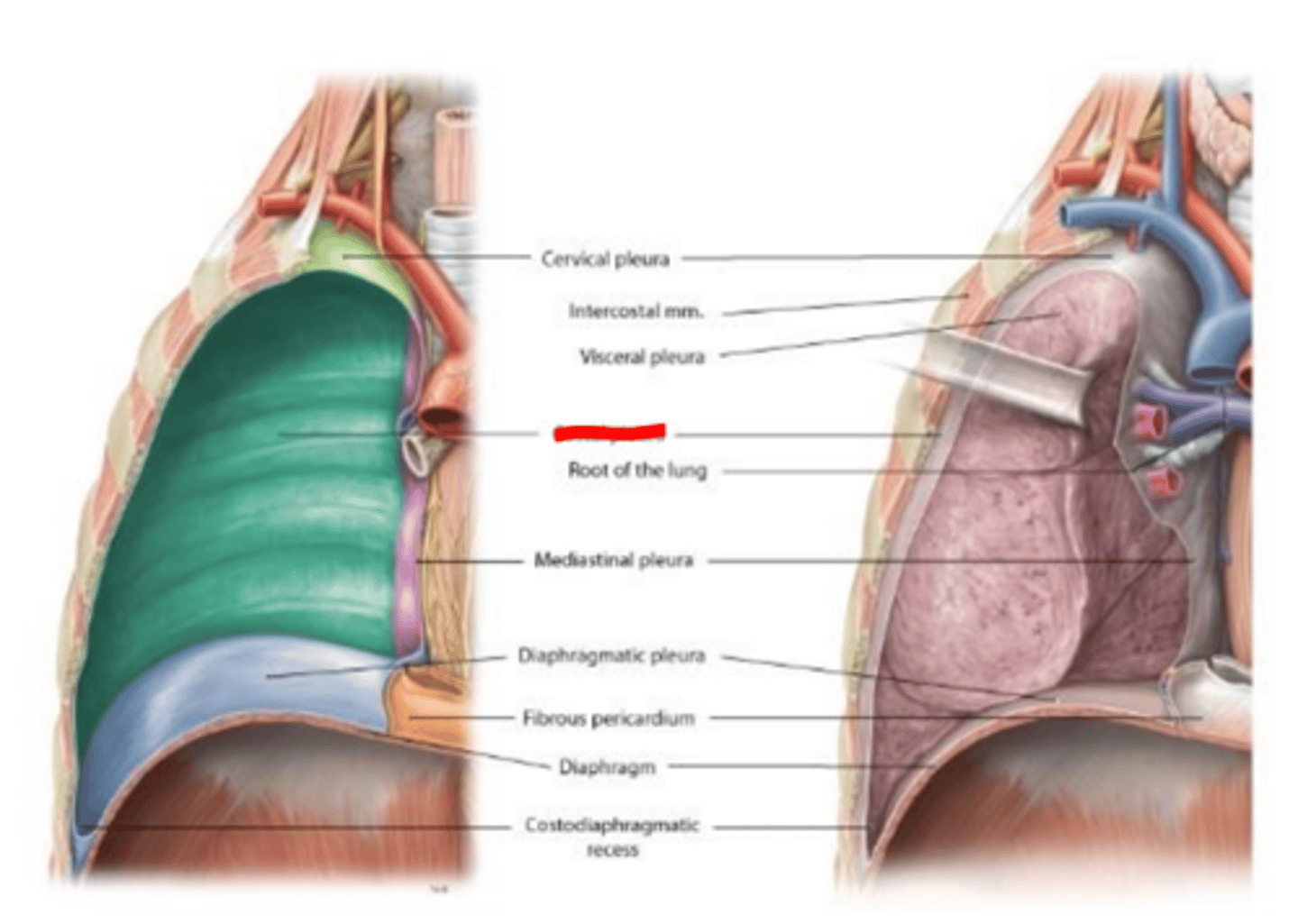
costal surface of the lung
pressed against the ribcage
Related to lateral thoracic wall.
Separated by costal pleura & endothoracic facia
Upper 6 ribs in midclavicular line, 8 in the midaxillary line, and 10 in midscapular line
costal pleura
covers the inner surface of the rib cage
endothoracic fascia
CT layer separating the parietal (costal) pleura from the internal surface of the thoracic wall.
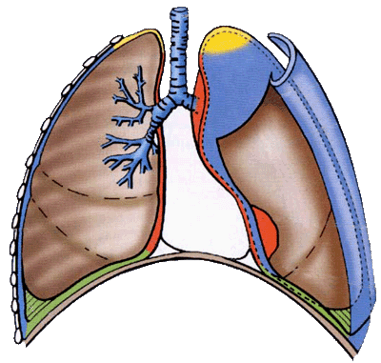
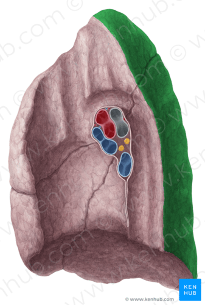
medial surface of lung
the concave, vertically oriented inner surface facing the mediastinum and heart. It contains the lung hilum
Forms root of the lungs.
Covered with pleura (parietal becomes visceral)
parietal pleura
outer layer of pleura lying closer to the ribs and chest wall
parietal pleura parts
costal pleura, mediastinal pleura, diaphragmatic pleura, cervical pleura
visceral pleura
inner layer of pleura lying closer to the lung tissue
lung hilum
on mediastinal surface; site for attachment of blood vessels, bronchi, lymphatic vessels, and nerves
.
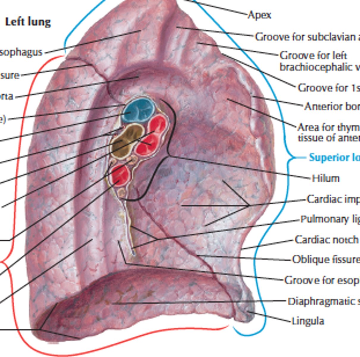
superior pulmonary vein.
The most anterior
the inferior pulmonary vein.
The most inferior
the bronchus
most posterior
The right pulmonary artery
anterior to the bronchus
the left pulmonary artery
superior to the bronchus.
Bronchopulmonary segment
Pyramidal portion of lung supplied by each tertiary bronchi
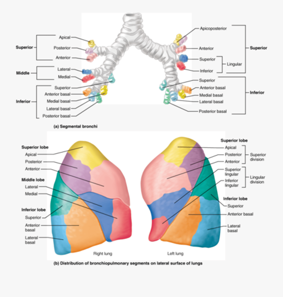
Bronchopulmonary segment right : upper lobe
Has apical , anterior & posterior segments.

Bronchopulmonary segment right: middle lobe
Has medial & lateral segments
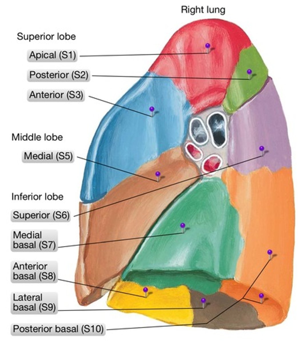
Bronchopulmonary segment right: lower lobe
Has superior, anterior basal, posterior basal, medial basal & lateral basal segments.

Bronchopulmonary segments left: upper lobe
Has apical, posterior, anterior , superior lingular & inferior lingular segments.
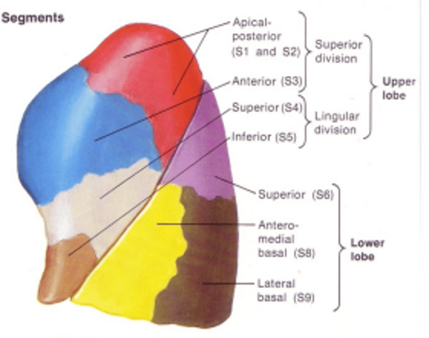
Bronchopulmonary segments left: lower lobe
Has superior , anterior basal, medial basal posterior basal & lateral basal segments.

blood flow of lungs
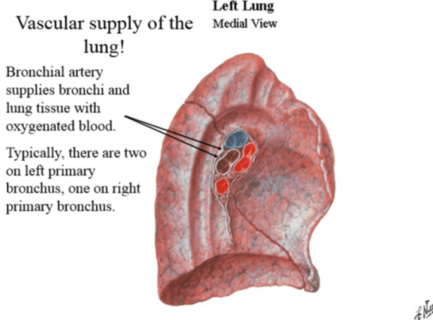
The pulmonary artery carries deoxygenated blood into alveoli (gas exchange) and then into pulmonary veins, which carry oxygenated blood into the left atrium, into the left ventricle, into the aorta, and into bronchial arteries to the lung tissue.
Blood supply of the lungs:
Pulmonary artery = major supplier of blood to the lungs
provides deoxygenated blood for gas exchange
Bronchial artery = supplies nutrients and removes waste from bronchi
provides collateral blood flow to remainder of lung parenchyma