1403- Anatomy-Mediastum
1/29
There's no tags or description
Looks like no tags are added yet.
Name | Mastery | Learn | Test | Matching | Spaced | Call with Kai |
|---|
No analytics yet
Send a link to your students to track their progress
30 Terms
What is the mediastinum
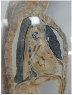
The mediastinum is the central compartment of the thoracic cavity, located between the lungs. It contains vital structures such as the heart, trachea, esophagus, and major blood vessels and includes the mediastinal pleura.
Boundaries of the mediastinum
Anteriorly- the sternum
Posteriorly→ Vertebral column
Superiorly→ Thoracic inlet
Inferiorly → Diaphragm
On each side → Mediastinal pleura
Importance of knowing mediastinum boundaries
To monitor infection which can move throughout the mediastinal space as it communictes with both the neck and the retroperitoneum.
Fascial planes connect the mediastinum to other regions( neck and retroperitoneum), allowing for the spread of disease and influencing surgical approaches.
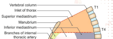
Subdivision- The superior mediastinum
Separated by imaginary plane at the sternal angle anteriorly.
Posteriorly- lower body of the fourth thoracic vertebra and extends to the thoracic inlet.
Subdivision - the inferior mediastinum
Separated into the middle, posterior and anterior mediastinum by the pericardium.
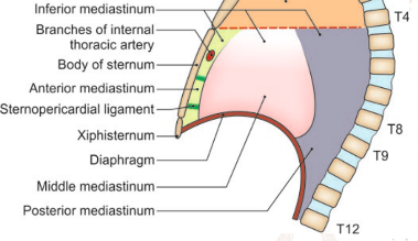
Anterior mediastinum
Subdivision of the inferior mediastinum.
Area in front of the pericardium, overlapped by thin anterior borders of both lungs.
Posterior mediastinum
Subdivision of the inferior mediastinum.
Area behind the pericardium.
Middle mediastinum
subdivision of the inferior mediastinum
The pericardium and its contents .
Boundaries of the superior mediastinum
Anteriorly- Manubrim sterni
Posteriorly- Upper four thoracic vertebrae
Superiorly- Plane of the thoracic inlet
Inferiorly- imaginary plan passing through the sternal angle in front, and the lower border body of the fourth thoracic vertebra behind.
On each side- Mediastinal pleura
Contents of the superior mediastinum
Trachea and oesphagus
Muscles
arteries
Veins
Nerves
Thymus
Thoracic duct
Lymph nodes
Muscles of the superior mediastinum
Origins of:
sternohyoid
sternothyroid
lower ends of the Longus colli
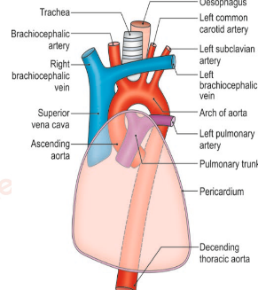
Arteries of the superior mediastinum
Arch of aorta
Branchiocephalic artery
Left common carotid artery
Left subclavian artery
Veins of the superior mediastinum
Left and right brachiocephalic veins
upper half of the superior vena cava
Left superior intercostal vein
Nerves of the superior mediastinum
Vagus
Phrenic cardiac nerves of both sides
Left recurrent laryngeal nerve
Lymph nodes of the suprior mediastinum
Paratracheal
Brachicephalic
Tracheobronchial
Boundaries of the anterior mediastinum
Anteriorly→ Body of sternum
Posteriorly→ Pericardium
superiorly- > Imaginary plane
Inferiorly→ Superior surface of the diaphram
on both sides → Mediastinal pleura
Contents of the anterior mediastinum
Sternopericardial ligaments
Lymph nodes with lymphatics
Small mediastinal branches of the internal thoracic artery
lowest part of the thymus
areolar tissue.
Boundaries of the middle mediastinum
anteriorly→ Sternopericardial ligaments
Posteriorly→ Esophagus, descending thoracic aorta and azygous vein
On each side → mediastinal pleuraa
Contents of middle mediastinum
Heart enclosed in pericardium
Arteries
Veins
Nerves
Lymph nodes
Arteries of the middle mediastinum
Ascending aorta
Pulmonary trunk
Two pulmonary arteries
Veins of the middle mediastinum
Lower half of the superior vena cava
Terminal part of the azygos vein
right and left pulmonary veins
Nerves of the middle mediastinum
Phrenic
Deep cardiac plexus
Lymph nodes of the middle mediastinum
Tracheobronchial nodes
Boundaries of the posterior mediastinum
Anteriorly→ Pricardium, bifurcation of trachea, pulmonary vessels and posterior part of the upper surface of the diaphragm.
Posteriorly→ lower eight thoracic vertebrae and intervening discs
on each side→ mediastinal pleura
Contents of the posterior mediastinum
Oesaphagus
arteries → descending aorta
Veins
nerves
Lymph nodes and lymphatics
Thoracis duct.
Veins of the posterior mediastinum
Azygos
Hemiazygos vein
Accessory hemiazygos vein
Nerves of the posterior mediastinum
Vagi
Splanchnic nerves → greate, lesser and least
Lymph nodes and lymphatics of the posterior mediastinum
Posterior mediastinal lymph nodes lying alongside the aorta
Thoracic duct.
Superior mediastinal mass
Most frequently caused by intrathoracic extension of the thyroid gland.
Middle mediastinal mass
Usually results from enlarged lymph nodes