Block 3 ALS Revision Questions
1/89
There's no tags or description
Looks like no tags are added yet.
Name | Mastery | Learn | Test | Matching | Spaced | Call with Kai |
|---|
No analytics yet
Send a link to your students to track their progress
90 Terms
What constitutes the upper GI tract?
Oesophagus
Stomach
Proximal duodenum
Liver
Gall bladder
Pancreas
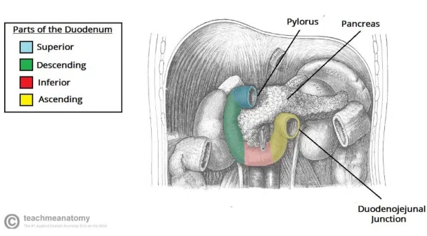
What is the junction between the foregut and the midgut?
Major duodenal papilla
What are the three main branches of the descending aorta that supply the GI tract?
Celiac trunk - foregut
Superior mesenteric artery - midgut
Inferior mesenteric artery - hindgut
What are the two cavities of the mouth?
Vestibule
Oral cavity
What structure separates the oral cavity and the oropharynx?
Palatoglossal fold
What muscle forms the floor of the oral cavity?
Mylohyoid muscle
What nerve innervates the mylohyoid muscle?
Mandibular nerve (V3), branch of the trigeminal
What is the function of the mylohyoid?
Depress the mandible to open the mouth
Pulls forward the pharynx during swallowing
What are the three salivary glands of the oral cavity?
Parotid gland
Submandibular gland
Sublingual gland
What nerve innervates the intrinsic muscles of the tongue?
Hypoglossal (XII)
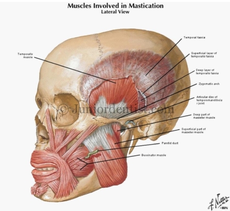
Which muscles of mastication elevate the mandible?
Temporalis
Masseter
Medial pterygoid
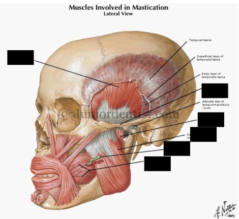
What are the muscles involved in mastication?
Temporalis
Zygomatic arch
Masseter (deep)
Masseter (superficial)
Parotid duct
Buccinator
Which nerve supplies sensation to the tongue and gums?
Lingual nerve
Where are the palatine tonsils?
Between the anterior and posterior palatoglossal folds
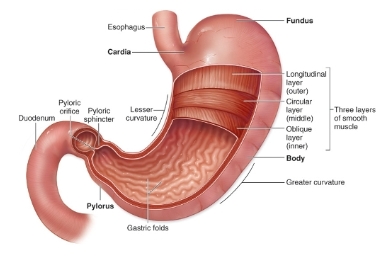
Which of the oesophageal sphincters is under voluntary control?
Upper - skeletal muscle
What is the principle function of the lower oesophageal sphincter?
Prevents gastro-oesophageal reflux
At which three points is the oesophagus constricted?
Pharangeal-oesophageal junction
Tracheal bifurcation
Gastro-oesophageal junction
Where in the GI tract is stratified squamous epithelium found?
Mouth
Oropharynx
Laryngopharynx
Oesophagus
What type of epithelium is found in the stomach?
Simple columnar
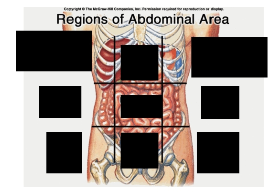
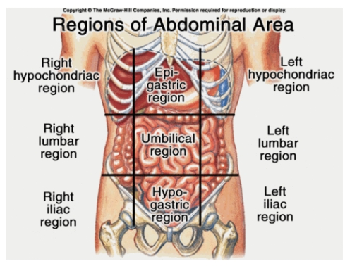
What are the nine abdominal regions?
Right hypochondriac
Epigastric
Left hypochondriac
Right lumbar
Umbilical
Left lumbar
Right iliac
Hypogastric
Left iliac
What is the main arterial supply of the oesophagus?
Oesophageal arteries
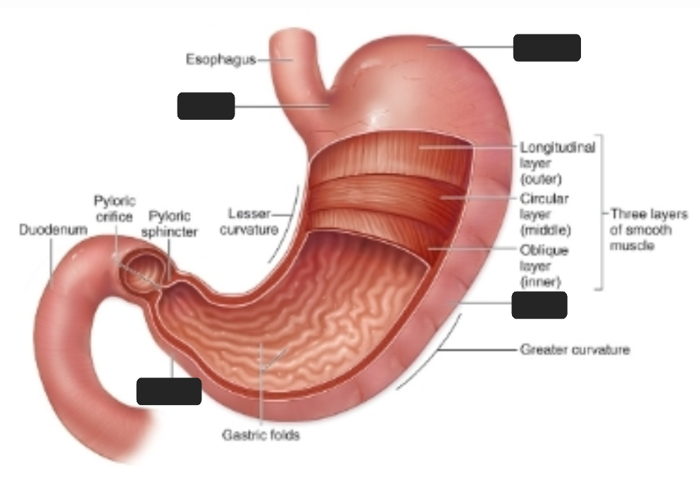
What are the four regions of the stomach?
Cardia
Fundus
Body
Pyloric
What are the three phases of swallowing?
Voluntary
Pharangeal
Oesophageal
What is peristalsis?
Contraction and relaxation of muscles in oesophagus
Produces wave down tube
Moves food down
Where is nausea regulated?
Medulla
Vomiting centre
Chemoreceptor trigger zone
What do chief cells secrete?
Pepsinogen
Gastric lipase
What is the physiology of gastric acid secretion?
Carbonic anhydrase catalyses formation of carbonic acid
This dissociates to give H+
Proton pump in parietal cell transports H+ into lumen and K+ into cell to balance out charge
Cl- diffuses out into lumen alongside K+ to balance the charge
H+ and Cl- combine to form HCl
What effect does gastrin have?
Stimulate secretion of gastric acid and pepsinogen
What chemicals inhibit gastric acid secretion?
Somatostatin
CCK (cholecystokinin)
Where are Peyer’s patches (clusters of lymphatic tissue) found?
Ileum
Where are Brunner’s glands (secrete alkaline mucus) found?
Duodenum (neutralise stomach acid)
In what form can the small intestine absorb sugars?
Monosaccharides
How are monosaccharides absorbed?
Facilitated diffusion or active transport
Which co-transporter enters mucosal cells with glucose?
2Na+
Which transporter allows the entry of glucose into capillaries from mucosal cells?
GLUT2
How are most peptides absorbed?
As amino acids via active transport
How are lipids absorbed?
Broken down into fatty acids
Surrounded by bile salts to form micelles
Move from lumen to brush border, fatty acids diffuse into absorptive cells
Recombine to form triglycerides
Aggregate with phospholipids & cholesterol, coated with proteins to become chylomicrons
Exocytosed into lacteals, travel through lymphatics, enter blood at left subclavian vein
Removed from blood at liver and adipose tissue by lipoprotein lipase
Where are most bile salts reabsorbed?
Ileum
What constitutes the midgut?
Distal duodenum
Jejenum
Ileum
Ascending colon
Proximal 2/5 transverse colon
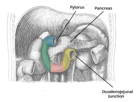
What are the four parts of the duodenum?
Superior
Descending
Inferior
Ascending
Is this the jejunum or ileum?
Jejunum
Long vasa recta (straight arteries)
Small arterial arcades (arterial loops)
What is the blood supply of the jejunum?
Jejunal arteries
Originate from superior mesenteric
What are the projections of the ileum into the large intestine called?
Ileocecal folds
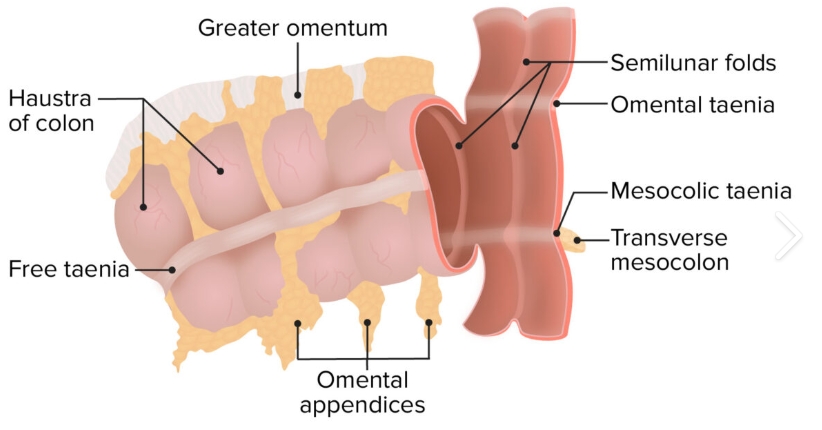
What are three identifying features of the large bowel?
Omental appendices (fat filled pouches of peitoneum attached externally)
Taeniae coli (three longitudinal bands of smooth muscle)
Haustra (sacculations created by semilunar folds on internal surface)
Is the colon peritoneal or retroperitoneal?
Retroperitoneal
At what level is the rectosigmoid junction?
Third sacral vertebra
What are the two types of smooth muscle in the muscalaris of the small intestine?
Outer - thin, longitudinal fibres
Inner - thick, circular fibres
What type of epithelial cells line the large intestine?
Simple columnar
What is the innervation of the GI tract?
Enteric nervous system
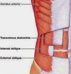
What are the three flat muscles of the anterior abdominal wall?
External oblique
Internal oblique
Transversus abdominus
What is formed at the midline by the aponeurosis of the flat muscles?
Linea alb
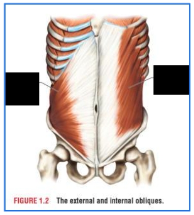
In what direction do the fibres of the external oblique run?
Inferomedial
What innervates the external oblique?
Anterior rami of lower thoracic spinal nerves (T7-12)
What are the primary functions of the external oblique?
Compression of abdominal contents
Flexion of trunk
Internal oblique
External oblique
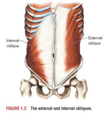
What is the function of the internal oblique?
Compress abdominal contents
Flex trunk
What is the innermost flat muscle?
Transversus abdominus
What innervates the transversus abdominus?
T7-12 and L1
What is the function of transversus abdominus?
Compress abdominal contents
What are the two vertical muscles?
Rectus abdominis
Pyramindalis
Which vertical muscle is sometimes absent?
Pyramidalis
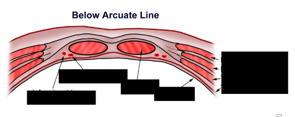
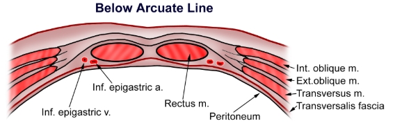
Which portion of the rectus abdominis is completely enclosed by the rectus sheath?
Upper ¾ (above arcuate line)
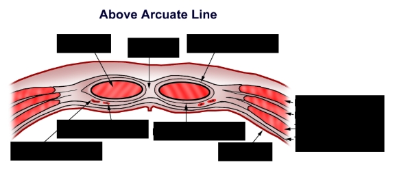
Rectus m.
Linea alba
Ant. rectus sheath
Inf. epigastric v.
Inf. epigastric a.
Post. rectus sheath
Peritoneum
Int. oblique m.
Ext. oblique m.
Transversus m.
Transversalis fascia
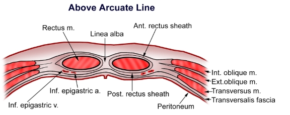
Inf. epigastric v.
Inf. epigastric a.
Rectus m.
Peritoneum
Int. oblique m.
Ext. oblique m.
Transversus m.
Transversalis fascia
What are the two divisions of the peritoneum?
Parietal (lines abdominal walls)
Visceral (covers the viscera)
What are the contents of the inguinal canal?
Genitofemoral nerve
Ilio-inguinal nerve
Spermatic cord (male)
Round ligament of the uterus (female)
What structures mark the start and end of the inguinal canal?
Deep inguinal ring
Superficial inguinal ring
What is a direct inguinal hernia?
Peritoneal sac that enters the medial end of inguinal canal directly through weakened posterior wall
What is an indirect inguinal hernia?
Peritoneal sac that enters inguinal canal through deep inguinal ring
Which structure formed from the peritoneum suspends the jejunum and ileum from the posterior abdominal wall?
Mesentery
What is the main problem with fat digestion and absorption?
Lipids are hydrophobic
How are fats digested in an aqueous environment?
Emulsification
How do bile salts aid fat digestion?
Stabilise small emulsion particles
How are bile salts formed?
Breakdown of cholesterol
What are the two primary bile acids?
Cholic acid
Chenodeoxycholic acid
How does cholecystokinin (CCK) stimulate the release of bile acids?
Contraction of gall bladder
Relaxation of sphincter of Oddi
Where in the abdomen is the liver located?
Right hypochondriac and epigastric regions
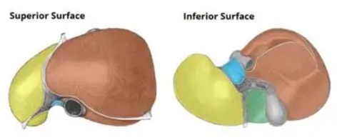
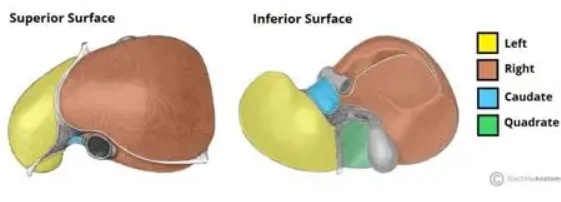
What are the four anatomical lobes of the liver?
Right
Left
Caudate
Quadrate
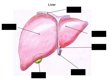
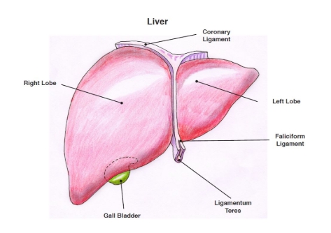
Right lobe
Coronary ligament
Left lobe
Falciform ligament
Ligamentum teres
Gall bladder
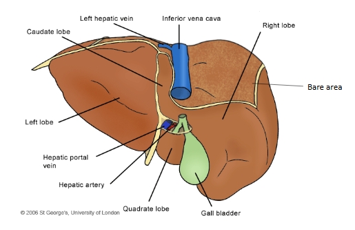
Caudate lobe
Left hepatic vein
Inferior vena cava
Right lobe
Bare area
Gall bladder
Quadrate lobe
Hepatic artery
Hepatic portal vein
Left lobe
What is the blood supply of the liver?
Common hepatic artery → hepatic artery proper → left and right hepatic arteries
How many functional lobes does the liver have?
8
What is the porta hepatis?
Point of entry into the liver for
hepatic ducts
hepatic arteries
hepatic portal vein
Where is the spleen located?
Left hypochondrium
Which surface of the spleen is notched?
Anterior
What is the blood supply to the spleen?
Splenic artery
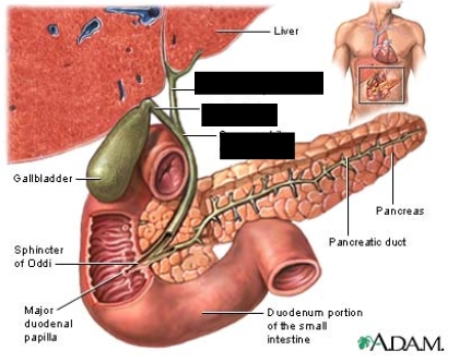
Common hepatic duct
Cystic duct
Common bile duct
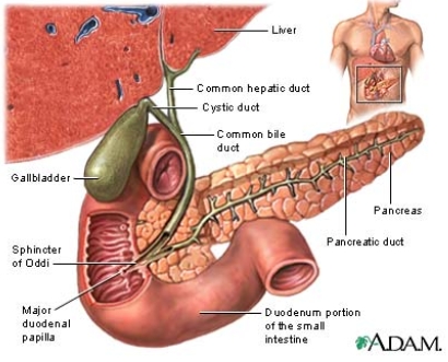
Where do the common bile duct and the pancreatic duct open into the duodenum?
Major duodenal papilla (hepatopancreatic ampulla)
What are the major functional cells of the liver?
Hepatocytes
How are hepatocytes arranged?
Into hepatic laminae