Module VI
 Call Kai
 Learn
Learn Practice Test
 Spaced Repetition
 Match
Match Flashcards
 Knowt Play
1/25

There's no tags or description
Looks like no tags are added yet.
Name | Mastery | Learn | Test | Matching | Spaced | Call with Kai |
|---|
No analytics yet
Send a link to your students to track their progress
26 Terms
Introduction
-Dental offices must regularly check radiographic equipment + document that x-ray is being used properly.
QA program: helps obtain the best image possible which in turns helps the dentist provide the best care possible + reduce unnecessary retakes + radiation exposure.
QA schedules
Daily
Weekly
Monthly
Yearly
Daily Schedules
➢ Check all computers and monitors to ensure they are working as expected
➢ Check the digital sensors visually paying close attention to the case and cable for any damage
➢ PSP system check the scanner and PSP plates visually for any malfunction or damage
➢ Extraoral imaging system start and perform a dry run to make sure all systems are working as expected.
➢ Review retake logs and identify potential issues that needs attention.
➢ Review infection control procedures as applicable to imaging, keep in mind digital sensors cannot be sterilized and special attention must be paid to barriers.
➢ All these must be done at the beginning of workday
Weekly Schedule
➢ Check the lead aprons and thyroid collars visually for any damages
➢ Check and clean all monitors used for viewing images
➢ Check all computer systems including keyboard and mouse to make sure they function as expected
➢ For PSP check the phosphor plates for scratches, damage
Monthly Schedule
Check that exposure charts are posted by x-ray machines
Review all radiographic equipment service records
Perform monitor calibration for all computers used for viewing images
Review image archive and storage systems
Review exposure monitoring records > Film badge
Yearly Schedule
Calibrate x-ray machine
This is usually done by service personal from the dental service companies.
Specialized equipments may be required.
• X-Ray Output
• Kilovoltage [kVp] Calibration
• Half value layer
• Timer
• Milliamperage [mA]
• Collimation
• Beam Alignment
• Tube Head Stability
• Any other manufacturer recommended annual services
Other Factors
Keep service records
Keep film badge logs
Follow manufacturer instructions
Follow local/ federal regulations
Don’t modify x-ray systems yourself
All imaging systems must be operated as specified by the manufactures of that system
Compliance with all regulatory agency guidelines
Radiology Regulations
-FDA and ADA issues guidelines for dental imaging
-ALARA—> a principle where images are taken with lowest possible radiation without compromise or diagnostic quality (As low As Reasonably Achievable)
Technical Errors
-Technical errors happen while taking the image
-Bisecting angle technique more prone for errors
-Errors in patient positioning
-Errors in positioning of the sensors
-Errors is in positioning of the x-ray source
-Selection of wrong exposure factors
Imaging Errors (Over exposure)
-Excess: mA, time, kVP
-Image is too dark
-Reduce exposure factors:
I.O: reduce exposure time
E.O: adjust time, mA, kVp
Imaging Errors ( Underexposure)
-Insufficient: mA, time, kVp
-Image is dull/washed out
-Increase exposure time:
I.O: adjust only exposure time
E.O: adjust time, mA, and kVp
Double exposure
-Using a flexible sensor without wiping out previous image or using the same plate more than once can lead to double imaging
-Error only happens with PSP/Flexible sensors or films/ error does not happen with hard sensor
Incorrect assembly of the Rinn Arm
-When looking through the aiming ring, only the teeth being image should be visible
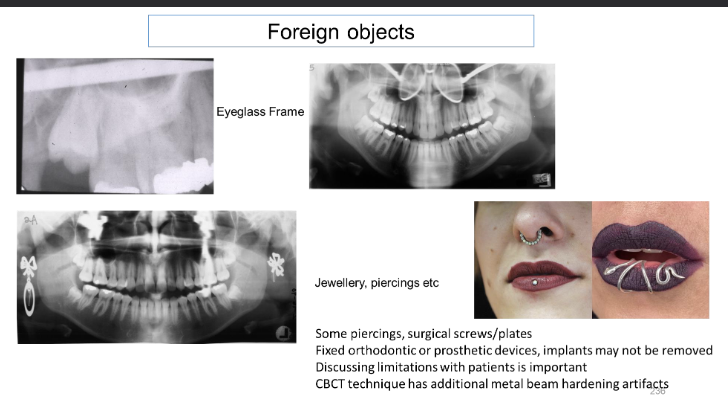
Metal objects left in mouth
-Must remove removable metal before imaging pr a retake may be needed
Thyroid Collar Cut
-Use in dental imaging
-For patient + operator confidence it is preferable to use the collar and shield, for children + pregnant patients and in CBCT imaging shielding is required
-If thyroid collar blocks part of the beam it covers anatomy on the image; thyroid collar cut
Cone Cut
-Occurs when PID ( Position-indicating device) is not centered over sensor
-Results in white unexposed area on image
Motion
Case 1
-Patient movement during exposure
-The resultant image will appear grainy with unsharpness
-the image may be undiagnostic
-The image may need retake
Case 2
-Film movement during exposure
-The patient had opened the mouth during the exposure and the sensor had moved
-The resultant image will have double edges structures
-Maybe undiagnostic
-The image may need to be repeated
Magnification Errors
-Receptor should be places as close as possible
-Increased distance between sensor + object will lead to magnification
Errors in Bisecting Angle Technique
-Primary controlling factors:
Angular relationship between: teeth, film, and beam
-Elongation + foreshortening:
Elongation: vertical angle too small + image shift apically ( insufficient beam = image is longer than object)
Foreshortening: vertical angle too great + image shift occlusally or incisally ( excessive beam = image is shorter than object)
Panoramic Errors
-Errors due to wrong exposure factors technical and processing errors are like intraoral imaging.
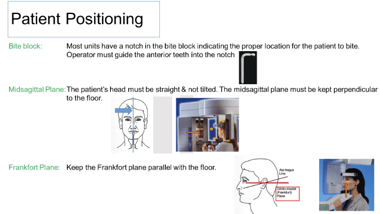
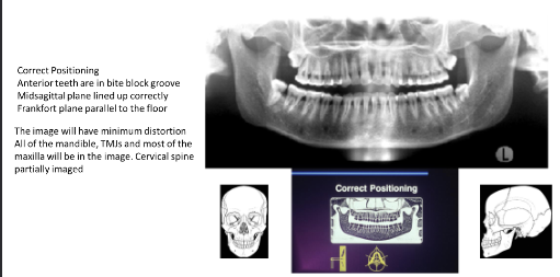
Correct postion
Rotational Error
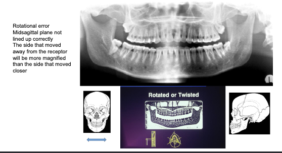
Bite block and teeth positioning error
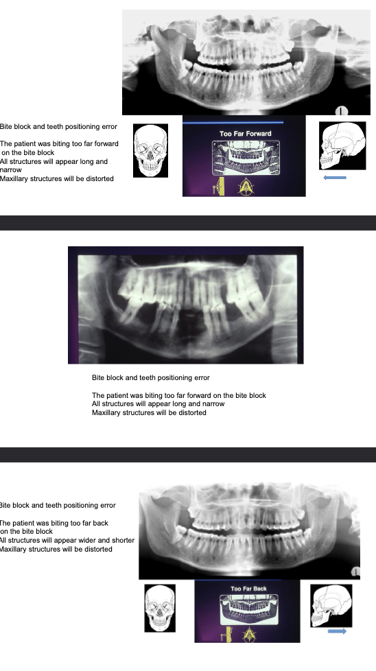
Frankfort Plane
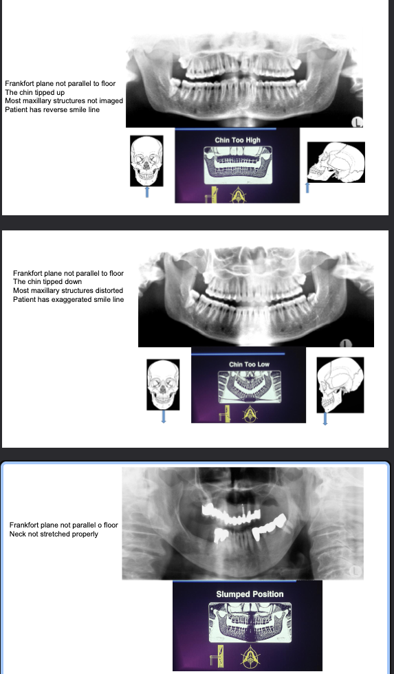
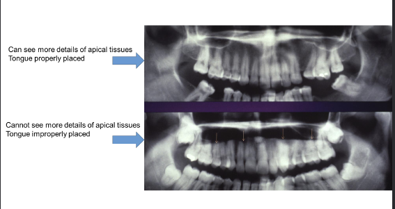
Tongue Improperly Positioned
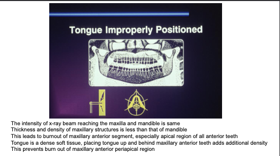
Tongue Improperly Positioned (part 2)