m Human Anatomy 100 Viva Exam
1/119
There's no tags or description
Looks like no tags are added yet.
Name | Mastery | Learn | Test | Matching | Spaced | Call with Kai |
|---|
No analytics yet
Send a link to your students to track their progress
120 Terms
acromioclavicular joint
synoivial joint between the acromion process of the scapula and the distal clavicle
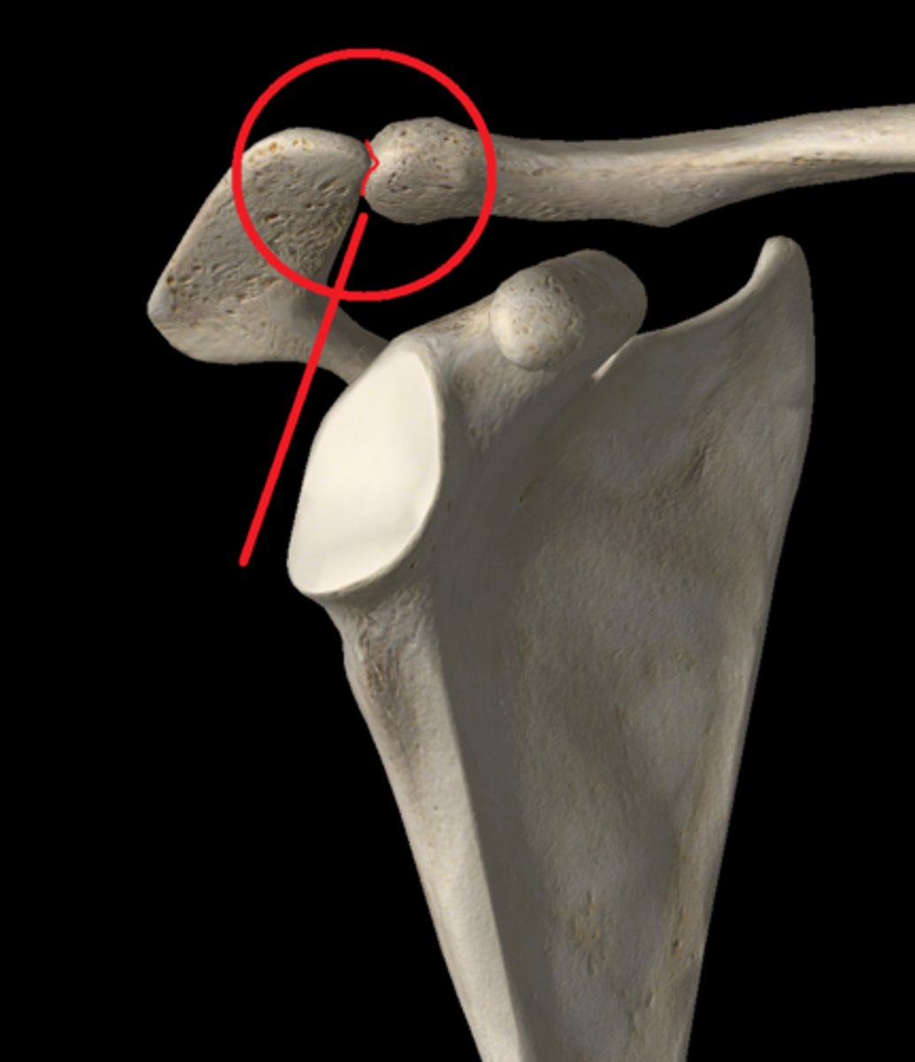
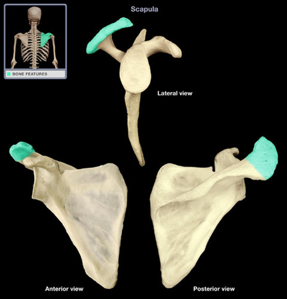
acromion process
located on the superior lateral aspect of the spine of the scapula, may be felt on the top of the shoulders
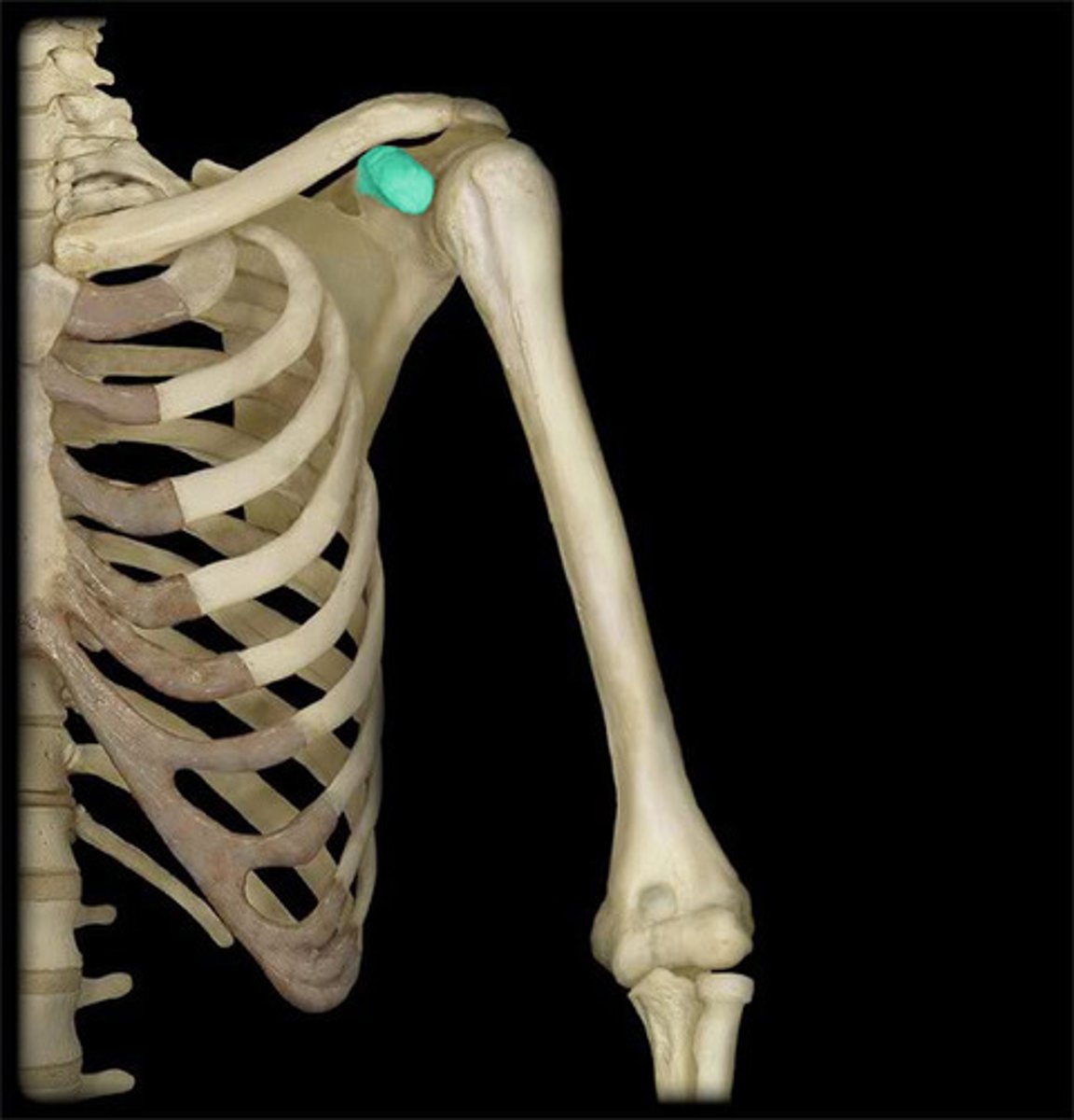
coracoid process
process above the glenoid cavity that permits muscle attachment
insertion of pectoralis minor
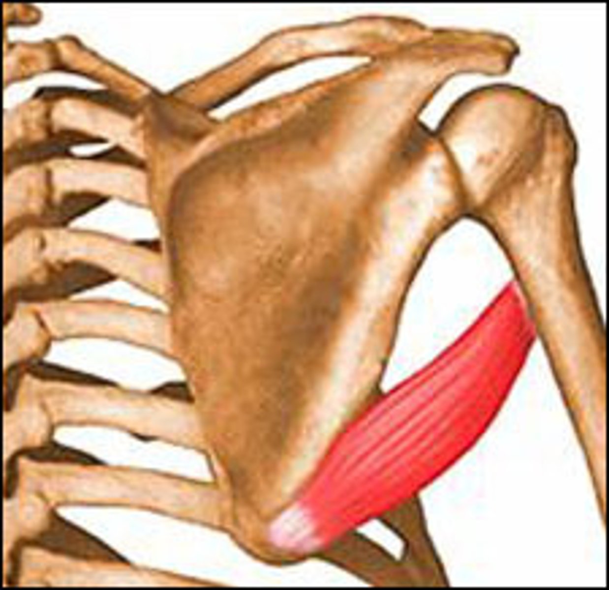
inferior angle of scapula
lowest point of the scapula
origin site for teres minor
can be palpated: medially rotate scapula
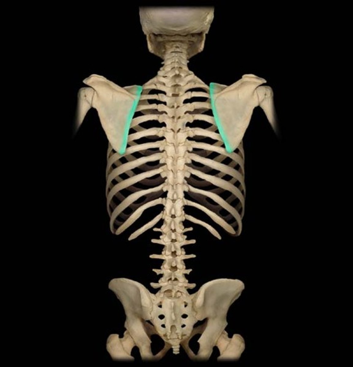
medial border of scapula
medial edge, next to vertebral column
insertion of serratus anterior
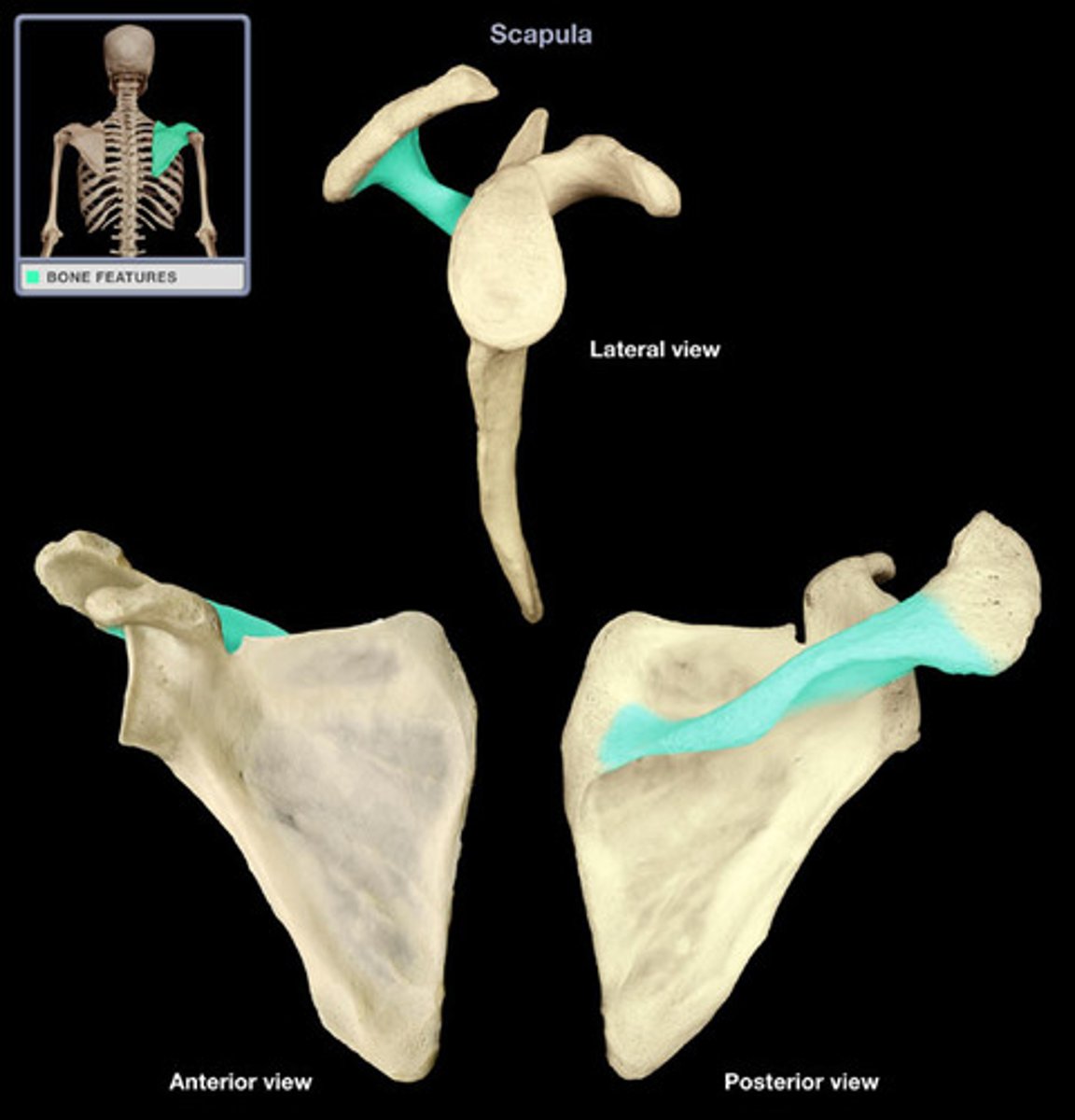
spine of scapula
a long projection dividing the lateral surface of the scapula.
articulates with acromion process, insertion for trapezius and origin of deltoid.
Superior to it: supraspinatus
Inferior to it: infraspinatus.
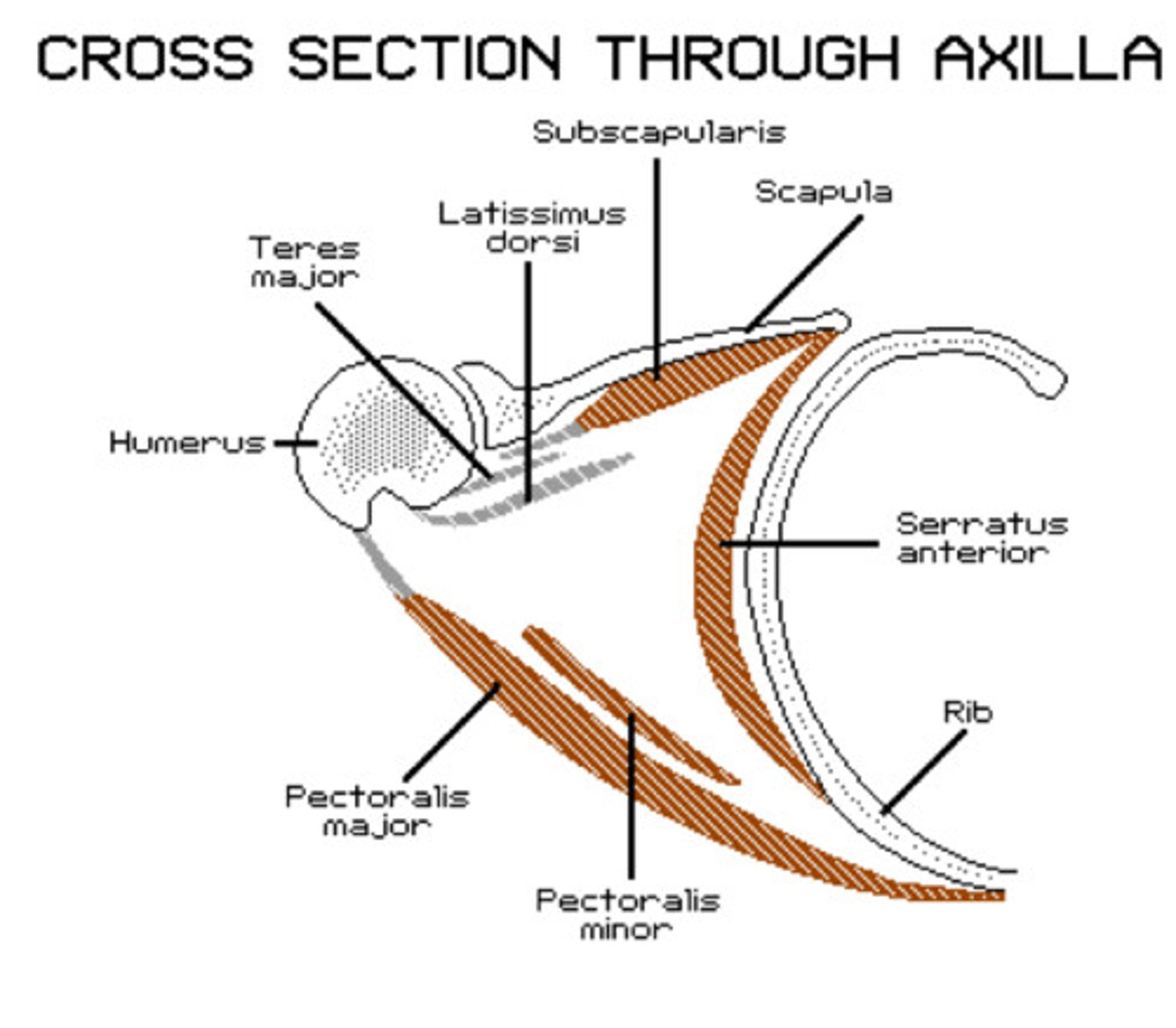
Axilla
Borders: Anterior: pectoralis major & Minor. Posterior: scapula, teres major, latissimus dorsi. Medial wall: serratus Anterior, ribs. Lateral wall: Humerus, Triceps.
Contents: axillary artery and vein, brachial plexus nerves, lymph nodes.
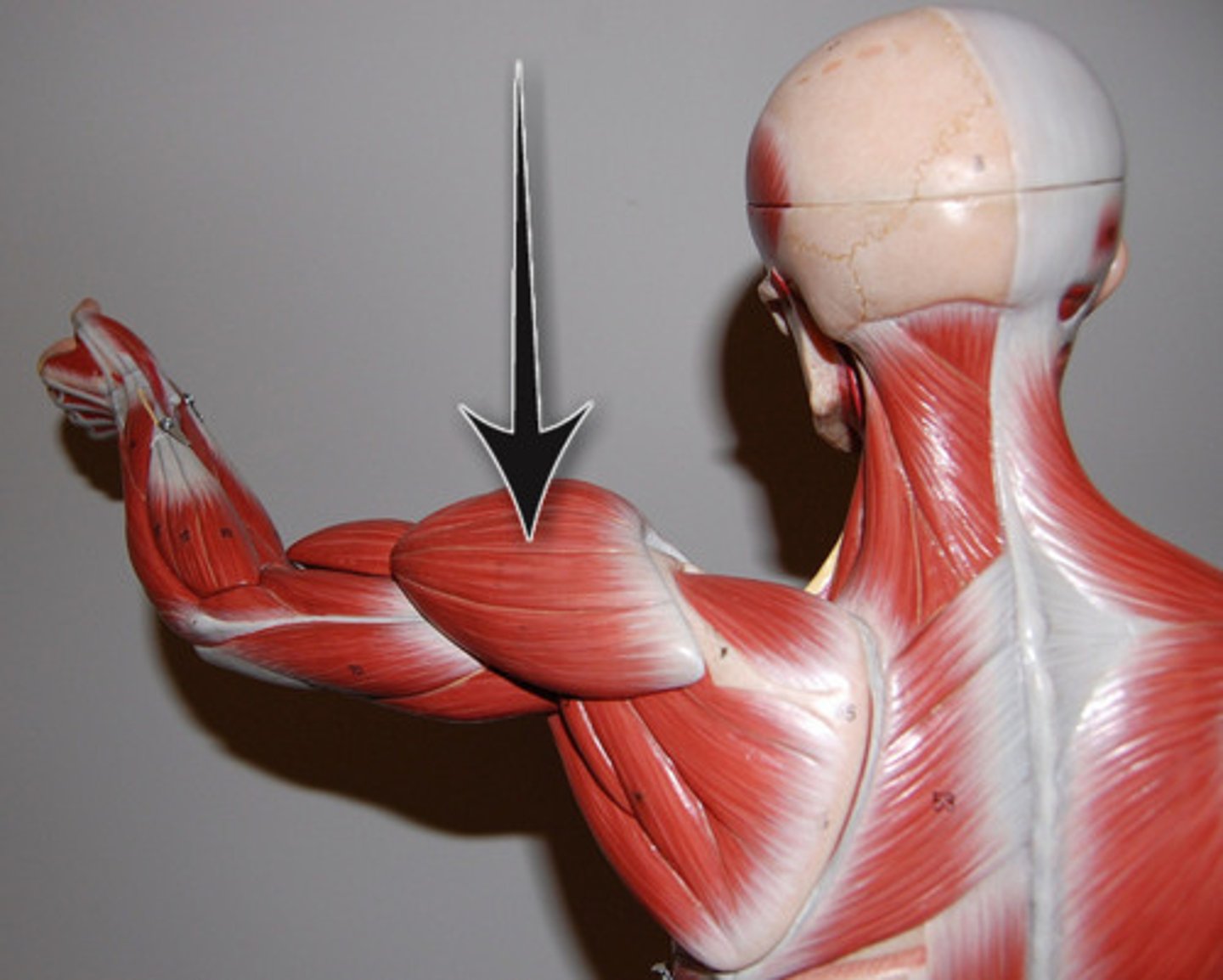
deltoid
-Origin: anterior border and upper surface of the lateral third of the clavicle, acromion, spine of the scapula
-Insertion: deltoid tuberosity of humerus
-Actions: flexion by anterior deltoid, shoulder abduction by middle deltoid, and extension by posterior deltoid
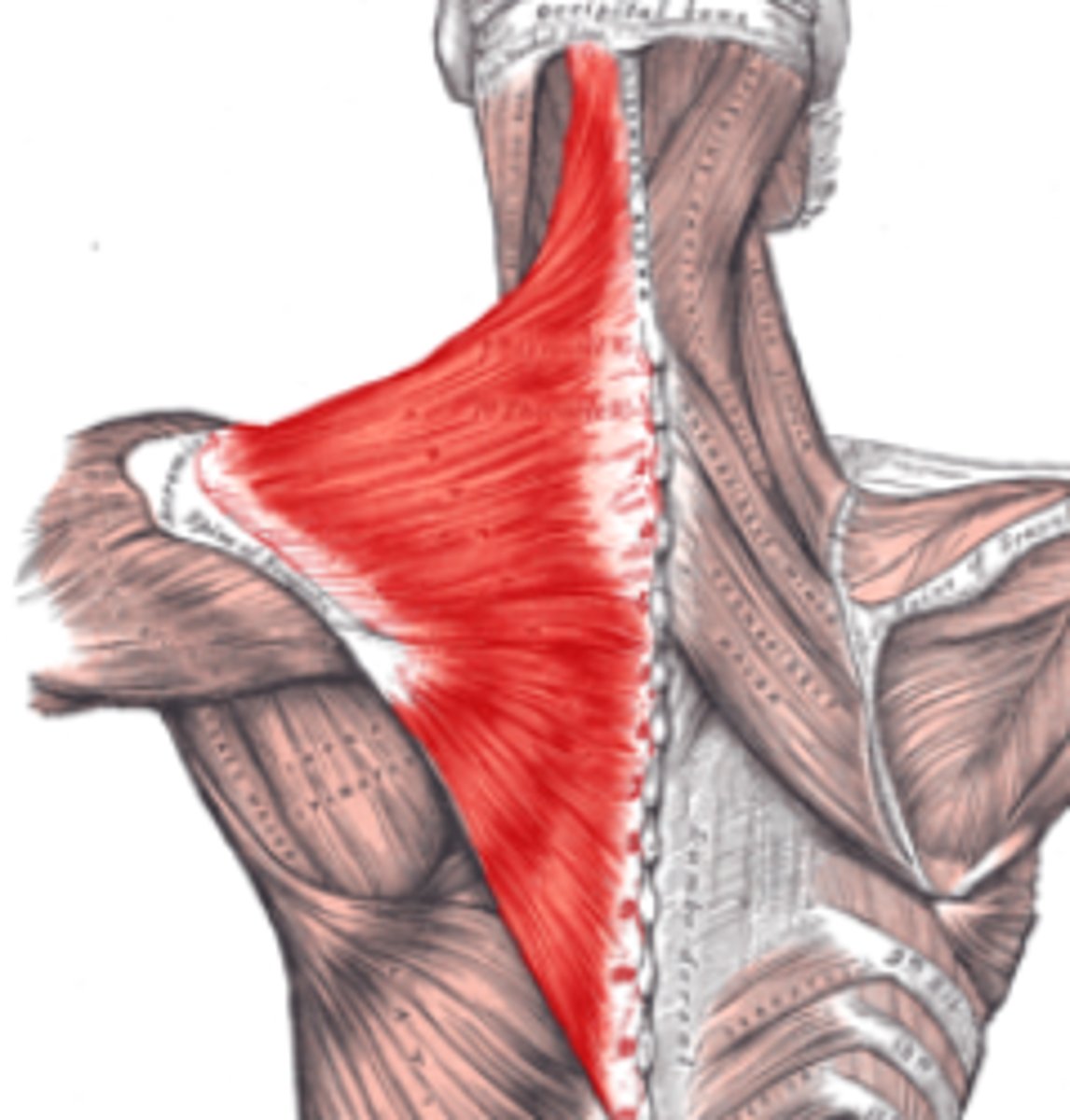
Trapezius
-Origin spinous processes of vertebrae C1-T12
Insertion: acromion process and spine of scapula
Elevates, depresses, retracts, and rotates the scapula; rotates the arm
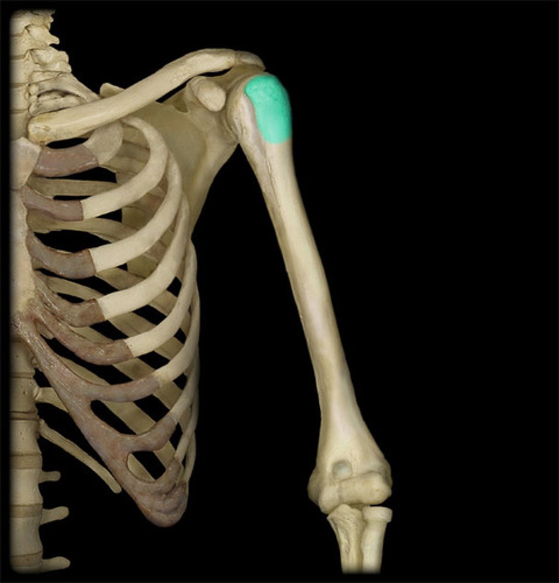
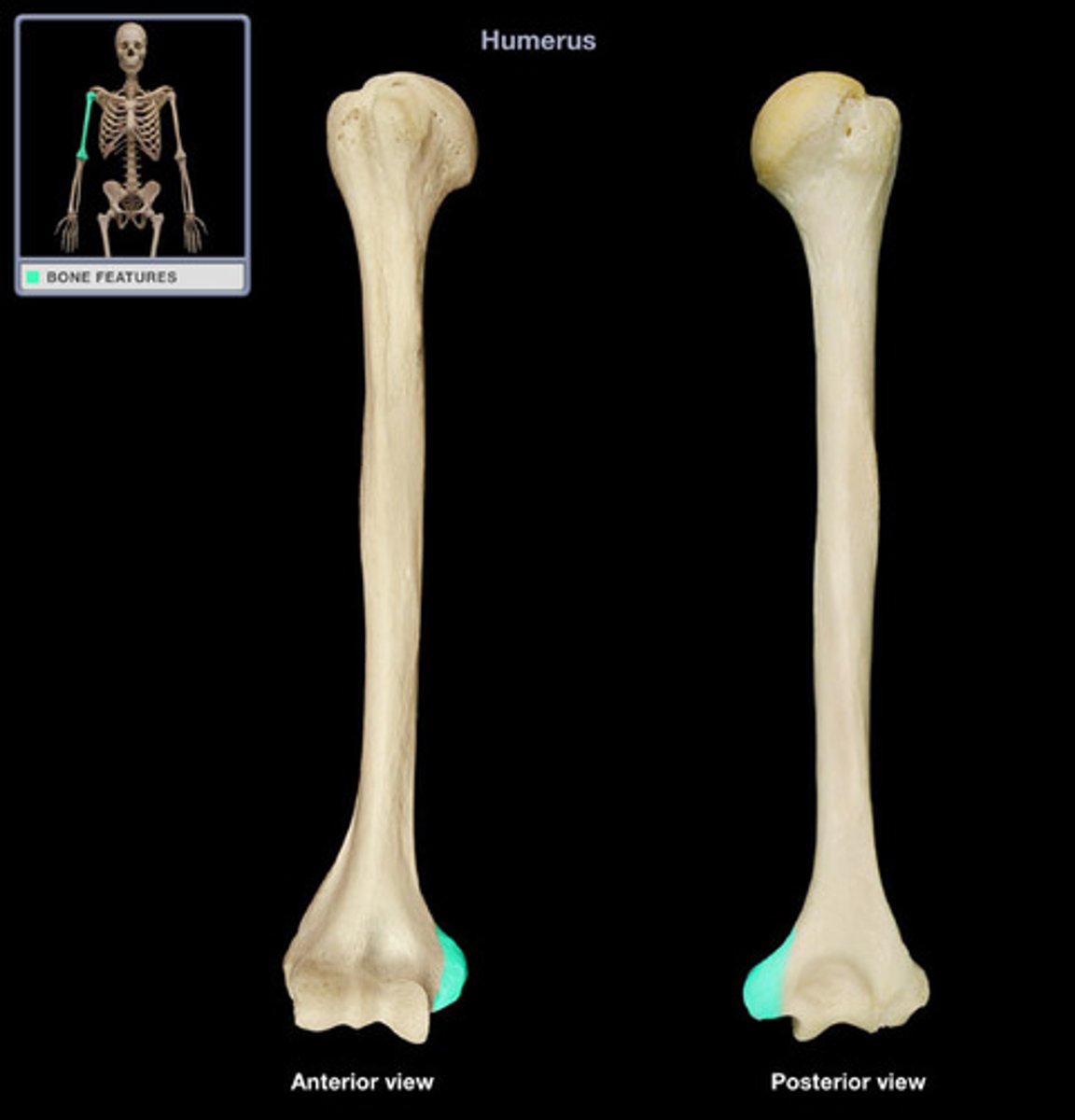
greater tubercle of humerus
located on the lateral surface just below the neck and is separated from the lesser by a deep depression called the intertubercular groove
attachment for supraspinatus, infraspinatus, and teres minor. confirm palpation by externally and internally rotating shoulder while elbow is flexed.
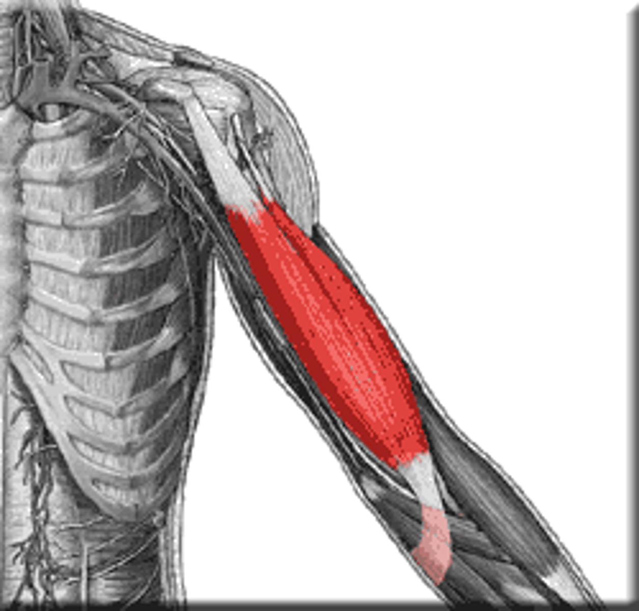
biceps brachii
-Origin
=Short head: coracoid process of the scapula.
=Long head: supraglenoid tubercle
-Insertion Radial tuberosity and bicipital aponeurosis into deep fascia on medial part of forearm
-Actions
=Flexes elbow,
=supinates radioulnar joint in the forearm
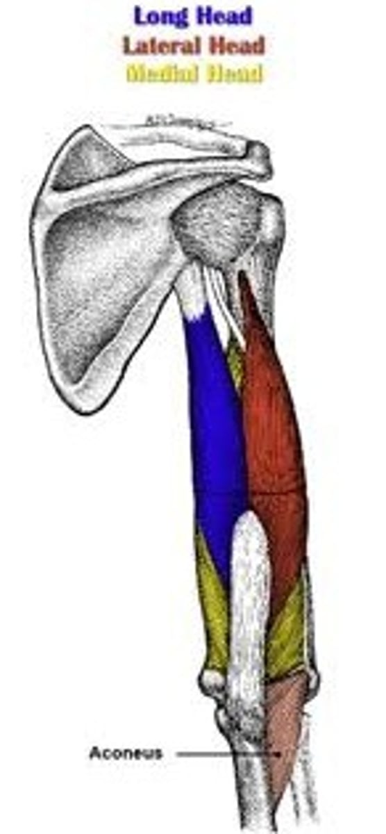
triceps brachii
-Origin=Long head: infraglenoid tubercle of scapula
=Lateral head: above the radial groove
=Medial head: below the radial groove
-Insertion Olecranon process of ulna
-Nerve: Radial nerve
Actions= Extends forearm,
= long head extends,adducts arm, Extends shoulder
lateral and medial epicondyles of humerus
Medial more prominent. Lateral is more rounded, medial more square.
Lateral epicondyle is the origin of the extensors of the forearm (extensor carpi radialis brevis, extensor carpi ulnaris, extensor digitorum). as well as the radial collateral ligament.
Medial is the attachment for the flexors of the forearm
(flexor carpi radialis, flexor digitorum superificialis, and flexor carpi ulnaris). as well as the ulnar collateral ligament.
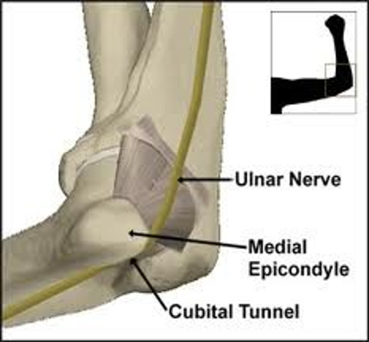
cubital tunnel
between medial epicondyle and olecranon process
ulnar nerve passes through
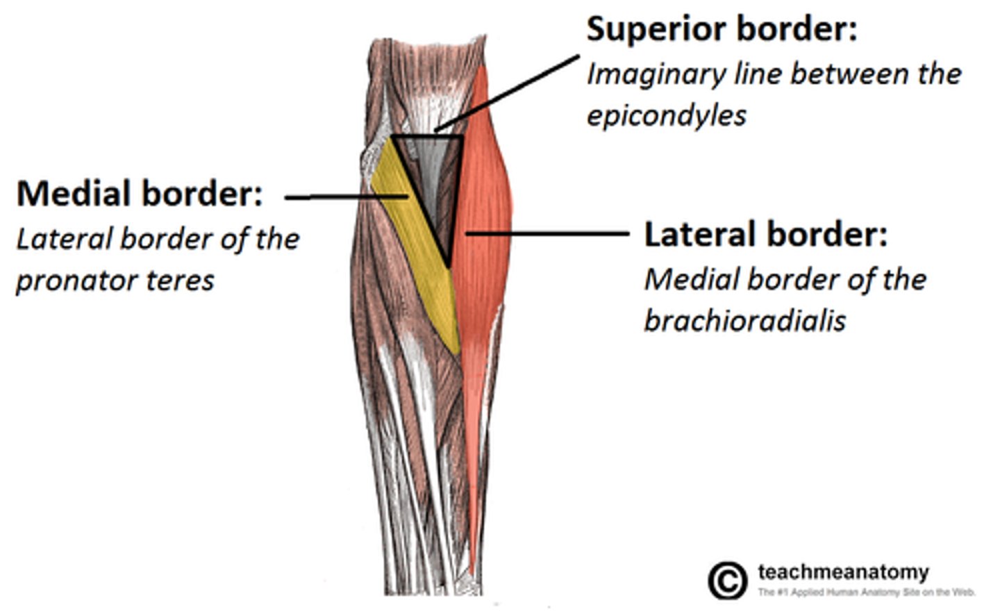
cubital fossa
boundaries: pronator teres, brachioradialis, line b/t epicondyles; contains: biceps tendon, brachial artery (which splits into radial and ulnar arteries), median nerve
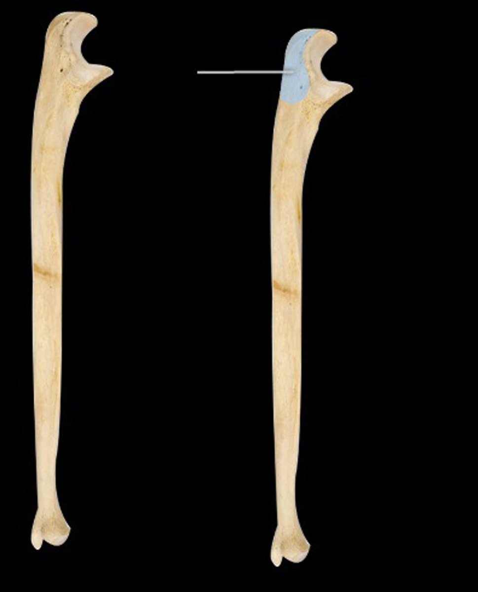
olecranon process of ulna
insertion of triceps brachii, can palpate: elbow bone.
Extensor compartment of forearm
Posterior forearm.
Superficial: extensor carpi radialis longus, extensor carpi radialis brevis, extensor carpi ulnaris, extensor digitorum.
deep: Supinator, abductor pollicis longus, extensor pollicis brevis, extensor pollicis longus.
innervated by radial.
extensor carpi radialis longus
Origin: lateral supracondylar ridge of humerus
Insertion: base of metacarpal 2
Action: extends and abducts hand
N: radial:
extensor carpi radialis brevis
Origin: Lateral epicondyle of humerus
Insertion: Base of 3rd metacarpal bone
Action: Extension and abduction at wrist
N: radial
extensor carpi ulnaris
Origin: Lateral epicondyle of the humerus
Insertion: 5th metacarpal
Action: Extends the hand
N: radial
flexor compartment of forearm
Superficial: Pronator teres, flexor carpi radialis, palmaris longus, flexor carpi ulnaris. (PFPF) Deeper: Flexor digitorum profundus, flexor digitorum superficialis. flexor pollucis longus. pronator quadratus
All medial nerve apart from flexor carpi ulnaris (both medial and ulnar)
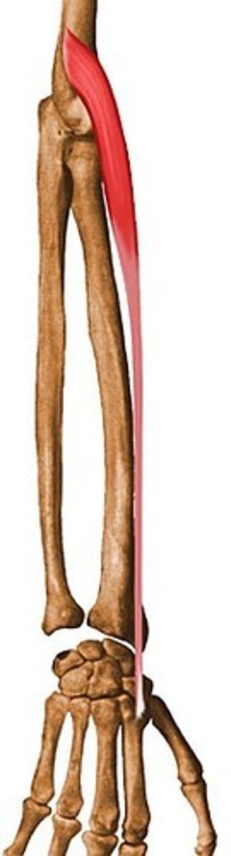
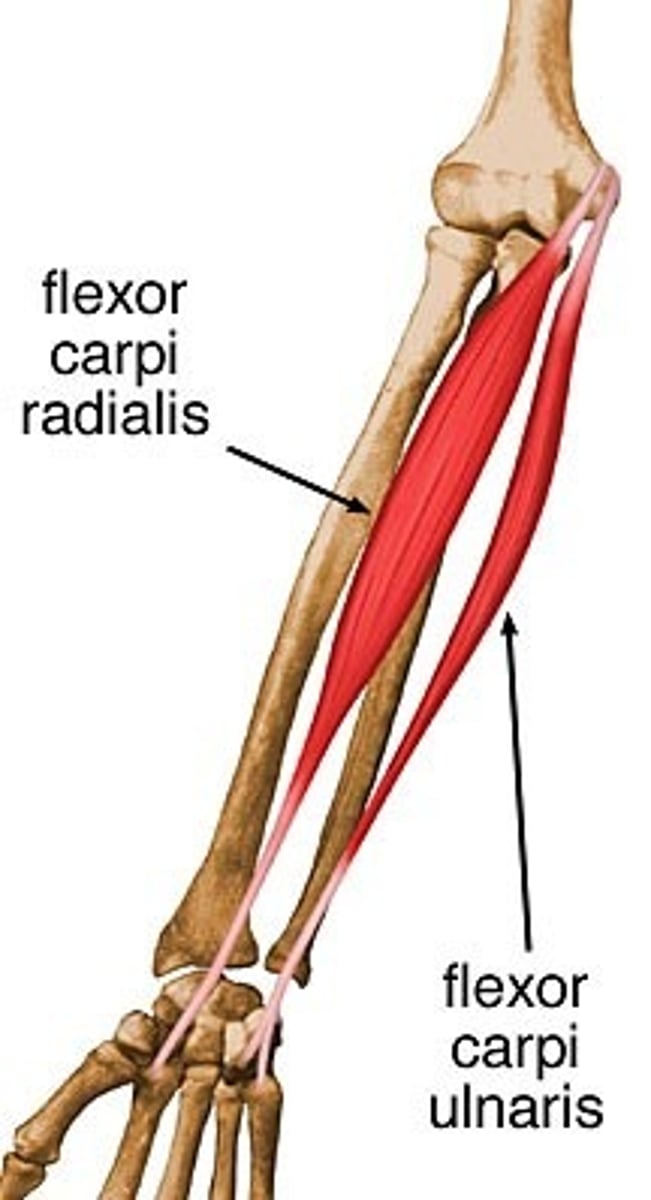
flexor carpi ulnaris
Origin: Medial epicondyle of the humerus
Insertion: 5th metacarpal
Action: Flexes the hand
N: ulnar

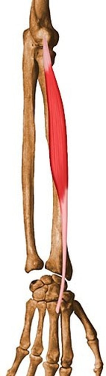
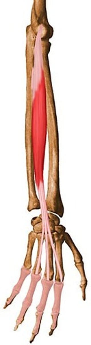
flexor carpi radialis
-Origin medial epicondyle of humerus
-Insertion Bases of second and third metacarpal bones
-Actions Flexion and abduction at wrist
N: median
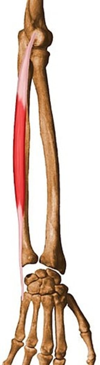
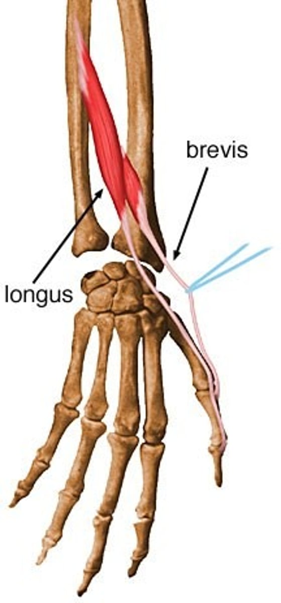
palmaris longus
-Origin medial epicondyle of humerus (common flexor tendon)
-Insertion palmar aponeurosis
-Actions wrist flexor
N: median
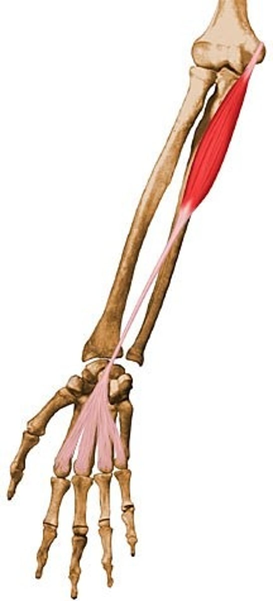
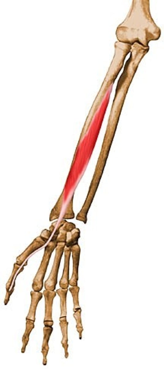
finger flexors (extrinsic)
flexor digitorum superficialis, flexor digitorum profundus, flexor pollicis longus
flexor pollicis longus
Origin: Anterior surface of radius, and interosseous membrane
Insertion: Distal phalanx of thumb
Action: Flexes thumb
N: median
flexor digitorum superficialis
Origin: Medial epicondyle of humerus, coronoid process of ulna, and shaft of radius
Insertion: Middle phalanges of fingers 2-5
Action: Flexes hand and middle phalanges of fingers 2-5
N: Median
flexor digitorum profundus
Origin: Anteromedial surface of ulna, interosseous membrane, and coronoid process
Insertion: Distal phalanges of fingers 2-5
Action: Flexes distal phalanges
N: Median and ulnar
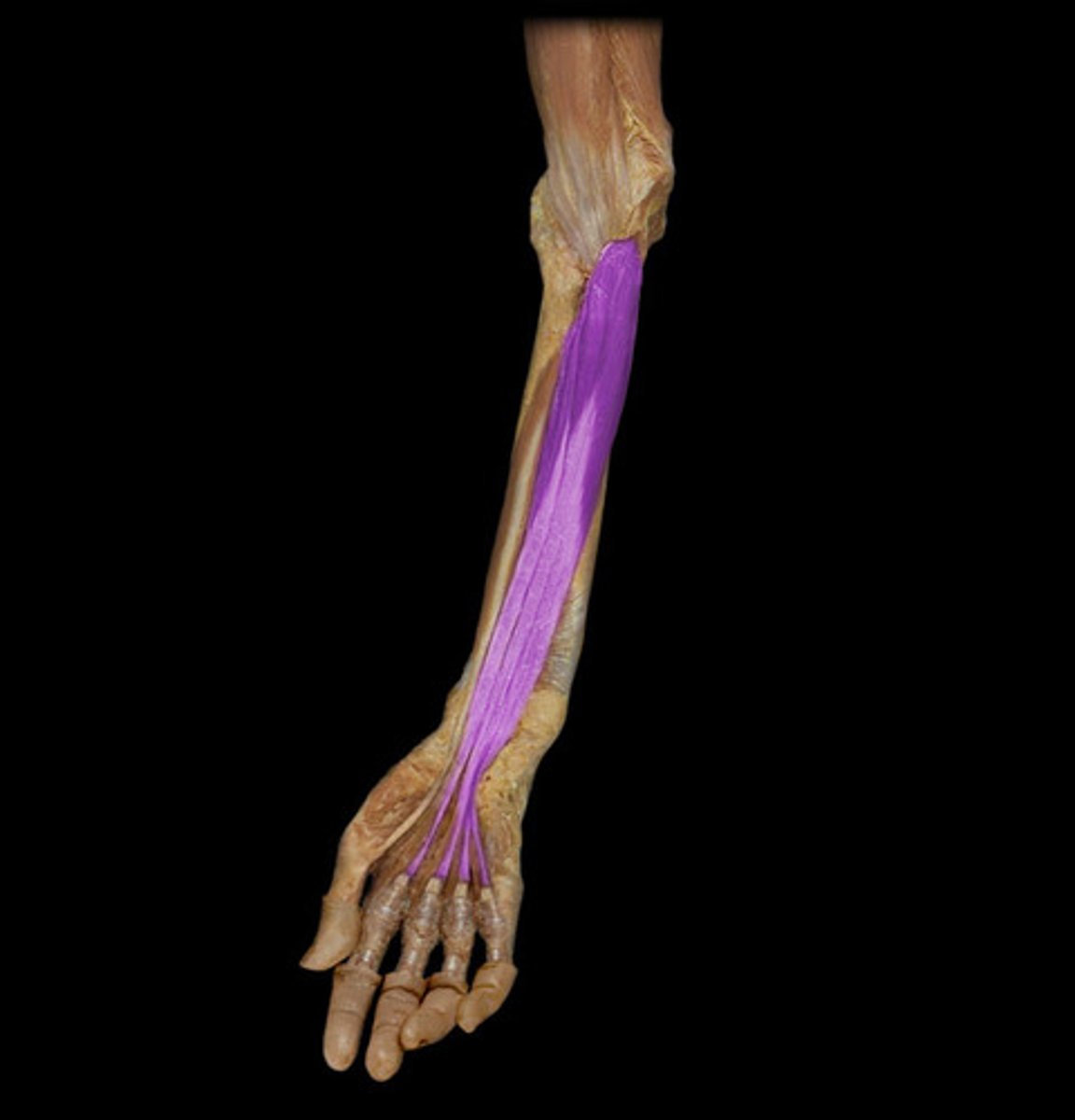
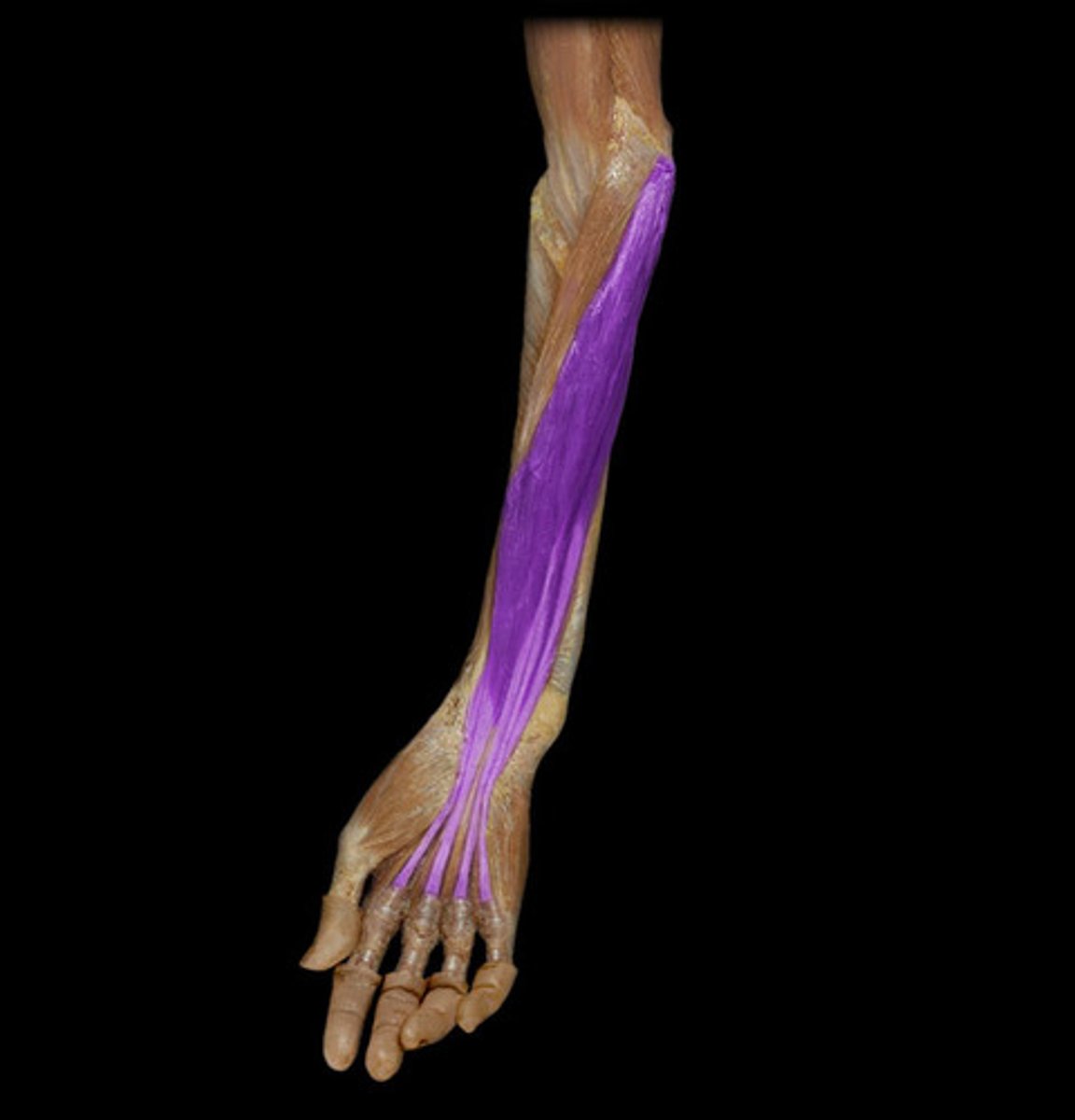
finger extensors
extensor digitorum, extensor pollicis longus and brevis
extensor digitorum
Origin: lateral epicondyle of humerus
Insertion: by four tendons into distal phalanges of fingers 2-5
Action: prime mover of finger extension
extensor pollicis longus
Origin: Posterior surface of middle of ulna and interosseous membrane
Insertion: Base of distal phalanx of thumb
Action: Extension of thumb

extensor pollicis brevis
Origin:
Posterior surface of middle of
radius and interosseous
membrane
Insertion:
Base of 1st proximal phalanx
Action:
Extension of thumb & radially deviate wrist
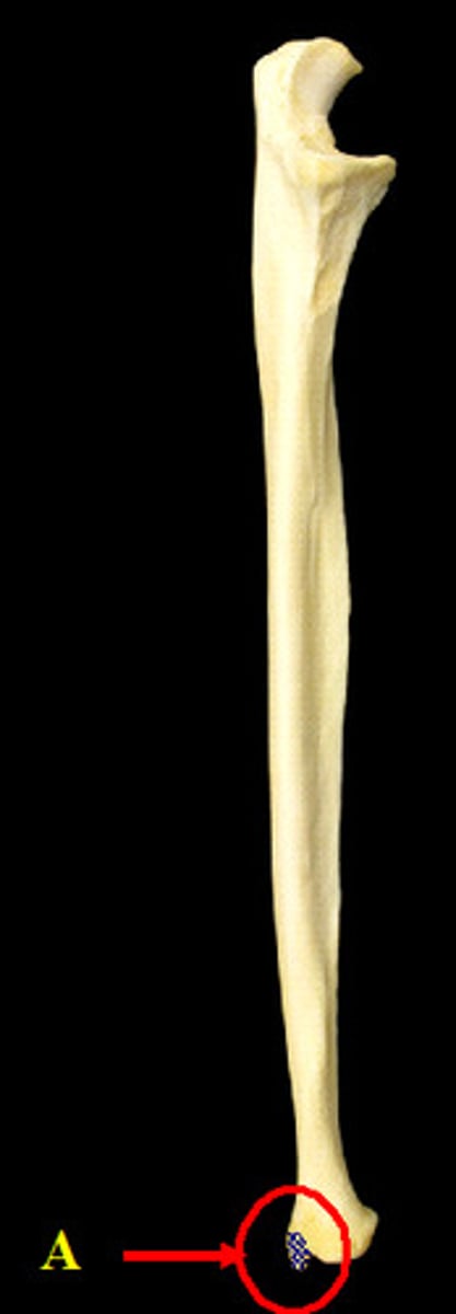
head of ulna
Distal bump of the ulna. Does not articulate with the carpal bones, unlike the head of the radius. Easy to find in pronation.
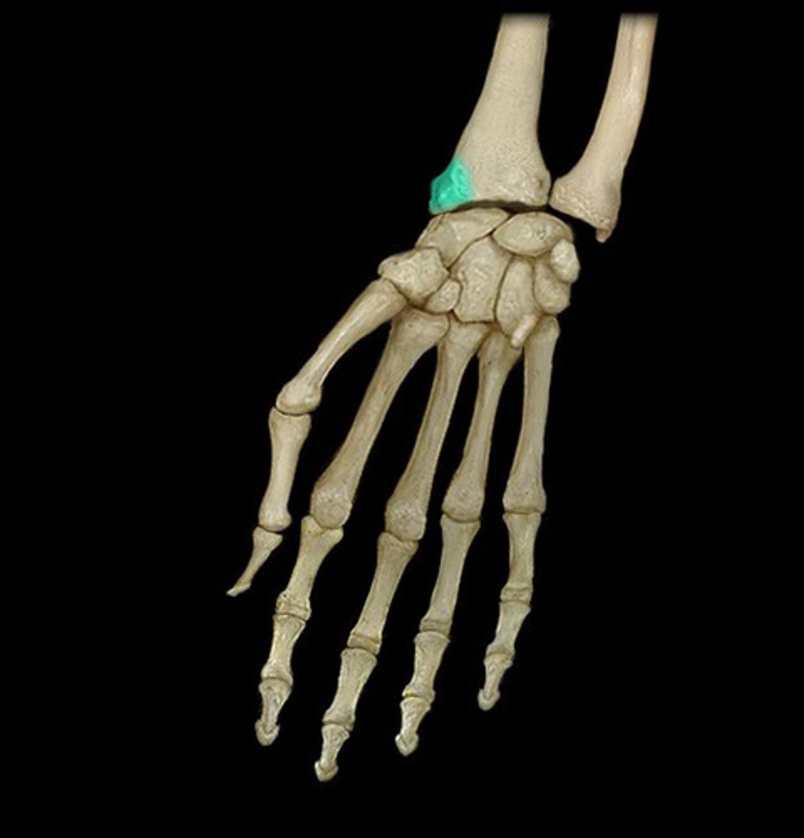
radial styloid process
distal prominence on head of radius; insertion of brachioradialias, origin of abductor pollicis longus and extensor pollicis brevis.
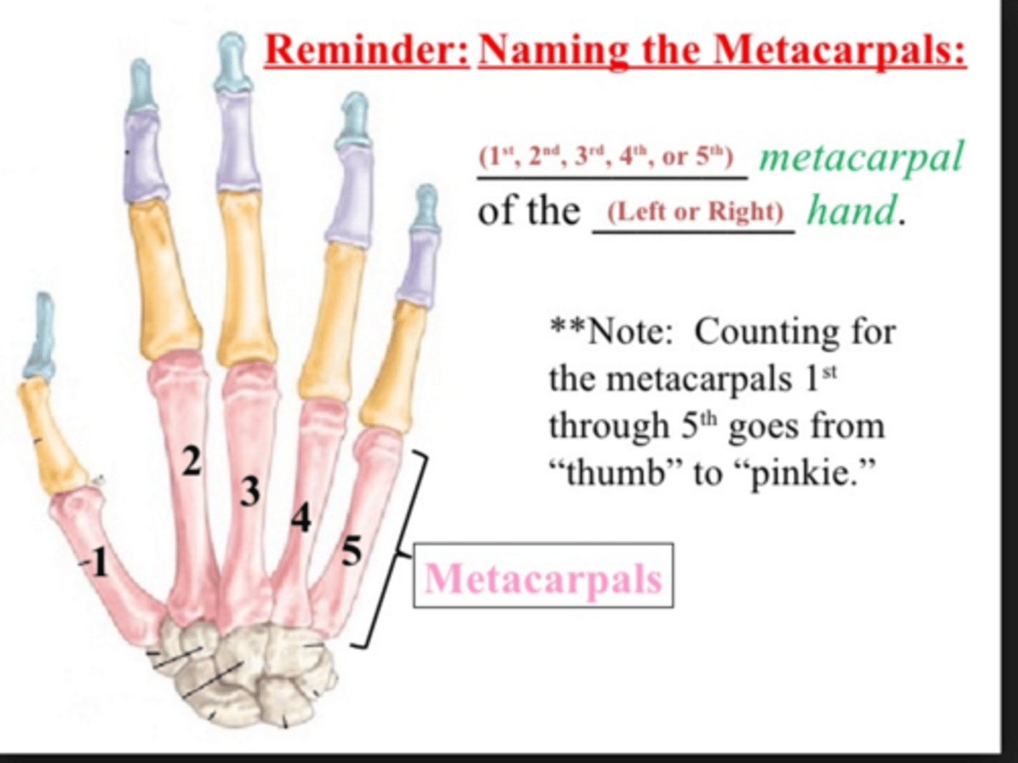
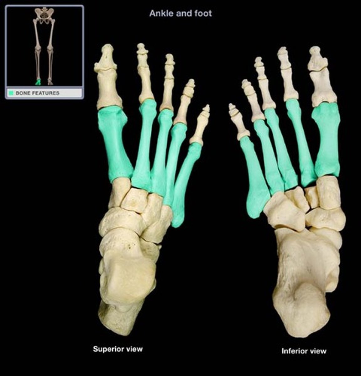
metacarpals 1,2,3,4,5
connect carpal bones to phalanges. attachement of wrist flexors (extensor carpi radialis longus to base of 2nd metacarpal)
Intrinsic hand muscles (opponens pollicis and opponens digiti minimi insert, adductor pollicis originates)
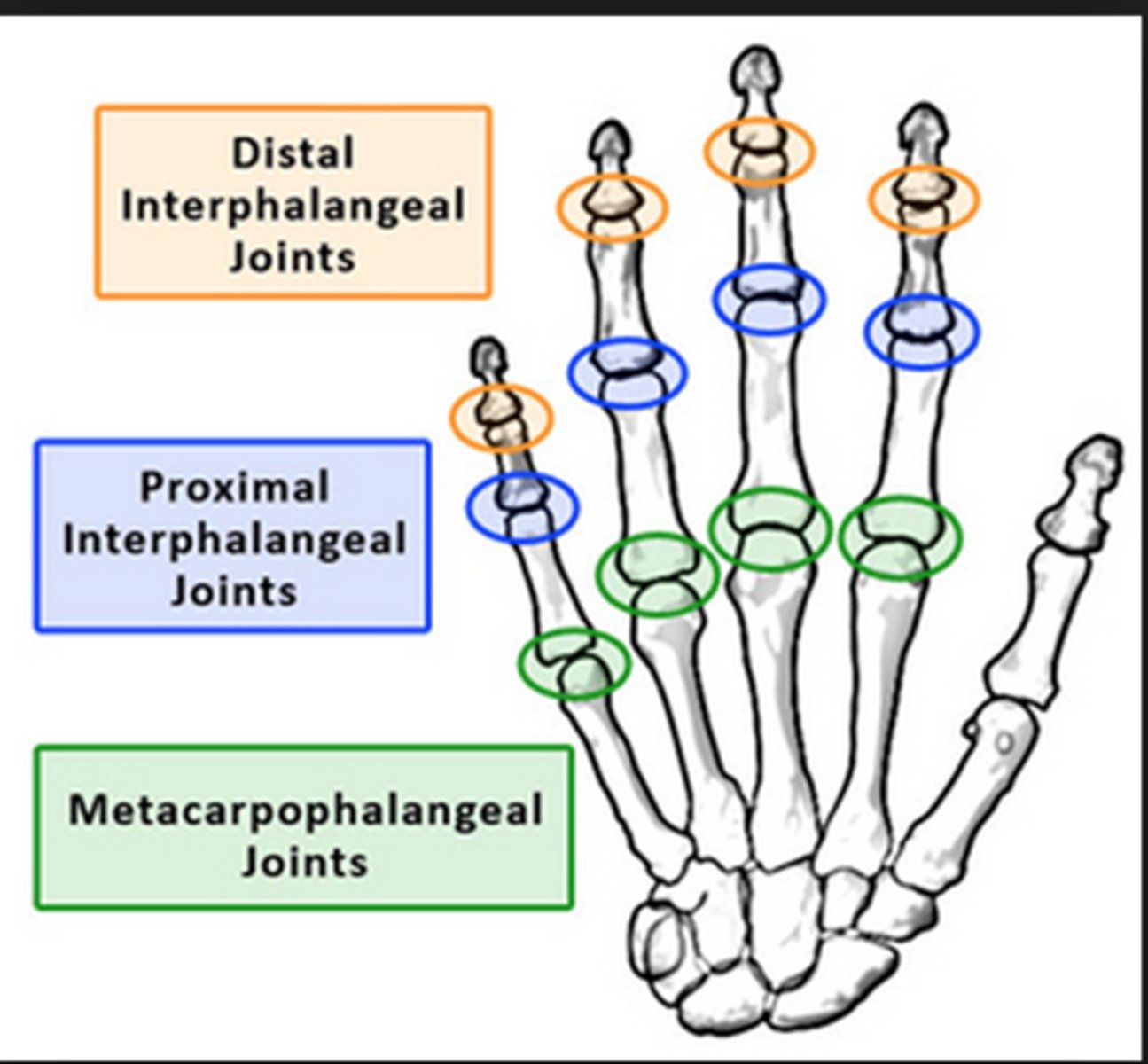
metacarpophalangeal joints
The synovial joints between the metacarpal bones and the phalanges. Knuckles.
Volar plates and transverse metacarpal ligaments prevent hyperextension at these joint.
Scaphoid
Distal crease and floor of anatomical snuffbox.
origin of flexor pollicis brevis. Articulates with trapezium carpal bone via intercarpal joints.
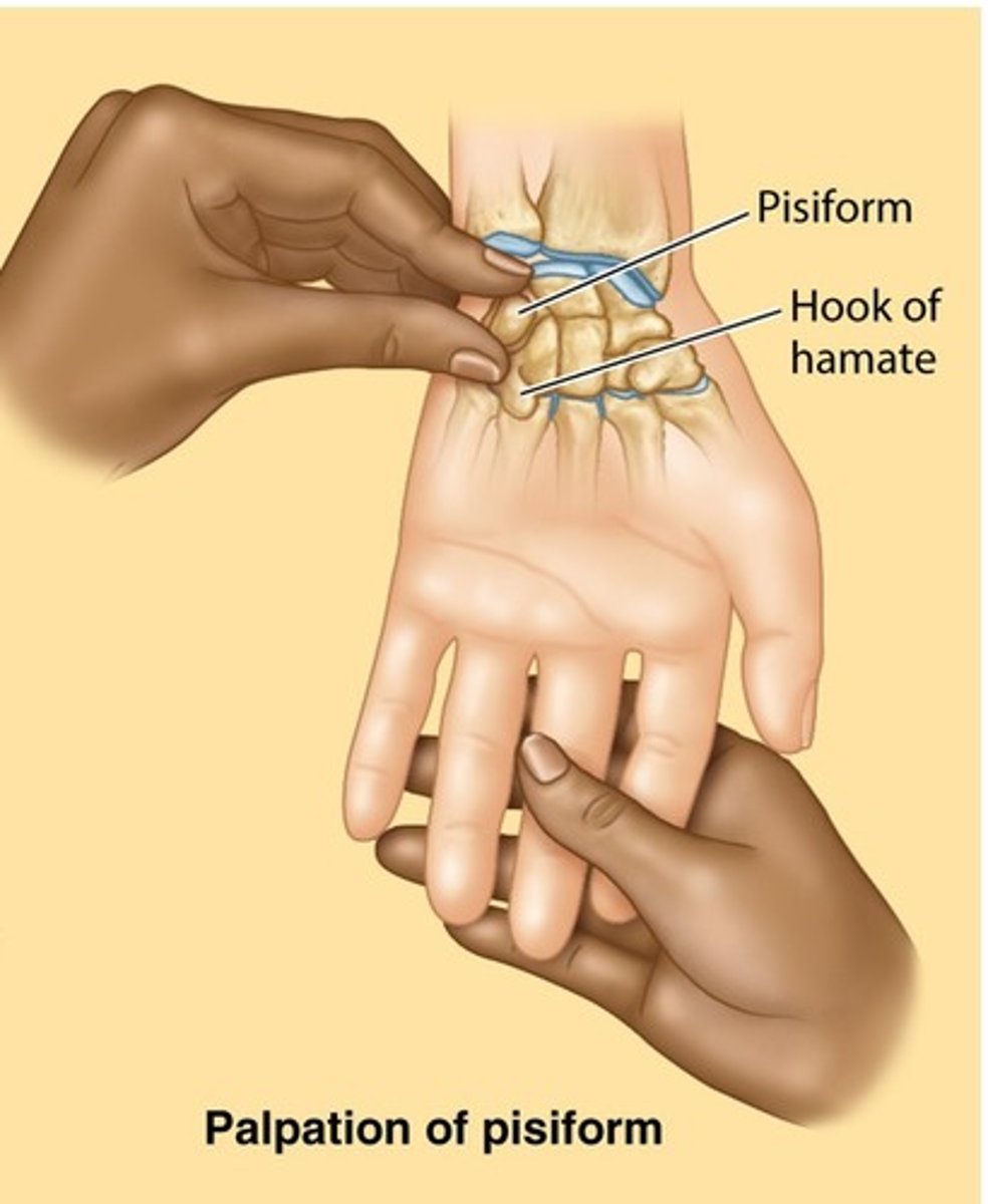
Pisiform
Located at medial base of hand. Sesamoid bone meaning it is inside a tendon.
Sits on the triquetrum carpal bone. origin of abductor digit minimi.
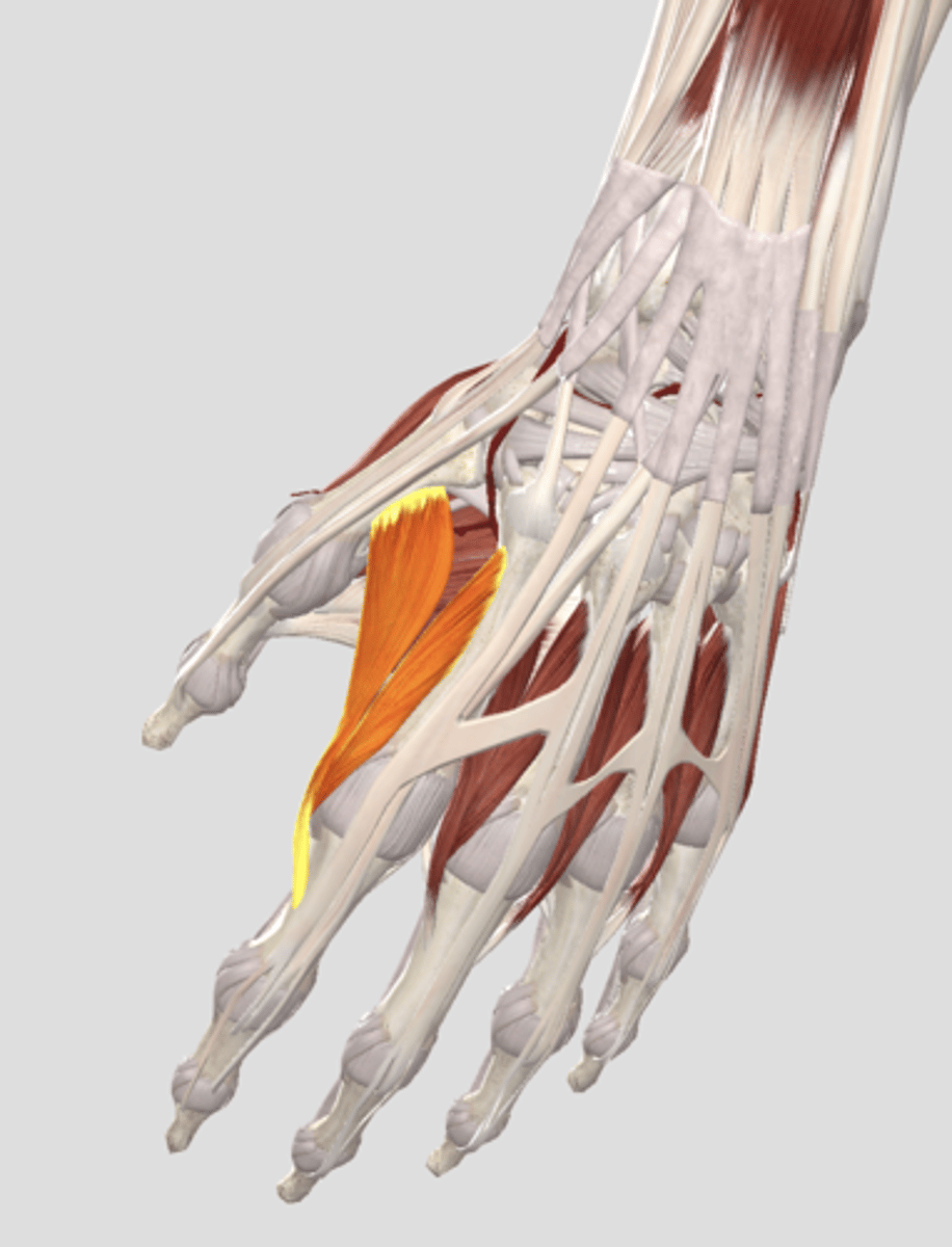
First dorsal interosseous muscle
Can see when abducting index finger. Located on the dorsal side of the hand. It abducts the fingers. Located inbetween metacarpals. PAD DAB.
Thenar muscles
opponens pollicis (median nerve), abductor pollicis brevis (median nerve), flexor pollicis brevis (median). adductor pollicis (ulnar nerve)
hypothenar muscles
opponens digiti minimi, abductor digiti minimi, flexor digiti minimi brevis.
All innervated by ulnar nerve.
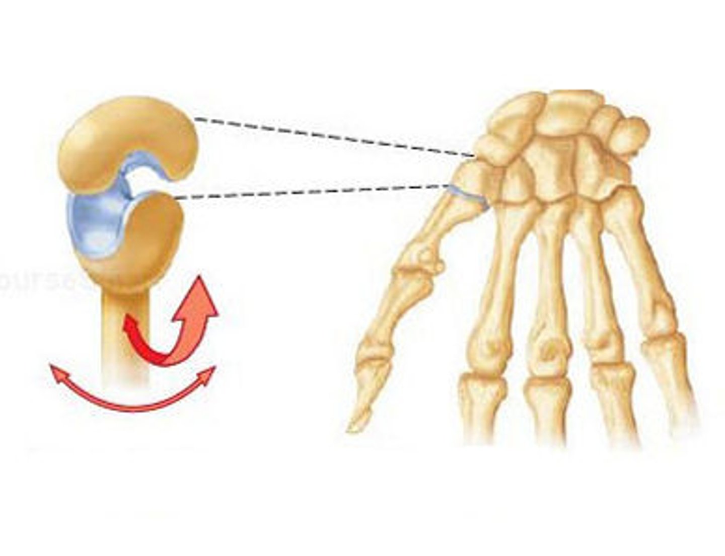
carpometacarpal joint of thumb
Connects carpal bones to the metacarpals. Synovial saddle joints with a concave and convex surface.
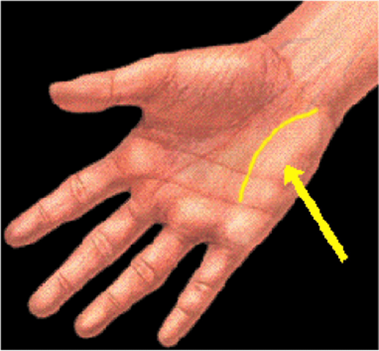
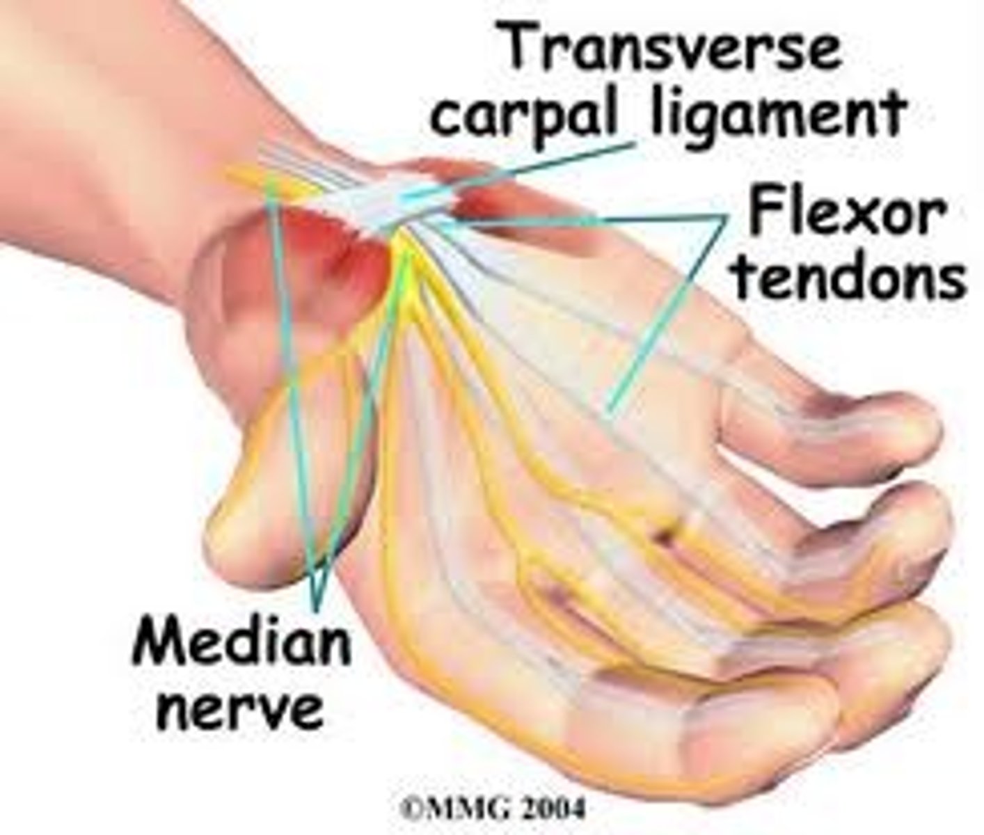
carpal tunnel
a passageway that runs from the forearm through the wrist
Consists of: flexor digitorum superficialis tendons, flexor digitorum profundus tendons, flexor pollicis longus tendon, median nerve
Borders: roof: flexor retinaculum. floor: carpal bones
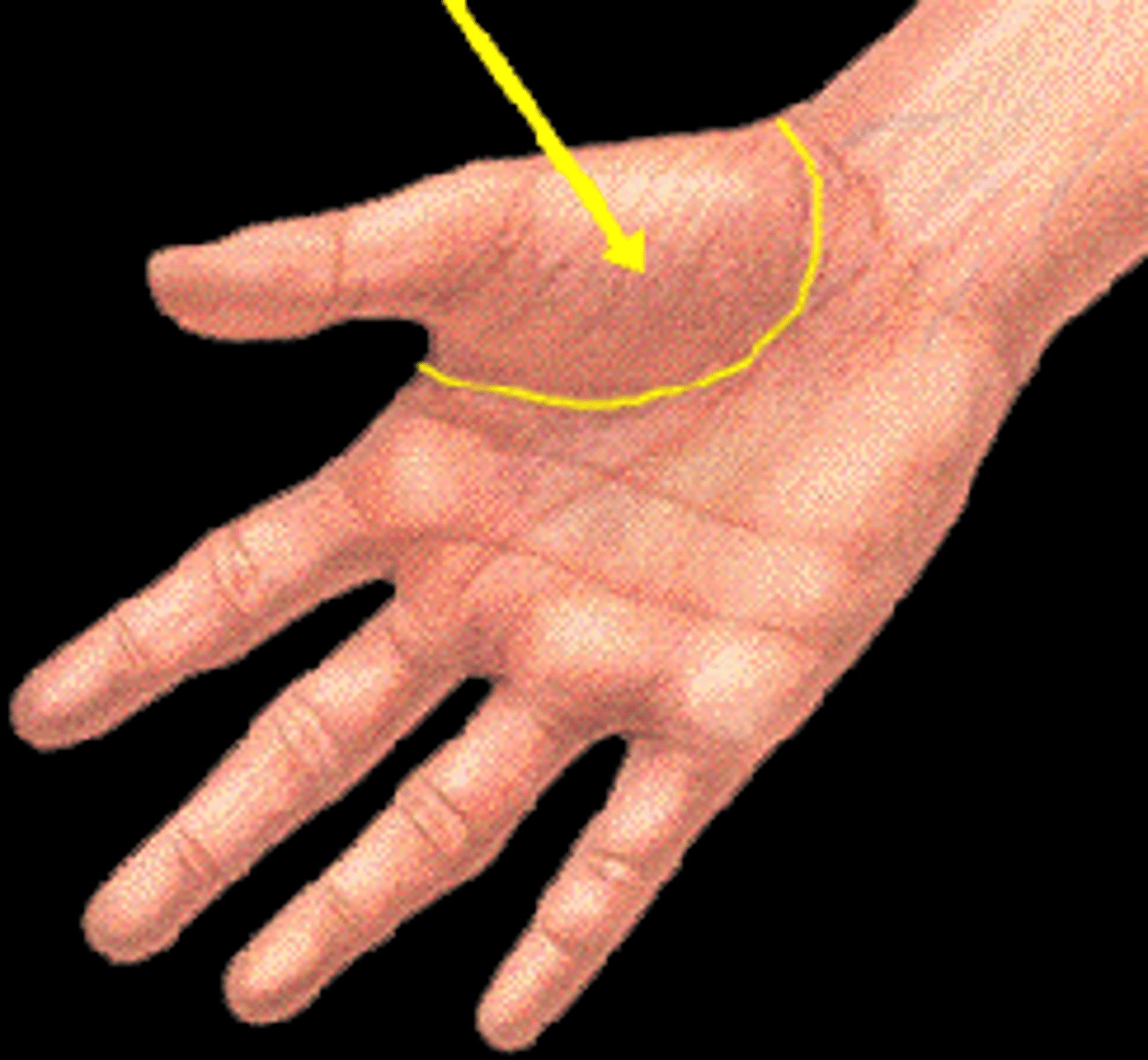
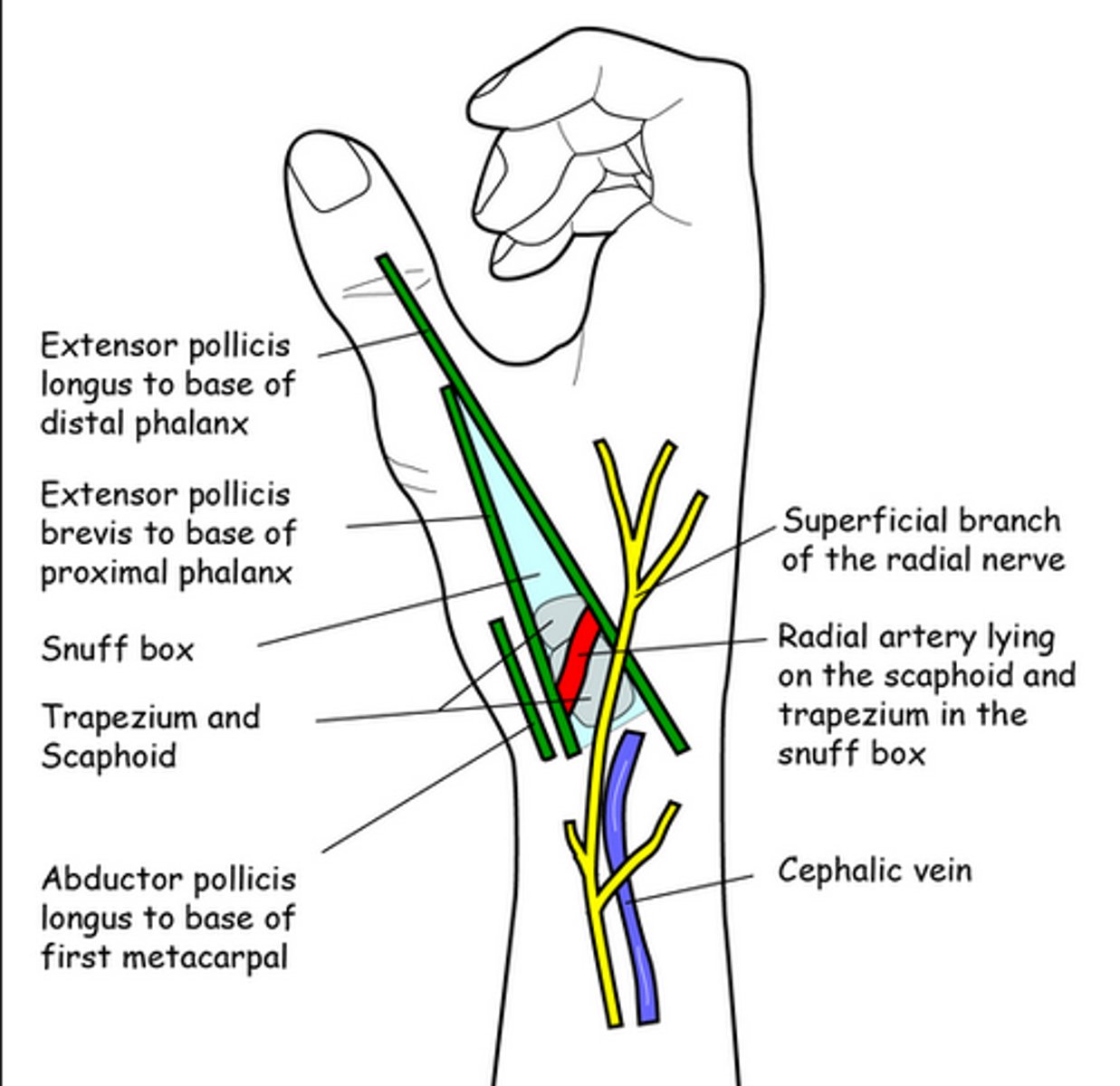
anatomical snuff box
Extend thumb to find it.
Scaphoid: base/floor.
abductor pollicis longus, extensor pollicis brevis, extensor pollicis longus.
Radial artery travels through floor.
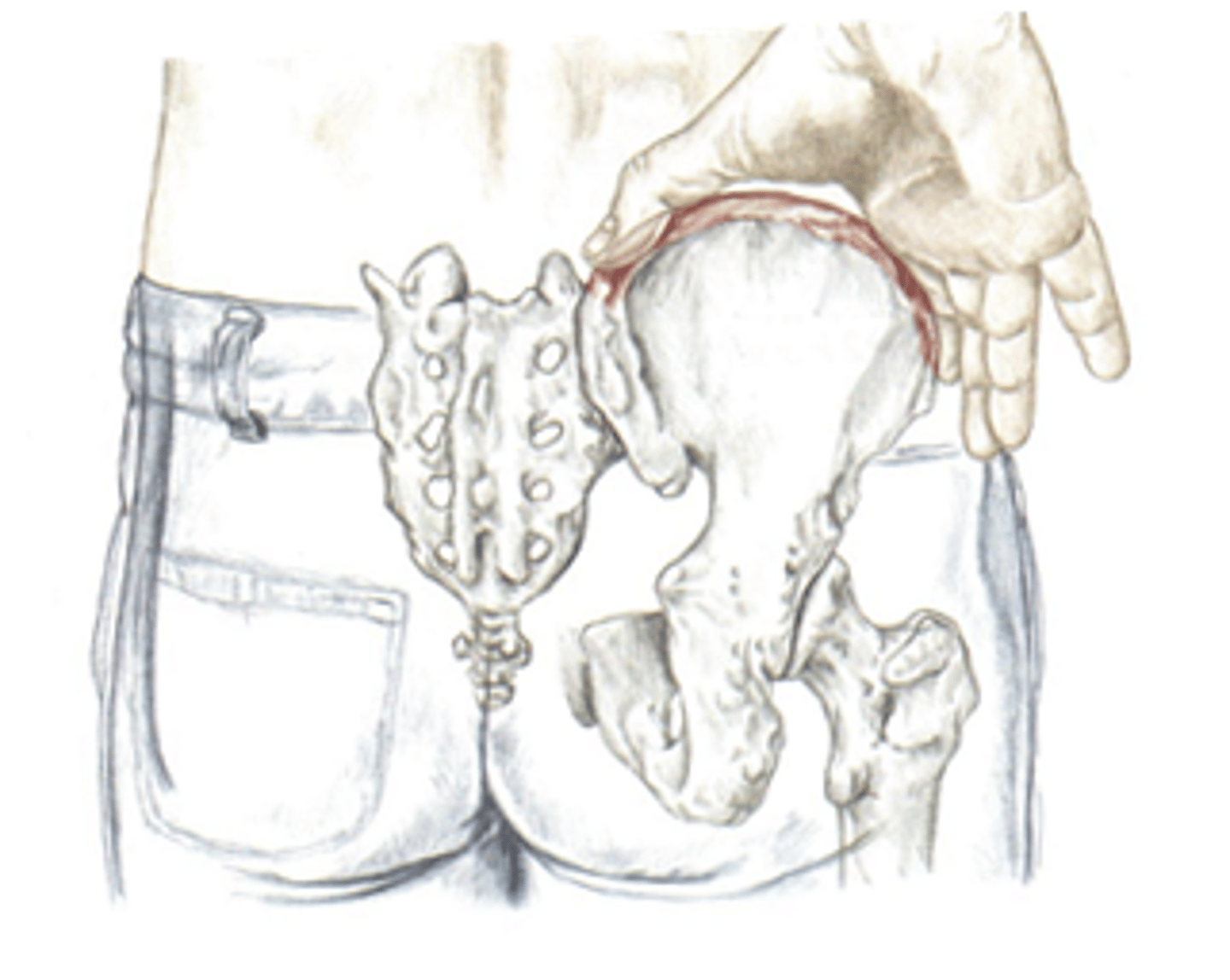
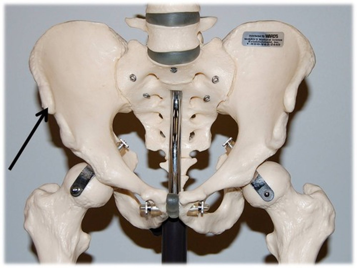
Iliac Crest (palpate)
Found on the top of the hip bone. pistol grip.
Site of attachment for erector spinae (extends spine) and latissimus dorsi (shoulder extension and adduction) and lumbar fascia.
Origin of gluteus maximus (hip extension)
Transverse abdominus attaches (compresses abdomen)
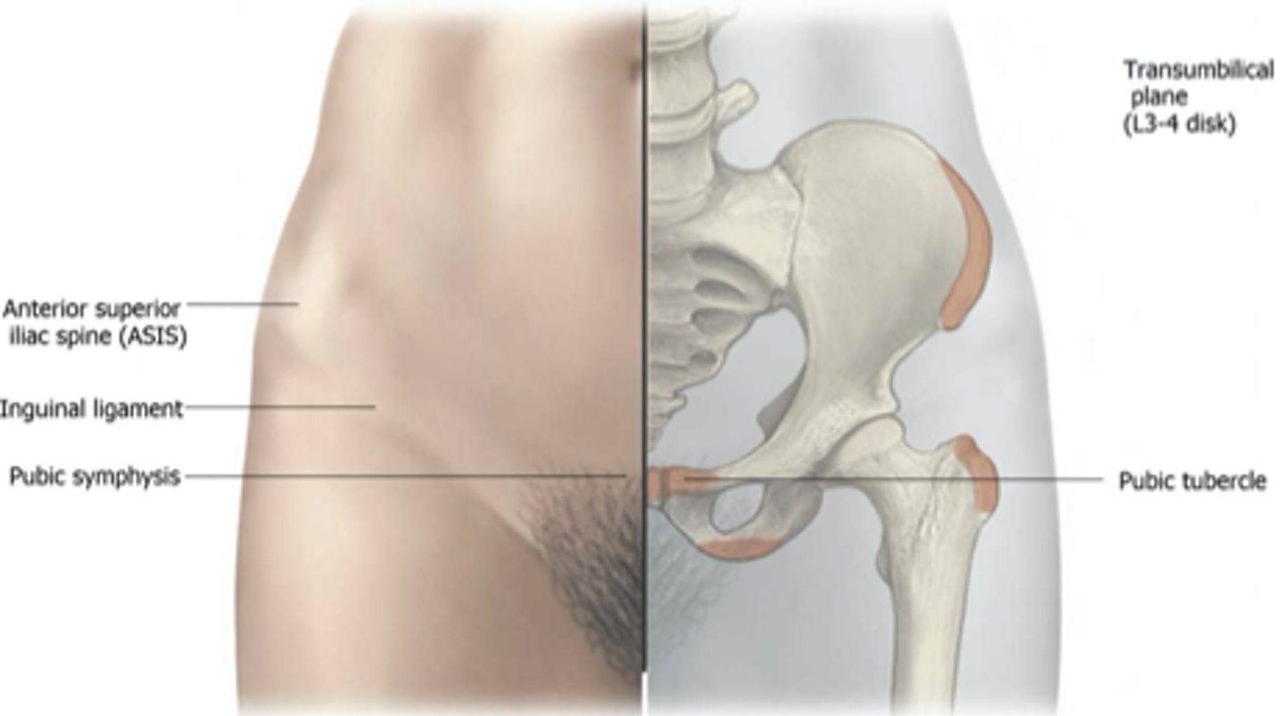
anterior superior iliac spine
Most anterior prominence of the ilium bone of the pelvis.
The attachment for the inguinal ligament and sartorius.
greater trochanter of femur
Palm on iliac crest, fingers on trochanter. Confirm by rotating hip interally/externally to feel it moving under hand.
Bony prominence on shaft of femur.
Insertion of medius and minimus (hip abduction).
Muscles that externally rotate the hip attach here: PGOGOQ: piriformis, gemellus superior and inferior, obturator internus and externus, quadratus femoris.
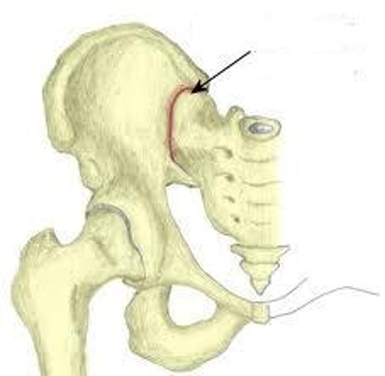
posterior superior iliac spine
find dimples of venus. the sharp posterior end of the iliac crest
gluteus maximus attaches here (hip extension) innervated by inferior gluteal nerve.
sacroiliac joint
the joint between the sacrum (5 fused vertebrae) and the ilium of pelvis. can palpate by finding ASIS dimples, as it is deep to that.
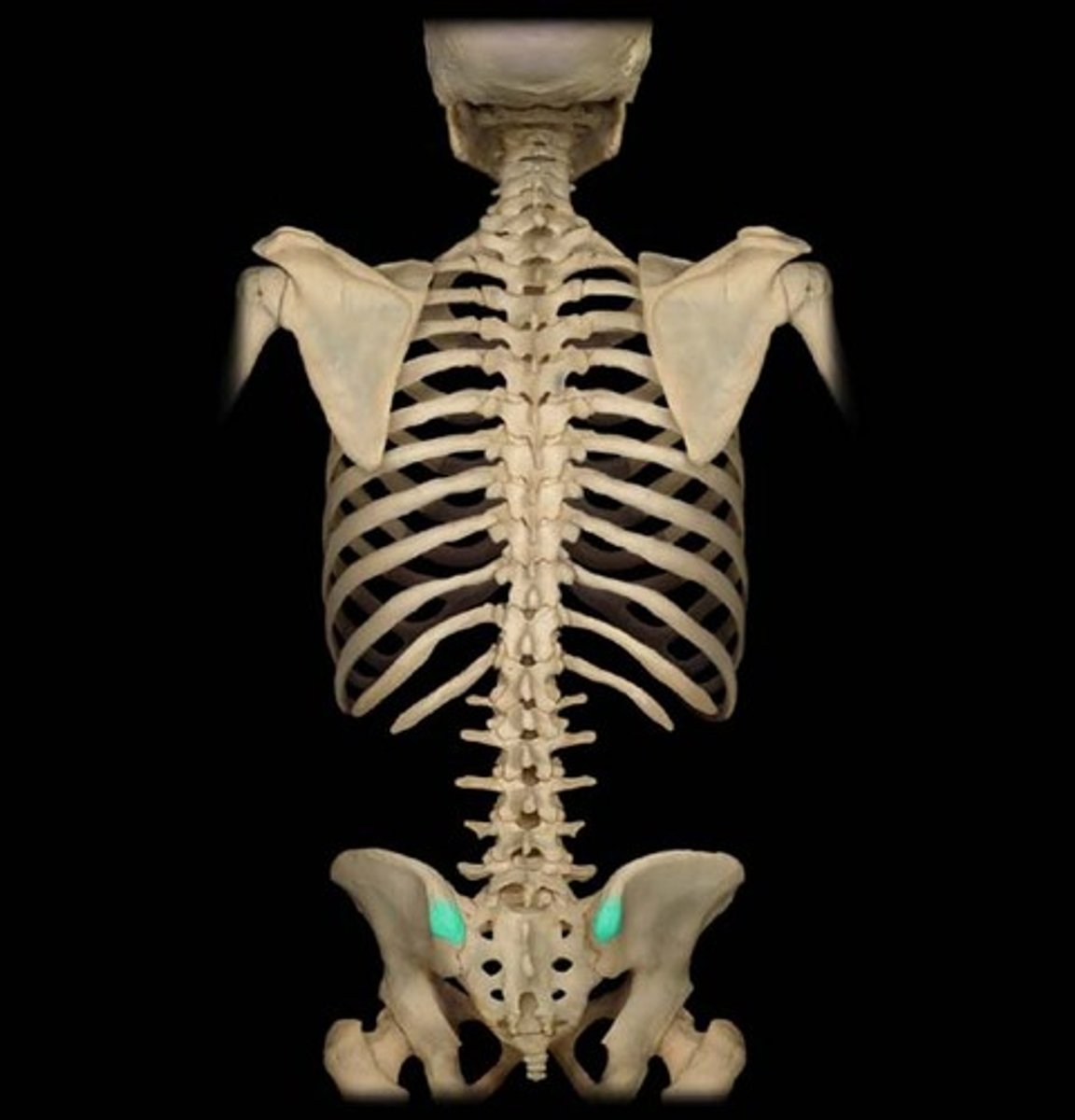
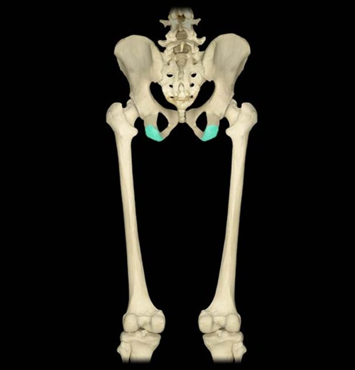
ischial tuberosity
Ischium bone. receives the weight of the body when sitting.
origin of hamstrings: biceps femoris, semimembranosus, and semitendinous (hip extension and knee flexion)
(sciatic nerve)
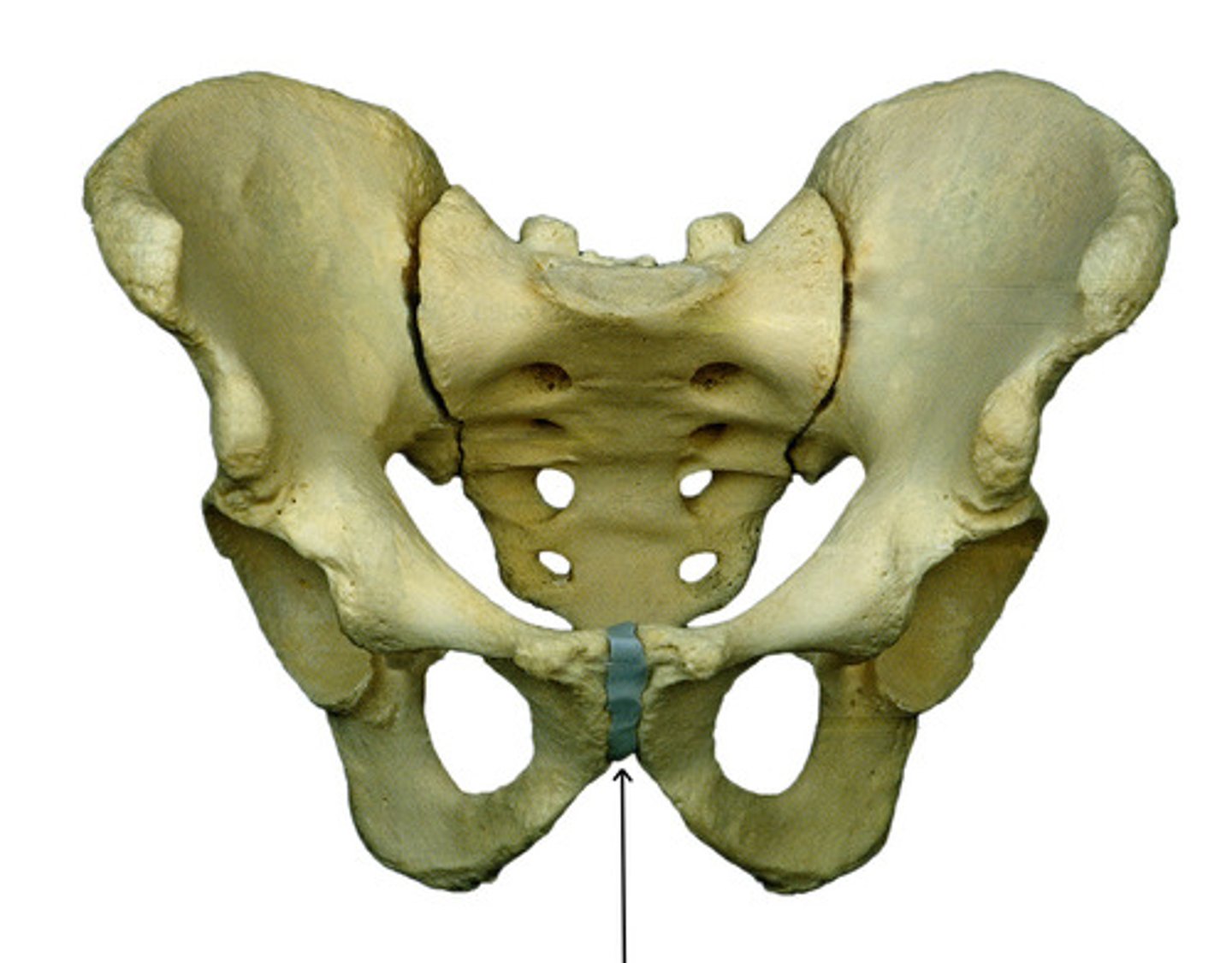
pubic symphysis
the cartilaginous joint known that allows some movement to facilitate childbirth
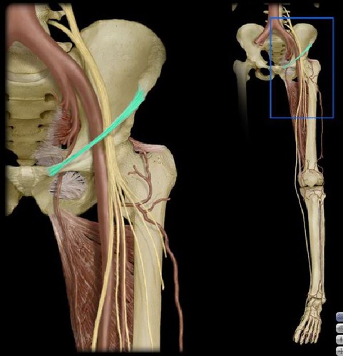
attachments of inguinal ligament
anterior superior iliac spine to pubis (tubercle). superior border to the femoral triangle: a transition zone containing femoral artery, nerve, and vein. Protects these structures passing over the hip joint.
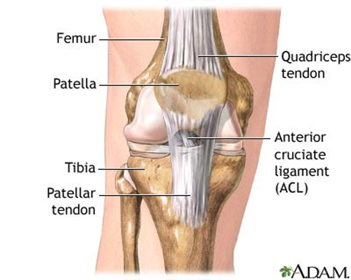
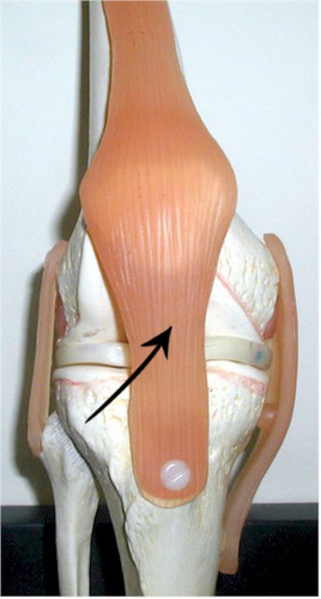
quadriceps tendon
common tendon for quadriceps group, attaches to patella to allow for knee extension
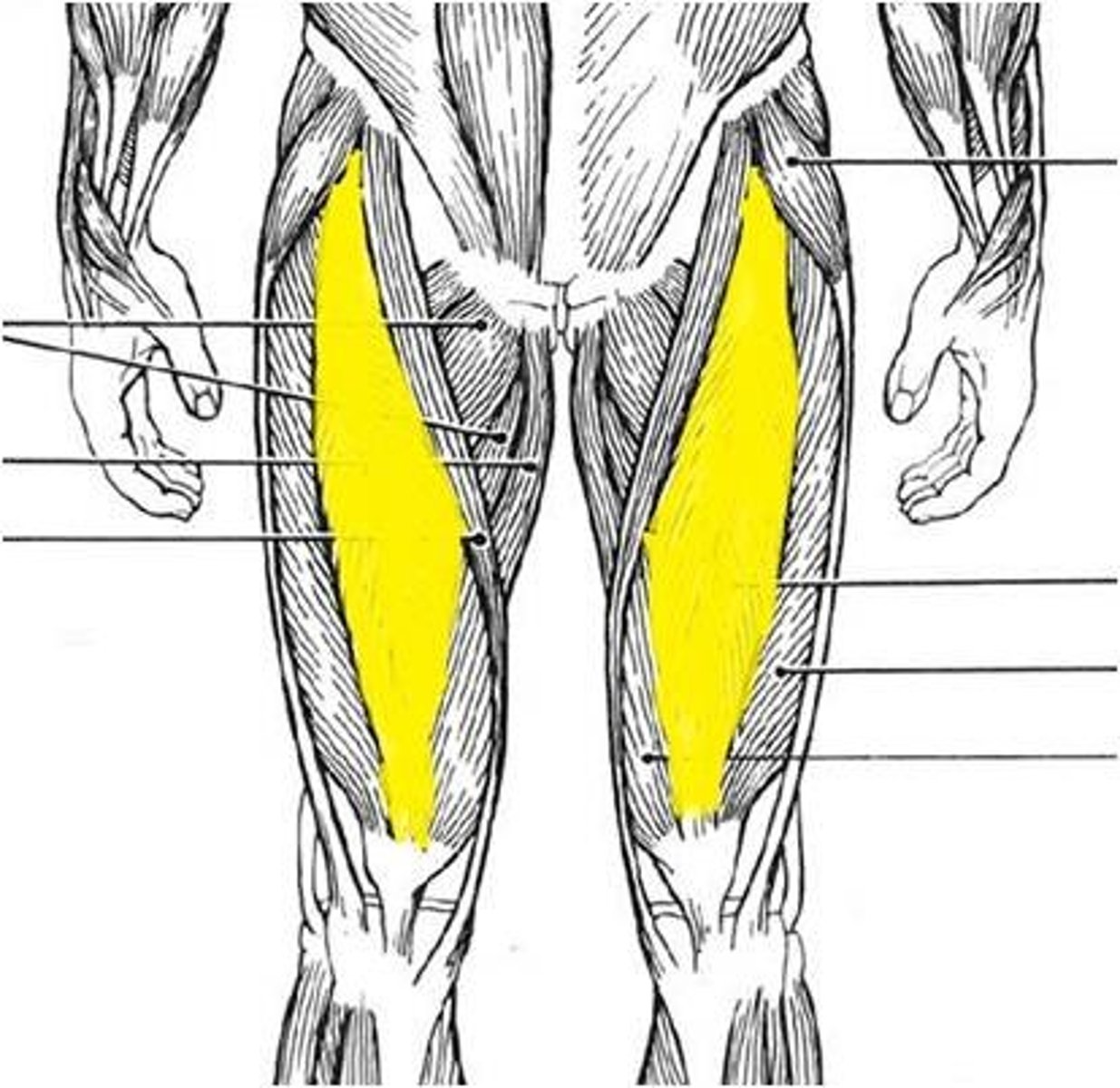
anterior compartment of the thigh
Contains quadriceps muscles and sartorius. all innervated by the femoral nerve.
Rectus femoris: Origin- ilium bone
Vastus lateralis, vastus intermedius, vastus medialis: Origin- femur bone.
Action = hip flexion and knee extension
Sartorius:
longest muscle in the body. Action: external rotation of hip, knee flexion, sitting cross-legged
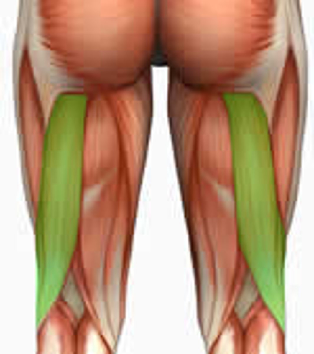
biceps femoris
-Origin: ischial tuberosity, femur
-Insertion: the head of the fibula which articulates with the back of the lateral tibial condyle
-Nerve long head: tibial nerve. short head: common fibular nerve
-Actions: flexes knee, extends hip joint (long head only)
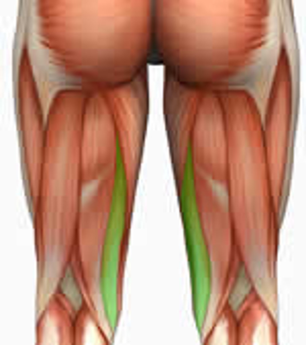
Semitendinosus
Hip extension, knee flexion; belongs to the hamstring group
Insertion: medial tibial condyle
Forms the medial border of popliteal fossa: transition zone behind knee: popliteal artery/vein, and tibial nerve.
N: sciatic
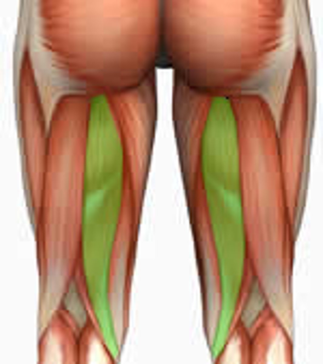
Semimembranosus
Origin: Ischial tuberosity
Insertion: medial tibial condyle
Action: Hip extension, knee flexion
Forms the medial border of popliteal fossa: transition zone behind knee: popliteal artery/vein, and tibial nerve.
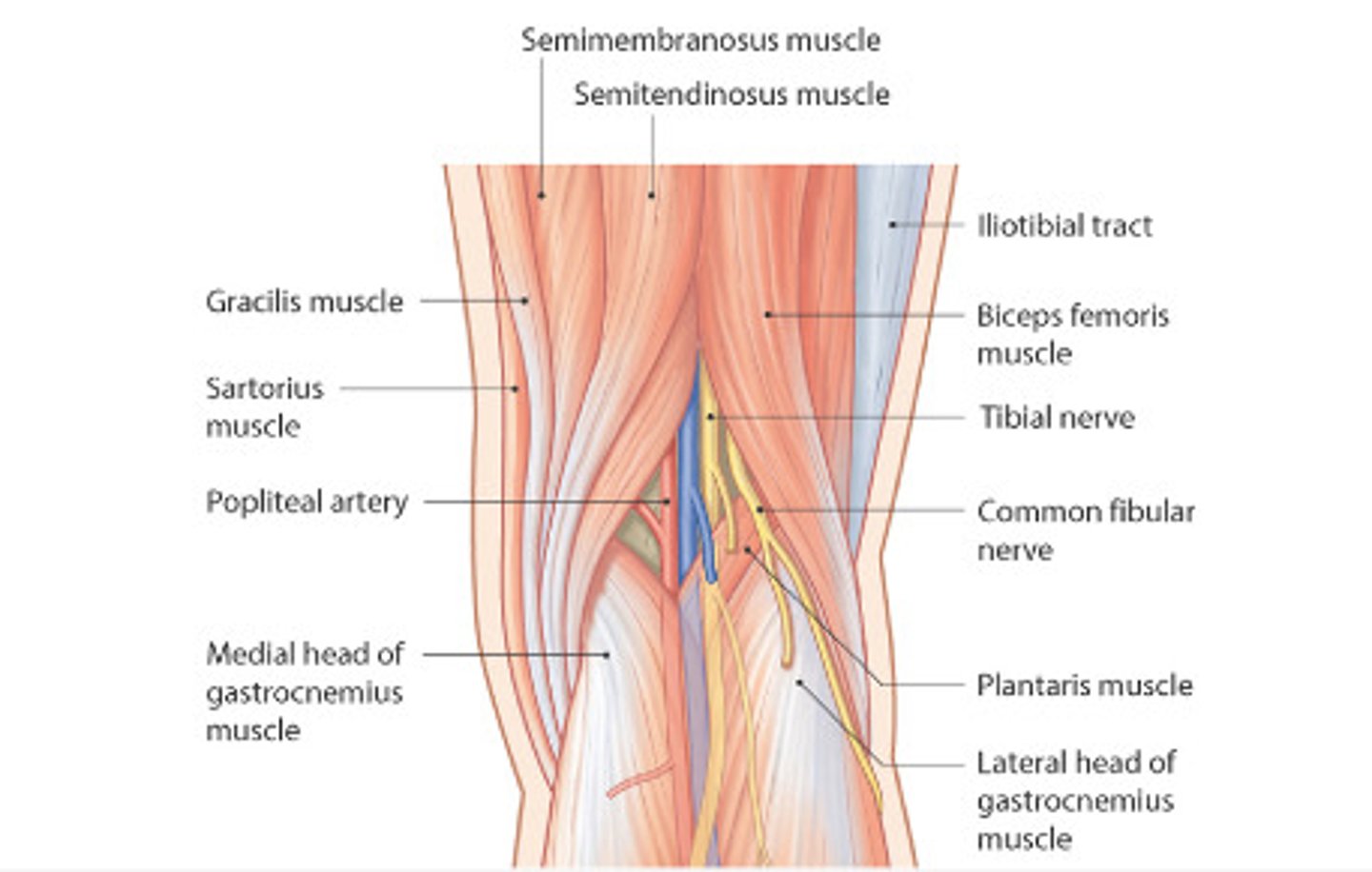
popliteal fossa
Border
superior/lateral: biceps femoris tendon
superior/medial: semimembranosus, semitendinosus
inferior/lateral: lateral head of gastrocnemius
inferior/medial: medial head of gastrocnemius
contents: popliteal Artery (from femoral A) Popliteal Vein (from femoral V), tibial nerve, and common fibular nerve
patellar ligament
connects the tibial tuberosity to the quadriceps tendon allowing for knee extension.
Gerdy's tubercle
superior and lateral to tibial tiberosisty.
Insertion of iliotibial band.
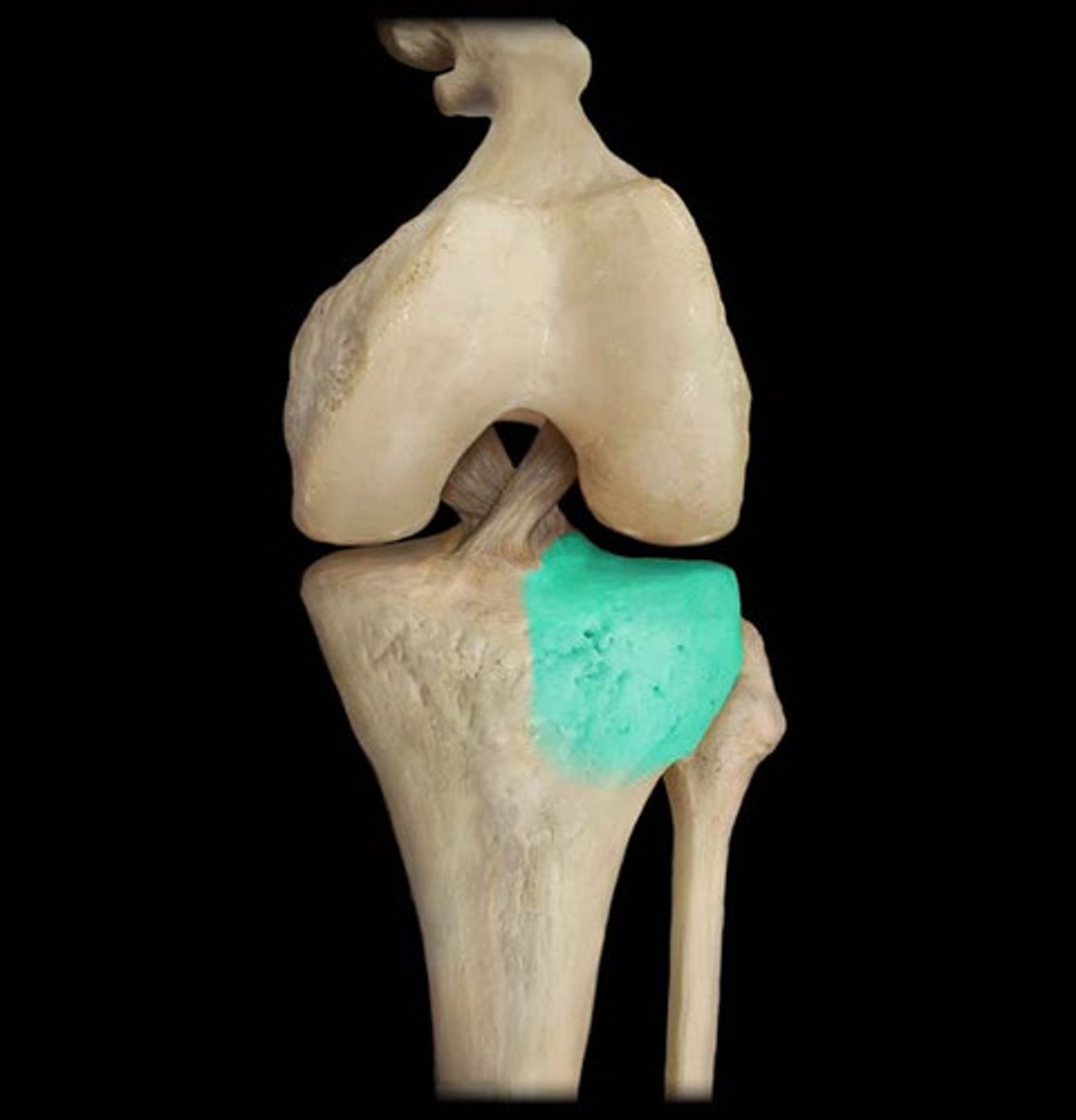
lateral tibial condyle
lateral plateau.
where tibia articulates with the lateral condyle of the femur via the lateral meniscus (fibrocartilagenous disc)
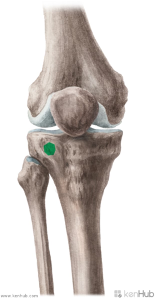
tibial tuberosity of tibia
Anterior aspect of tibia below patella.
-rough textured prominence located on midanterior surface of the tibia just distal to condyles
- distal attachment of patellar tendon- which connects to large muscle of quads for knee extension.
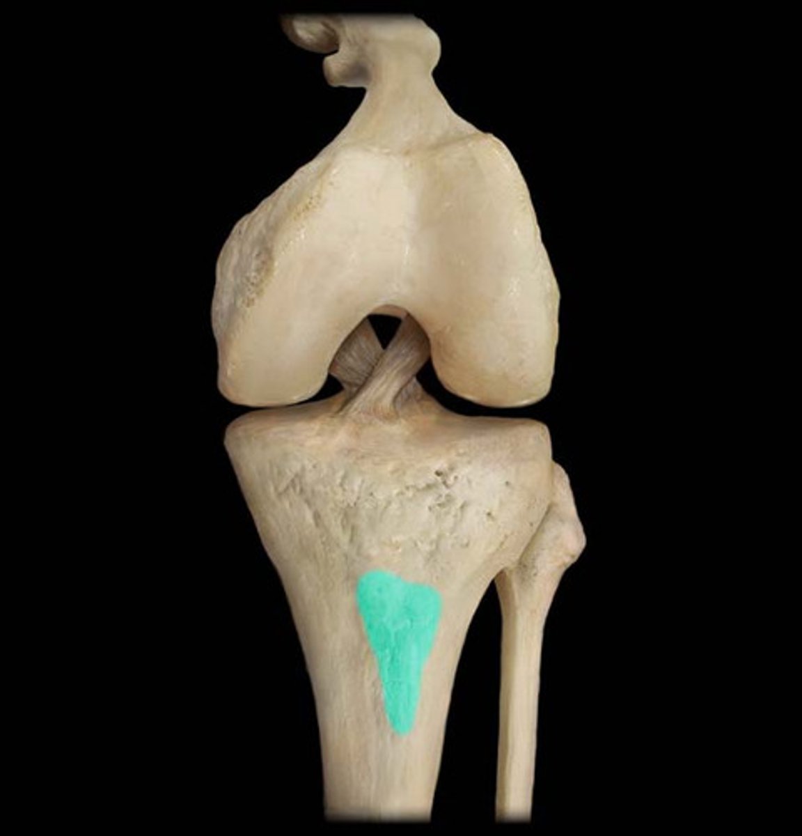
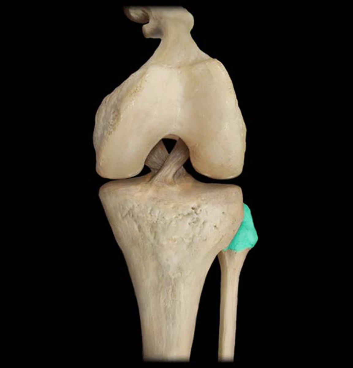
head of fibula
inferior and lateral to patella. Find tibial tuberosity and gerdy’s tubercle first
insertion of biceps femoris
attachement of fibular collateral ligament.
posterior, lateral knee
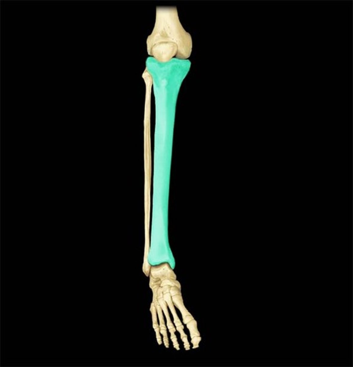
tibia bone
shin bone
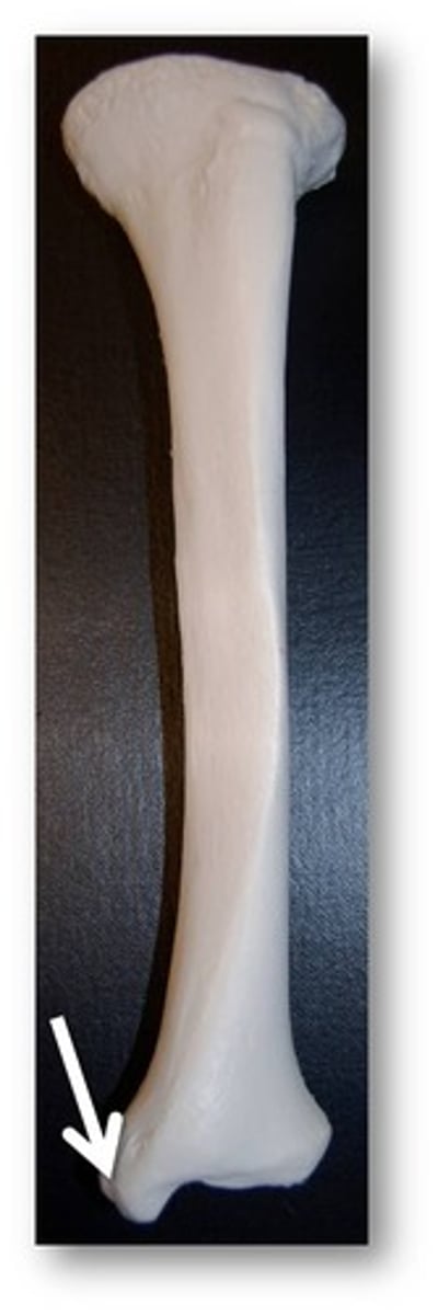
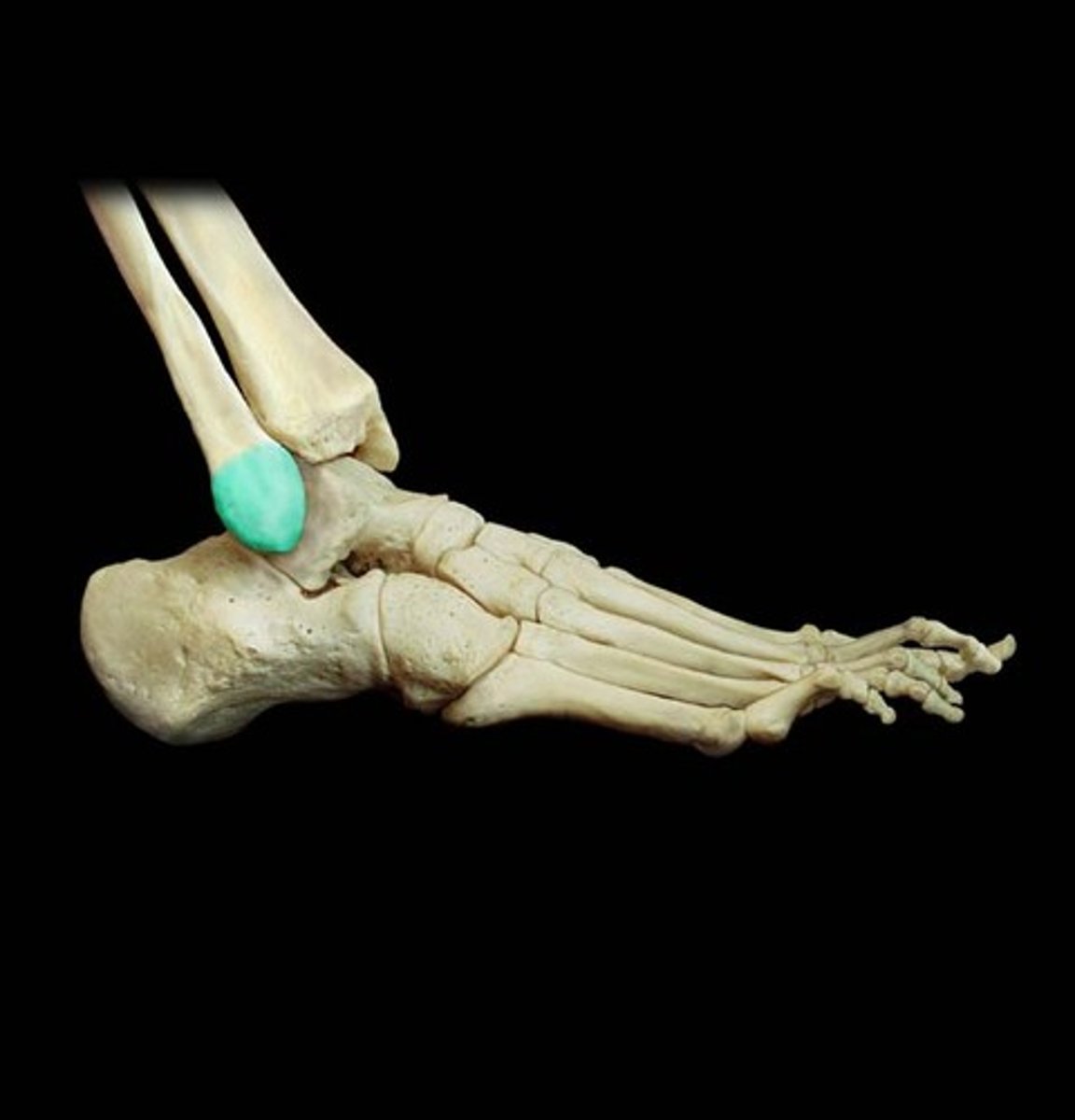
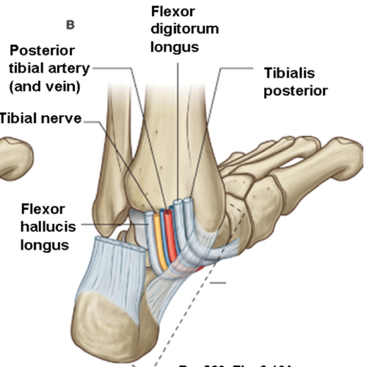
medial malleolus
medial process on distal end of tibia, forms medial bump of ankle
articulates with talus. forming part of ankle joint.
forms tarsal tunnel with flexor retinaculum (connective tissue band on medial ankle): transition zone (TDAVNH)
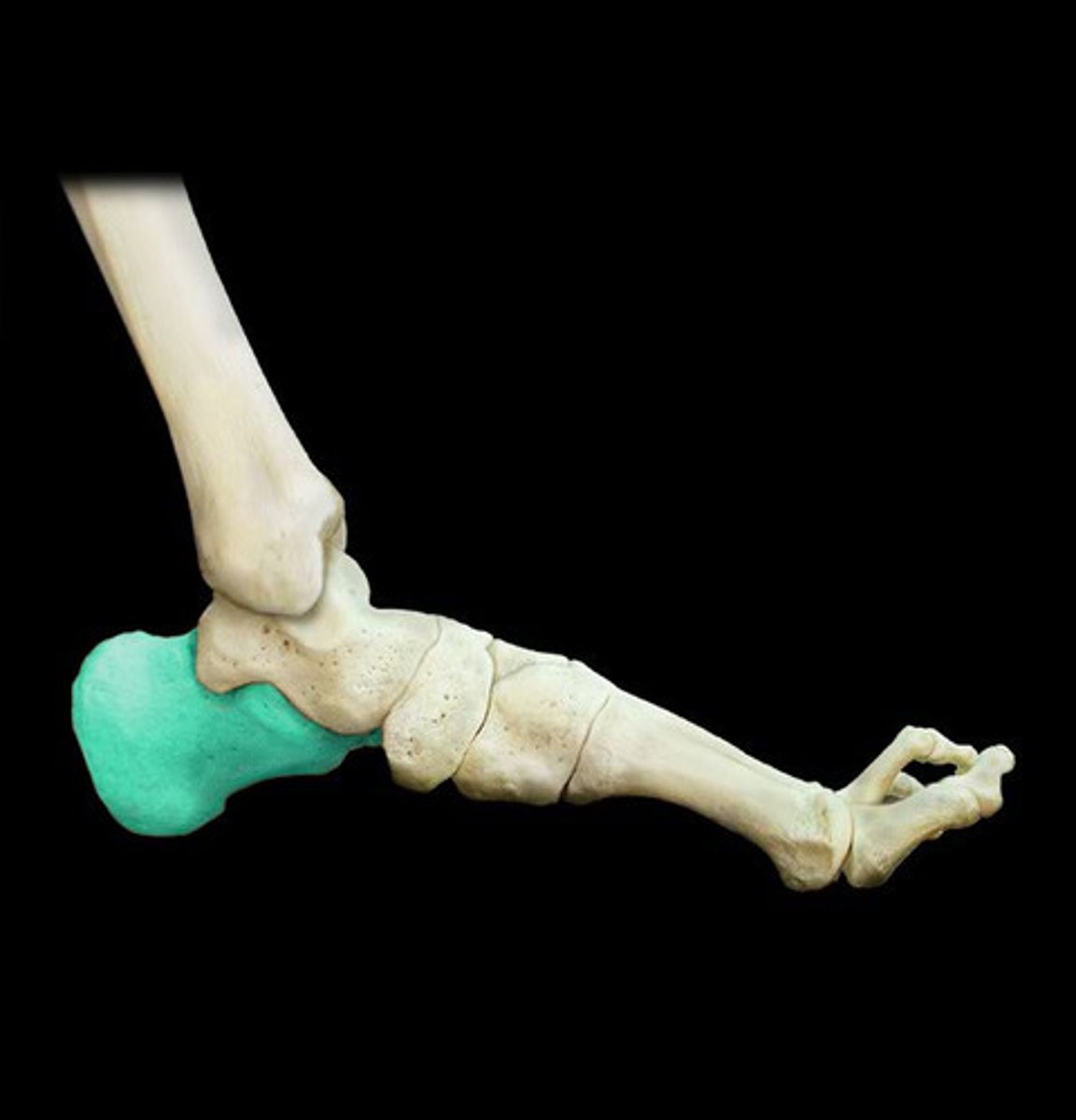
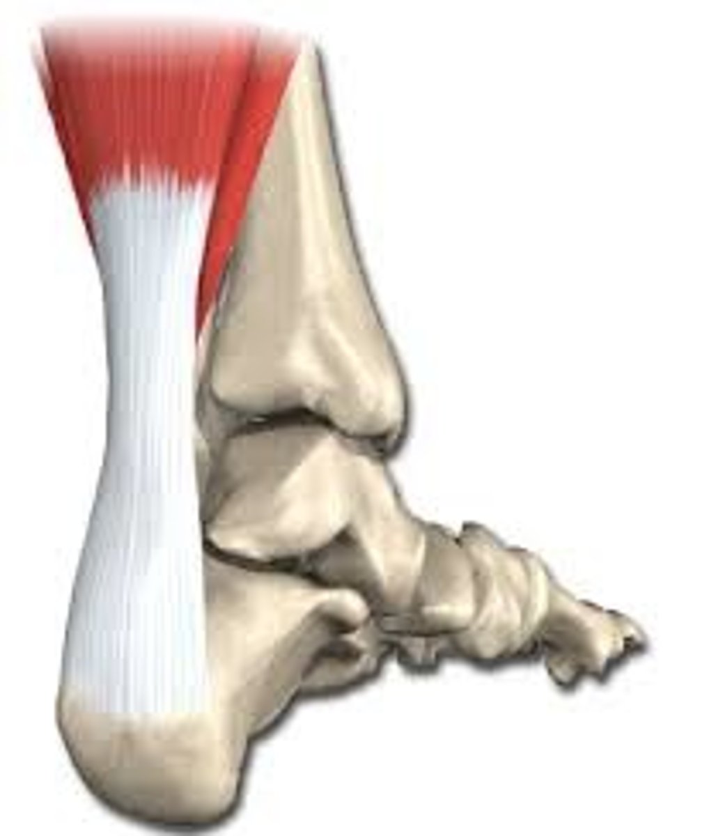
calcaneus
posterior heel bone
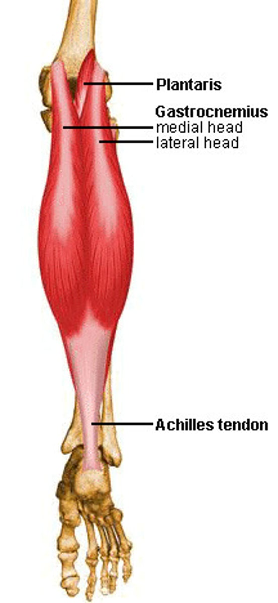
insertion of gastrocnemius, soleus, and planatris via achilles tendon.
Largest tarsal bone. origin of extensor hallucis brevis, extensor digitorum brevis.
lateral malleolus
Distal fibula. forms the lateral bulge of the ankle and articulates with the talus
Contains groove for fibularis longis and brevis muscles (ankle everters)
achilles tendon
attaches the gastrocnemius muscle to the heel bone
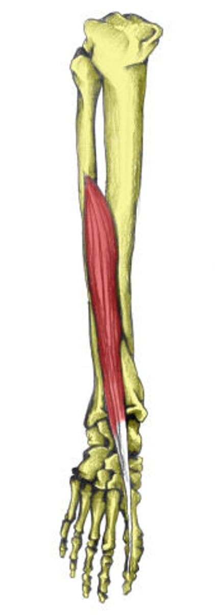
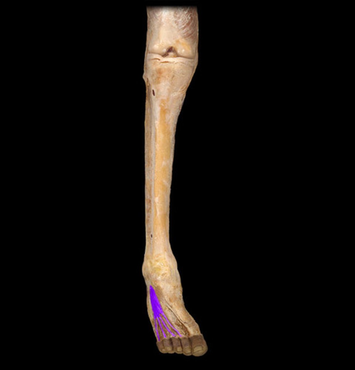
extensor hallucis longus
-Origin: Arises from the middle portion of the fibula on the anterior surface and the interosseous membrane between fibula and tibia.
-Insertion: on the dorsal side of the base of the distal phalanx of the big toe
-Actions: Extends the big toe and assists in dorsiflexion of the foot at the ankle.
N: deep fibular
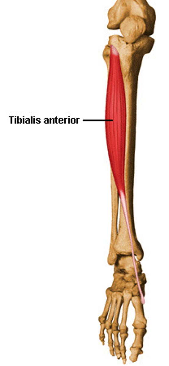
tibialis anterior
Origin: lateral surface of tibia
Insertion: medial cuneiform and 1st metatarsal
Action: dorsiflexes and inverts foot
N: deep fibular
extensor digitorum longus
-Origin: Anterior lateral condyle of tibia, anterior shaft of fibula and interosseous membrane
-Insertion: Dorsal surface; middle and distal phalanges of toes 2-5
-Actions: extension of toes, dorsiflexion of ankle
N: deep fibular
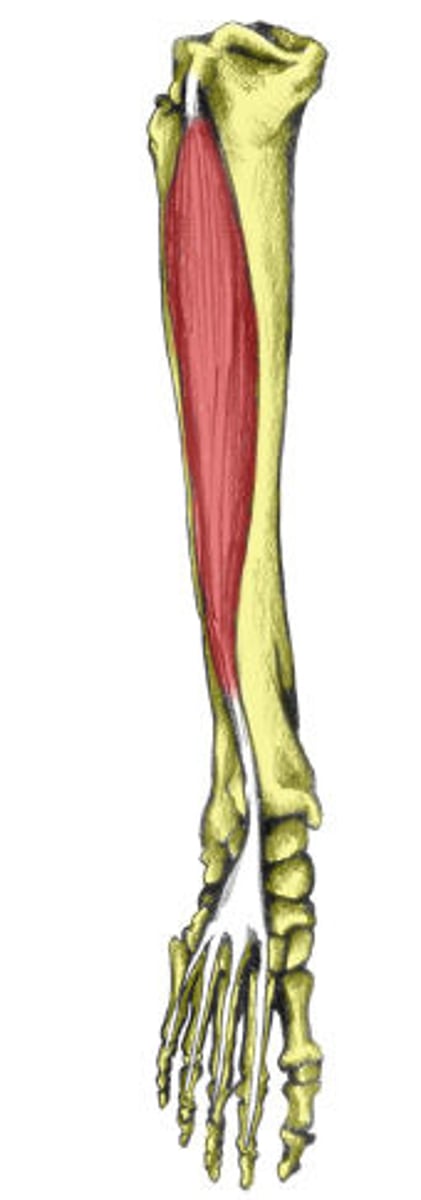
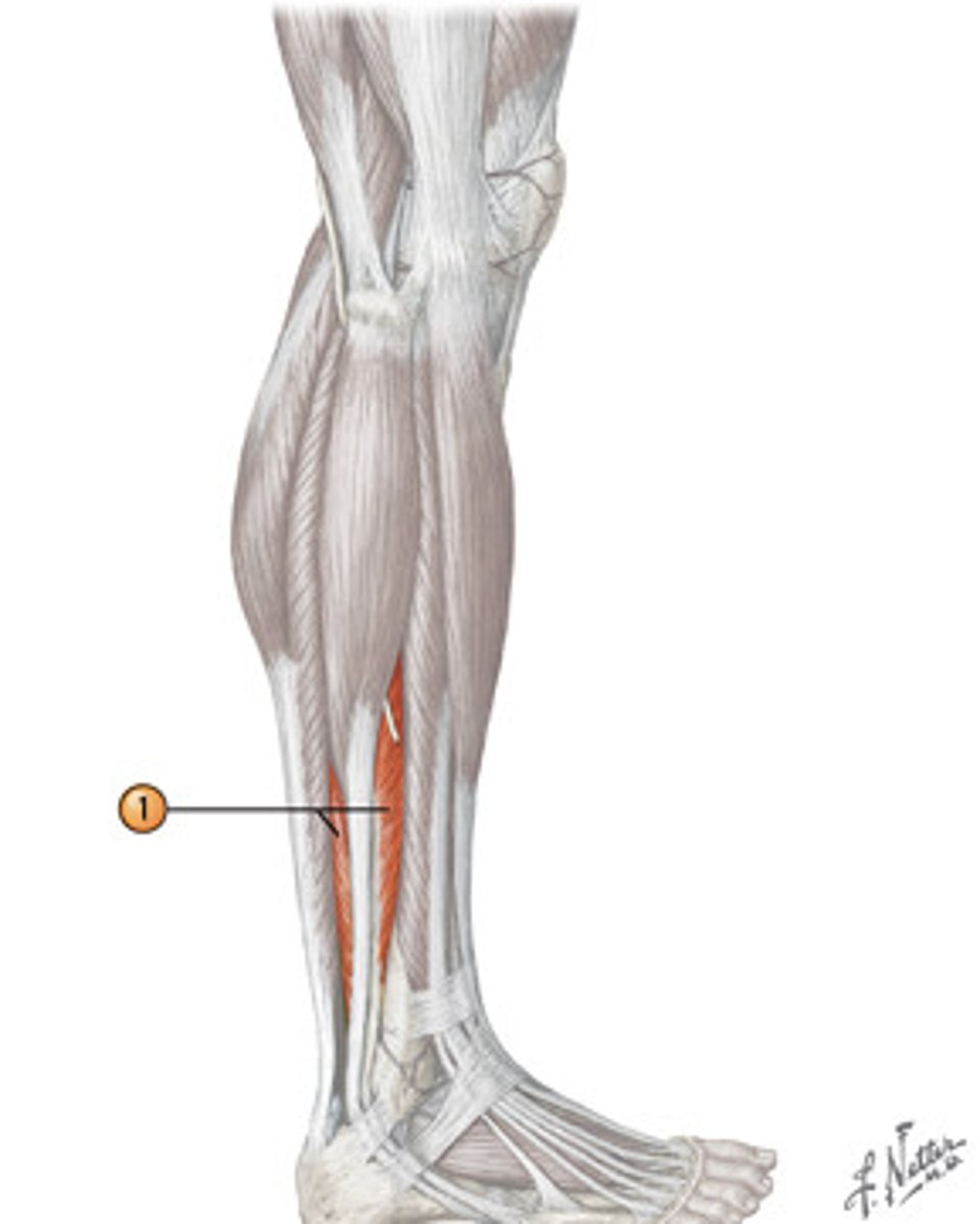
Gastrocnemius
Two heads.
Origin: lateral and medial condyles of femur
Insertion: calcaneus (achilles tendon)
Action: plantar flexion
N: tibial nerve
Can feel achilles tendon attaches to calcaneus.
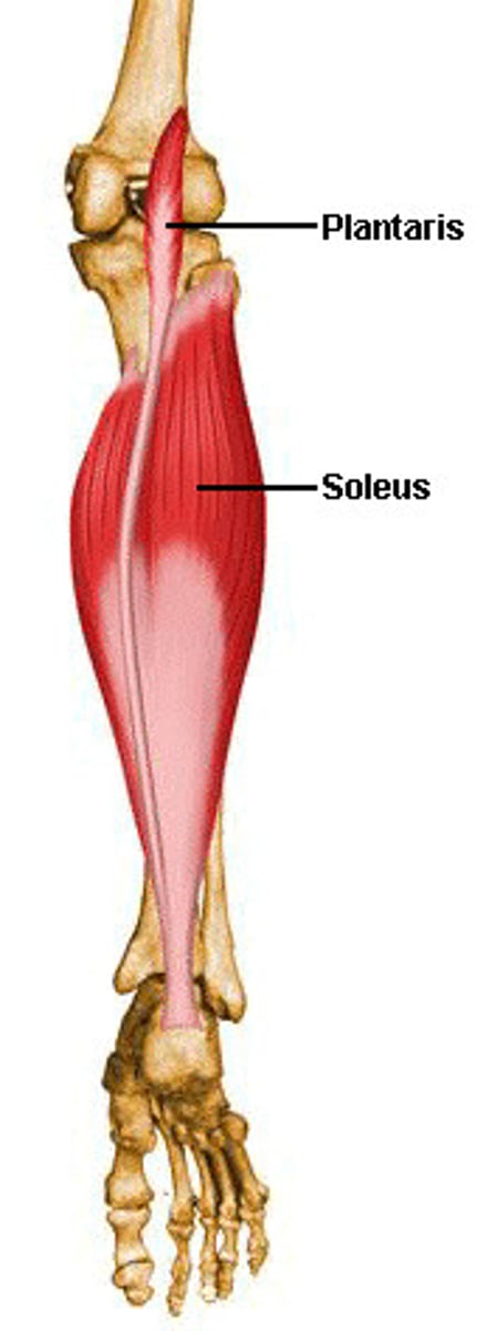
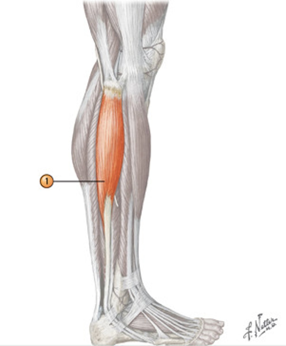
soleus
deeper to gastrocnemius
-Origin: fibula, tibia (soleal line)
Insertion: achilles tendon on calcaneus
Action: plantar flexion
fibularis longus
origin: lateral surface of fibula
insertion: medial cuneiform and 1st metatarsal
action: plantar flexes and everts foot
fibularis brevis
Origin: Distal fibula.
Insertion: 5th metatarsal
Action: Plantar flexes and everts foot.
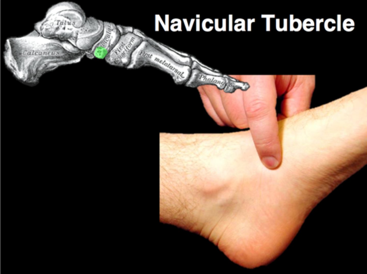
navicular
Medial aspect of foot above arch.
A tarsal bone. Boat shaped.
tibialis posterior attaches to it.
cuboid
tarsal bone in lateral midfoot.
Locate the 5th metatarsal tuberosity. Run your finger down the outside edge of your foot until you feel a prominent bony bump, then depression.
Attachement: tibialis posterior (travels behind medial malleolus to attach on the plantar side)
base of 5th metatarsal
enlarged & prominent to serve as insertion for fibularis brevis & tertius
The heads of the metatarsals palpation
tarsal tunnel
Transtion zone. protects underlying structures.
Borders: medial malleolus. floor and the roof composed of the flexor retinaculum.
Contents: Tibialis posterior, flexor digitorum muscles, flexor hallucis longus, tibial nerve, artery, vein (TDAVNH)
extensor digitorum brevis
Origin: dorsal surface of calcaneus
Insertion: middle phalanges of digits 2-4
Action: extends digits 2-4
N: deep fibular
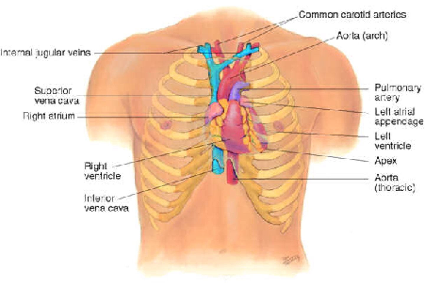
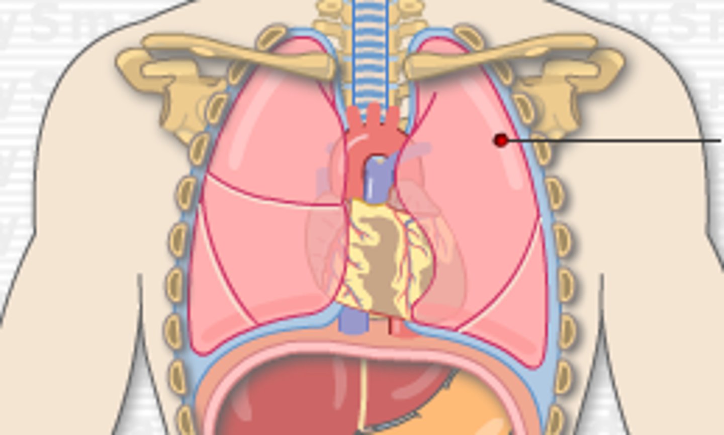
heart
About fist-sized. The base lies behind the sternum, extending slightly into the right side. The superior part lies in the 2nd intercostal space and the apex lies in the 5th intercostal space in the left.
Lies within a double-layered membrane called the pericardium, which is attached inferiorly to the diaphragm.
Position of lungs in the body
The superior lobes of the lungs rise above the clavicle.
The left lung moves laterally at the 4th rib to make the cardiac notch. The right lung moves laterally at the 6th rib.
In the mid-axillary line, the lungs extend down to the 8th ribs.
Posteriorly, the lungs extend down to the 10th ribs.
The parietal pleura extend down two ribs further than the lung tissue. The lungs sit on top of the diaphragm and so does the heart.
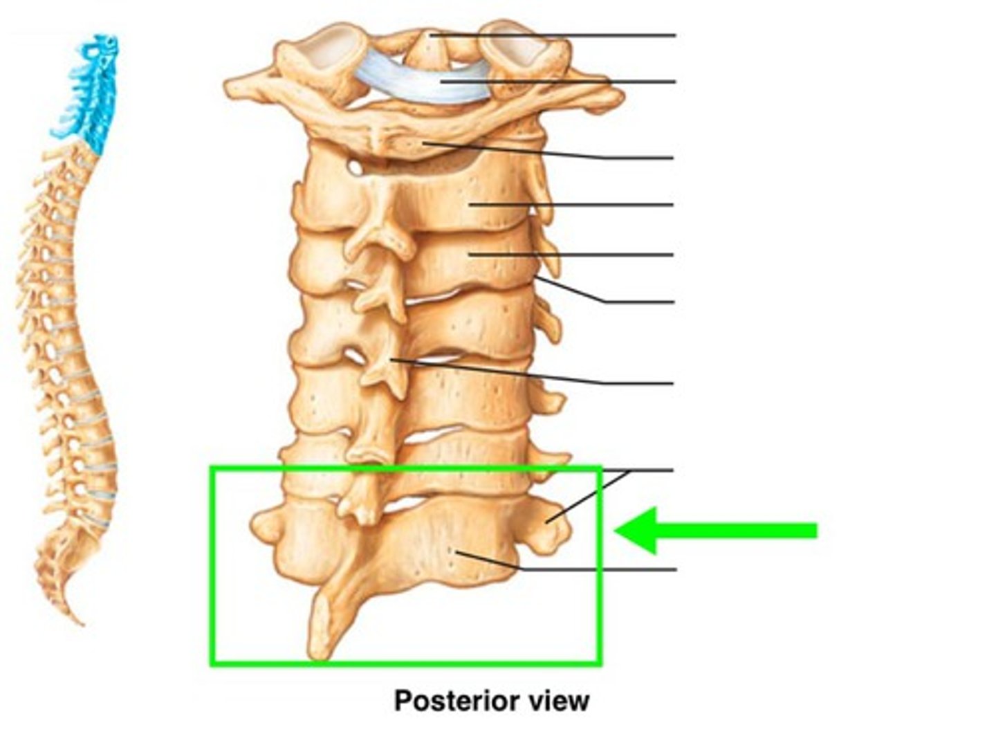
spinous process of C7
bow down head and feel most prominent bone on neck.
Unlike the other cervical vertebrae, this one has a large spinous process that protrudes posteriorly at back of neck.
last cervical vertebrae before thorax.
ligamentum nuchae (thick ligament on back of head) and muscles such as trapezius attach here.
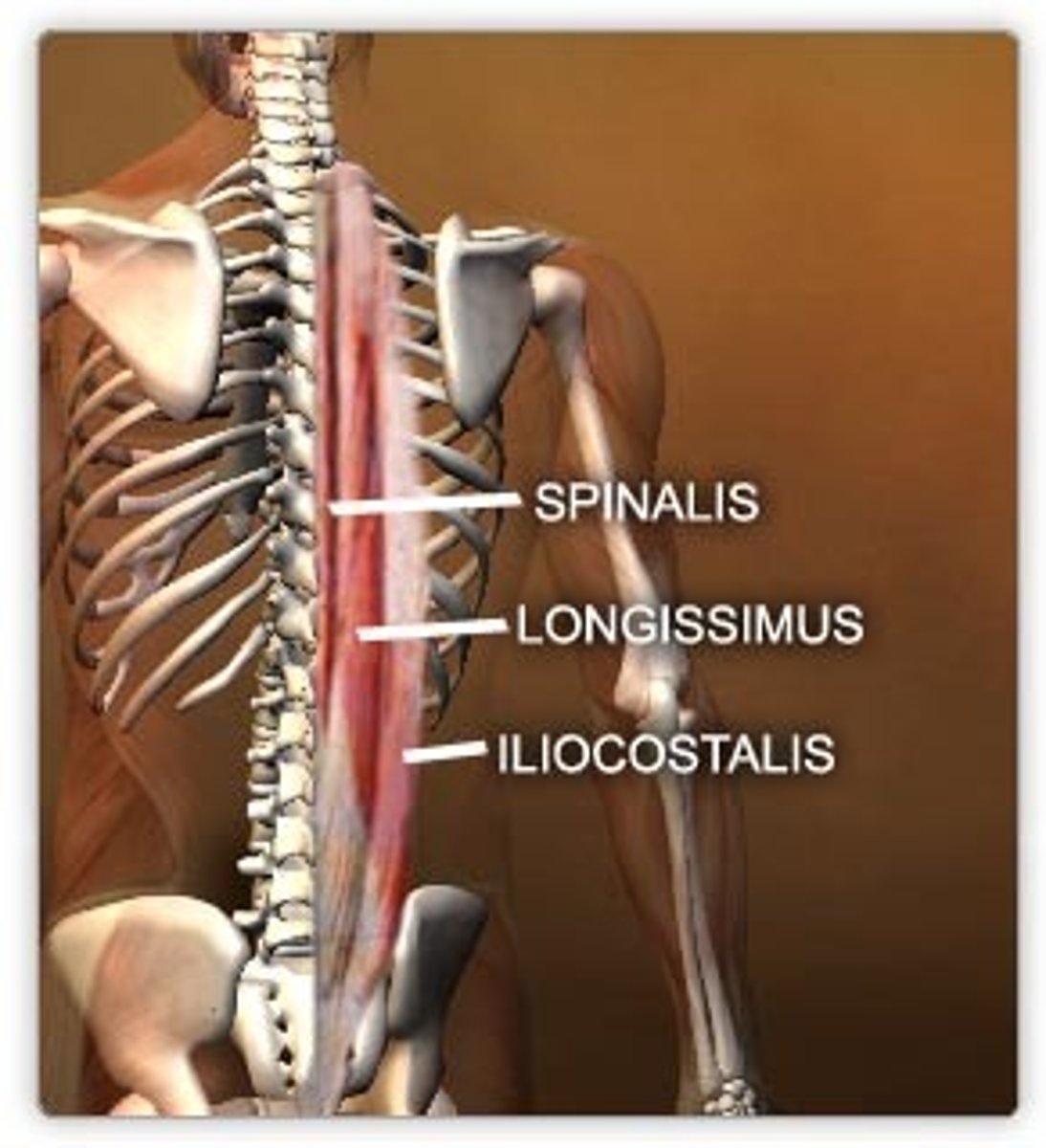
erector spinae
A: extend the vertebral column bilaterally. Unilateral: lateral flexion or rotation of trunk
Iliocostalis: O: the posterior iliac crest and lumbar fascia. I: the ribs.
Longissimus: O: Sacrum, posterior iliac crest, transverse process of vertebrae. I: Mastoid process of temporal bone.
Spinalis: O: Spinous process of vertebrae. I: spinous processes and occipital head bone.
Innervation: Posterior rami of spinal nerves
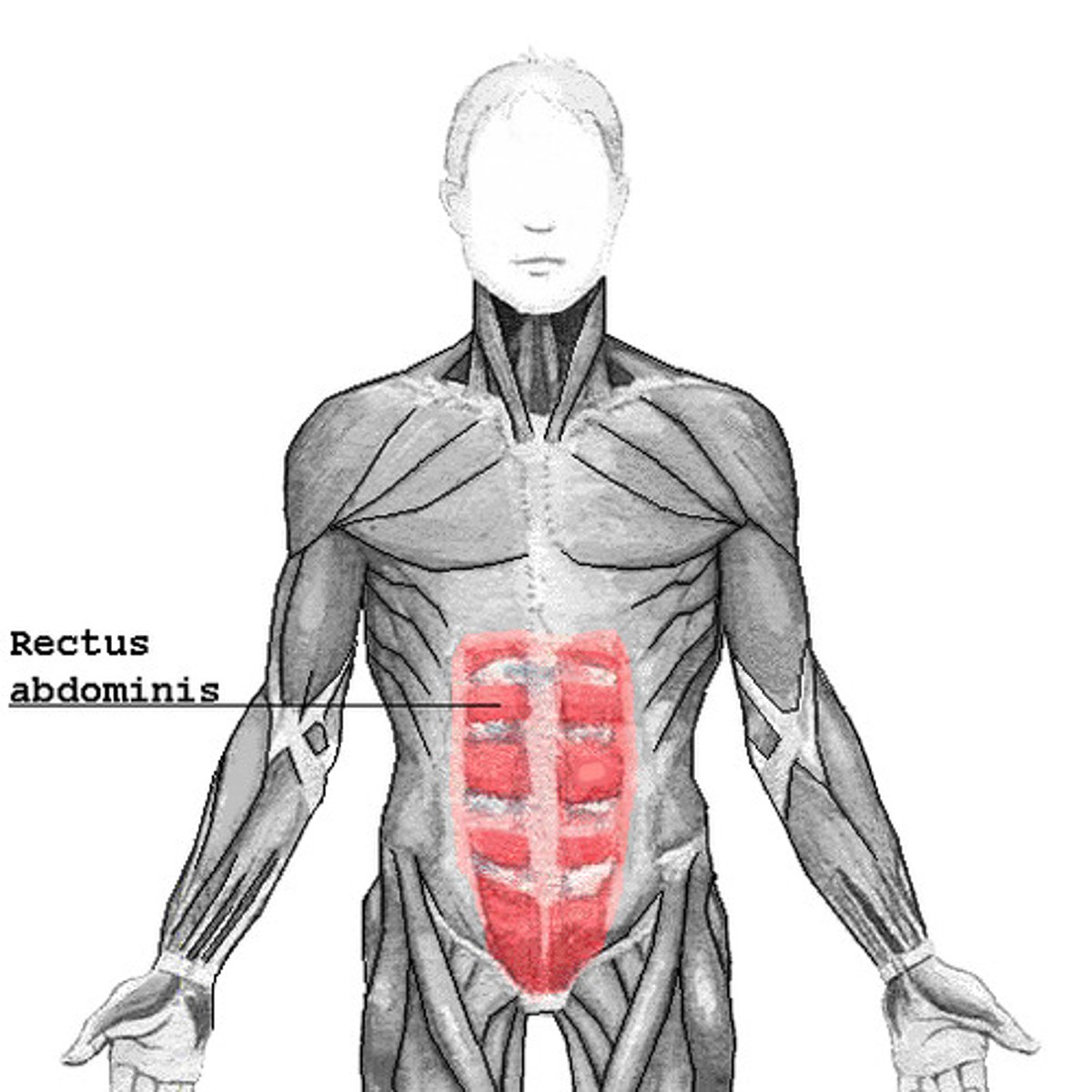
rectus abdominis
-Origin: pubis.
-Insertion: costal cartilage of ribs 5-7, xiphoid process of sternum
-Actions: Trunk flexion.
external oblique
O: ribs 5-12. I: iliac crest.
A: compresses abdomen, flexes trunk (bilateral), rotates trunk (unilateral).

internal oblique
O: iliac crest, inguinal ligament.
I: ribs 8-12. A: compresses abdomen, flexes trunk (bilateral), rotates trunk (unilateral).

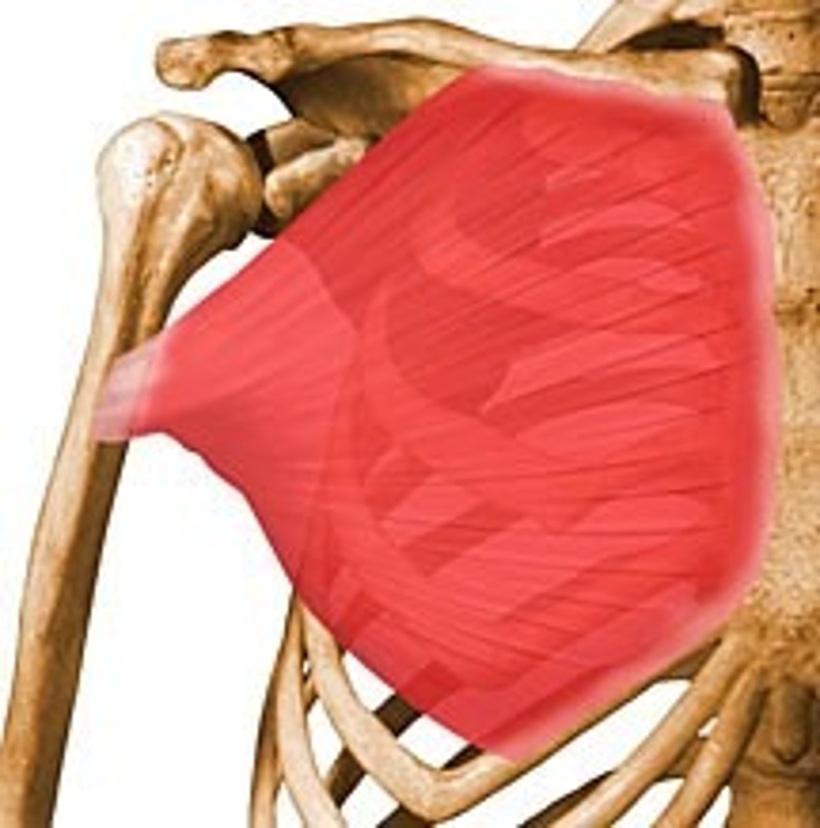
pectoralis major
-Origin: clavicle, anterior surface of the sternum, the superior six costal cartilages.
-Insertion: Intertubercular groove of the humerus
-Actions: adduction of the shoulder.
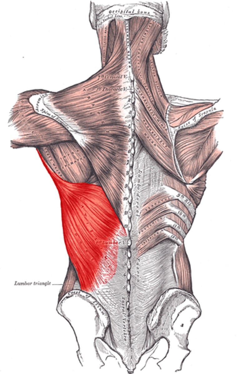
latissimus dorsi
-Origin: Spinous processes of vertebrae., thoracolumbar fascia, iliac crest, inferior ribs and inferior angle of scapula
-Insertion: intertubercular groove of the humerus
-Actions: Scapula depression, shoulder extension
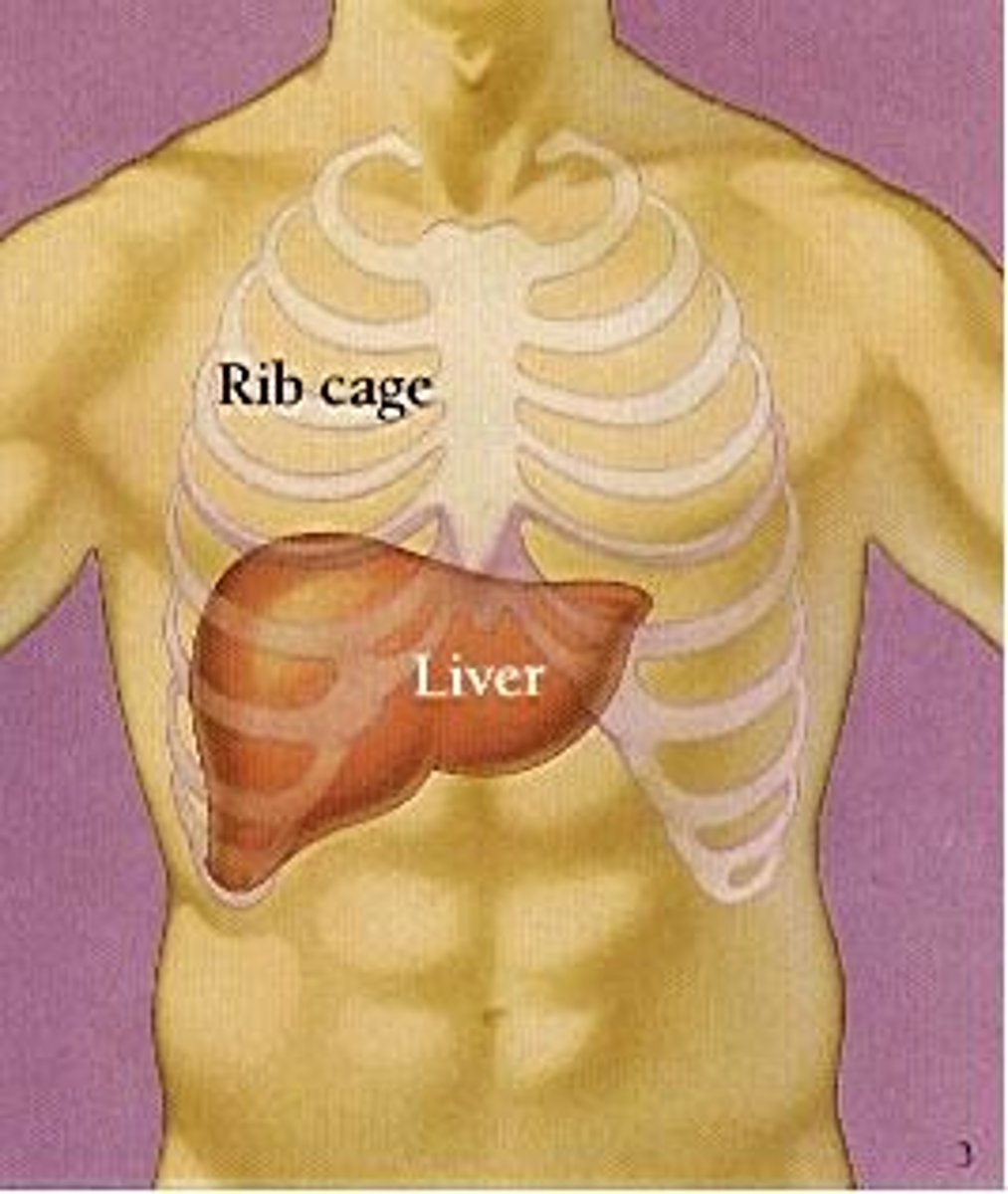
Liver
Upper right quadrant of the abdominal cavity. inferior to diaphragm
largest internal organ.
lesser omentum anchors liver to stomach.
Blood supply: recieves blood from heart via hepatic artery (supports liver tissue), recieves deoxygenated nutrient-rich blood from digestive organs via portal vein.
takes detoxified blood to heart via hepatic vein.
sternum
manubrium, body, xiphoid process.
Manubrium of sternum
Two fingers below sternal notch: Articulates with ribs 1, 2, & clavicles. Sternocleidomastoid muscles attach here.
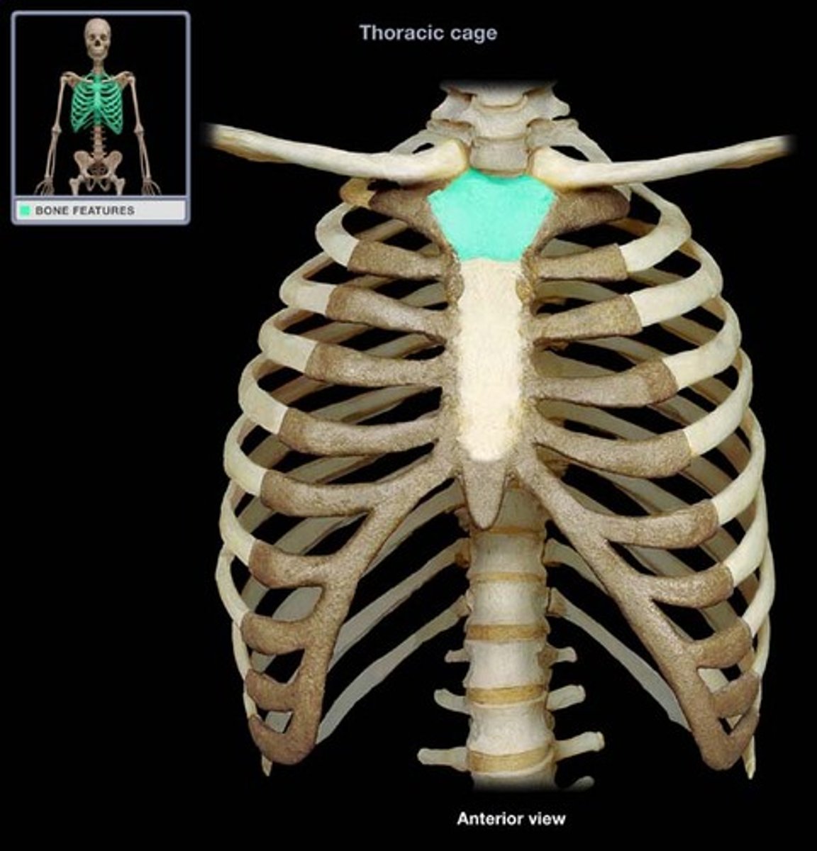
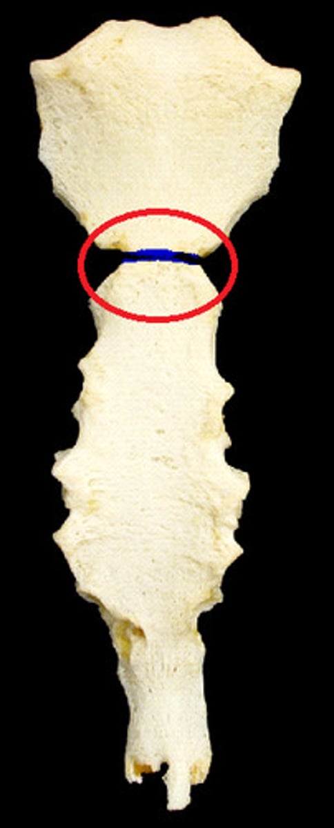
sternal angle
Ridge between manubrium and body at second rib.
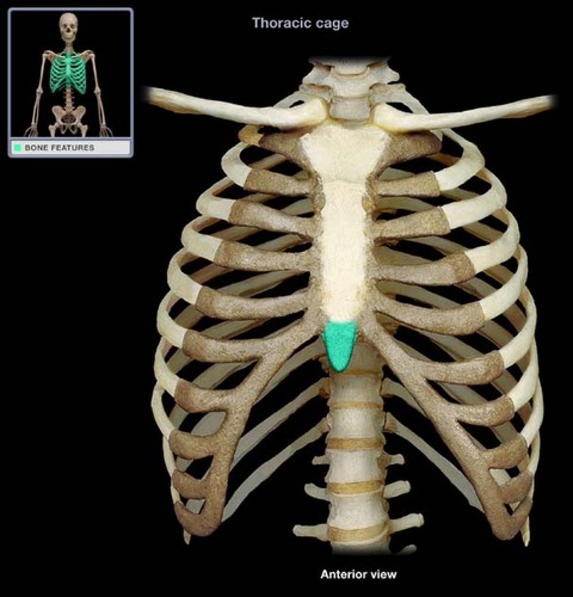
xiphoid process
Find sternum, move inferiorly. Small bony process. Attachment for diaphragm and rectus abdominus.
Pelvic floor
Group of muscles that resist intra-abdominal pressure and support internal organs. Attaches to pubic bone anteriorly, coccyx posteriorly, and ischial spines.
Made up of two muscle groups: Levator ani (puborectalis, pubococcygeous, and iliococcygeous) and coccygeus.
Passes through: females: urethra, vagina, anus. males: anus and urethra.
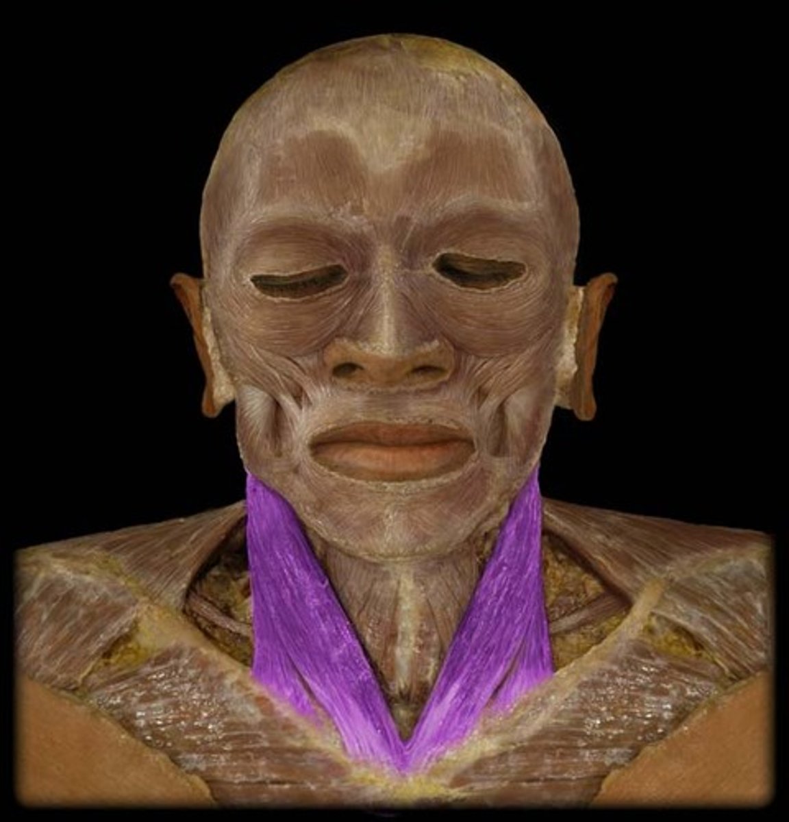
Sternocleidomastoid
Palpate: rotate head, feel most prominent muscle behind ear and follow inferiorly.
Inserts onto mastoid process behind ear.
Action: flexion of neck forward, rotates the head to opposite shoulder. assists with active inhalation as it pulls the ribcage up, creating space in thoracic cavity
proximal phalanges 1-5 hands
located between metacarpophalangeal joints and proximal interphalangeal joints.
Insertion of interossei. Base of proximal phalanges: abductor digiti minimi, abductor pollicis, flexor pollicis brevi.
Interphalangeal joints
Volar plate prevents hyperextension
Middle phalanges 2-5
Between proximal and distal interphalangeal joints.
Insertion of flexor digitorum superificialis.