ch 3 - blood
 Call Kai
 Learn
Learn Practice Test
 Spaced Repetition
 Match
Match Flashcards
 Knowt Play
1/23

There's no tags or description
Looks like no tags are added yet.
Name | Mastery | Learn | Test | Matching | Spaced | Call with Kai |
|---|
No analytics yet
Send a link to your students to track their progress
24 Terms
tissue type & elements that make up blood
connective tissue
formed elements (cellular)
erythroctyes (rbc)
leukocytes (wbc)
platelets (cell fragments)
extracellular matrix (plasma)
water-based fluid suspending formed elements in blood, allowing blood to flow through vessels
plasma
functions of blood (3)
transportation: delivering absorbed nutrients, diffusing oxygen, releasing hormones
defense: leukocytes protecting body from external pathogens, internal body cell malfunctions
homeostasis: blood dissipating/conserving heat through blood flow
hematocrit, centrifuge
blood test measuring erythrocyte percentage in a blood sample
centrifuge: equipment spinning blood to separate cellular elements from lighter plasma - heaver elements sink to bottom of tube
which element has the greatest quantity in blood: leukocytes, erythrocytes, or platelets?
erythrocytes / RBC
describe the components of a blood sample spun in a centrifuge:
plasma
buffy coat
hematocrit
plasma: water, proteins, nutrients, hormones
buffy coat: leukocytes, platelets
hematocrit: erythrocytes
normal blood hematocrit %’s (male & female)
male: 42-52%
female: 37-47%
anemia
depressed hematocrit %
polycythemia
elevated hematocrit %
describe the following characteristics of blood:
colour
viscosity
temperature
pH
body weight (% & L for males and females)
colour:
oxygenated = bright red
deoxygenated = darker red (hemoglobin in blood is rich in iron, which reacts to oxygen when exposed)
viscosity: 5x thicker than water (formed elements & plasma proteins)
temperature: 38 degrees C (friction and resistance)
pH: avg 7.4 (slightly basic)
body weight: 8% of adult body weight
males = 5-6L
females = 4-5L
plasma
% of water
names of plasma proteins
other substances
extracellular matrix in blood → 92% water
plasma proteins
albumin
globulins
alpha, beta
gamma
fibrinogen (antibodies)
hormones (insulin, oxytocin)
electrolytes (sodium, potassium, calcium)
dissolved gases (o2, co2, N)
organic nutrients (vitamins, lipids, glucose, amino acids)
metabolic wastes (co2, urea)
albumin (% of plasma protein, organ producing it, main functions)
54% of plasma proteins - produced by liver
allows transport in blood (binds to fatty acids, steroid hormones)
maintains osmotic pressure (holds water in blood vessels)
globulins (% of plasma protein, types → organ producing it, main functions)
38% of plasma proteins
aplha, beta globulins
produced by liver
transport substances, maintain osmotic pressure
gamma globulins
produced by leukocytes
also known as antibodies (for immunity)
fibrinogens (% of plasma protein, types → organ producing it, main functions)
7% of plasma proteins - produced by liver
essential for blood clotting
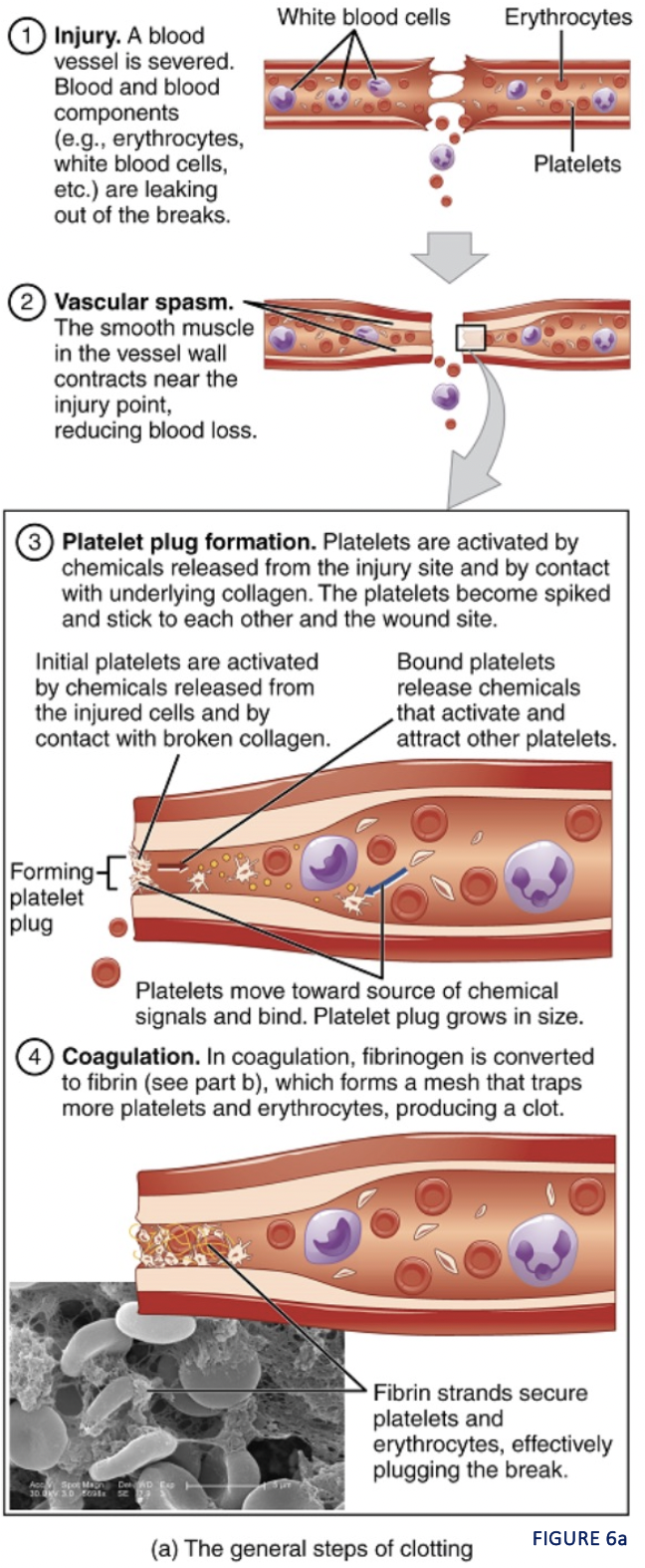
hemostasis (def + name the steps)
process of body sealing a ruptured blood vessel
vascular spasm
platelet plug formation
coagulation
vascular spasm
(hemostasis → step 1)
blood vessel damaged → smooth muscle wall severely contracts
contraction brought on by endothelin chemicals
blood flow is constricted
~ 30 mins
platelet plug
(hemostasis → step 2)
platelets clump at damaged site
become spiky, sticky as they bind to exposed collagen and endothelial lining
forms platelet plug using plasma’s glycoprotein = von willebrand vactor
chemicals released into blood plasma by platelets
ADP (adenosine diphosphate) reinforces the plug
serotonin maintains vasoconstriction
prostaglandins, phosphilipids maintain vasoconstriction
coagulation
(hemostasis → step 3)
blood clot made of fibrin protein begins to form
fibrin network traps formed elements of blood as a clot
fibrin
protein involved in coagulation step of hemostasis
insoluble protein derived from blood plasma protein fibrinogen (ALSO KNOWN AS ANTIBODIES)
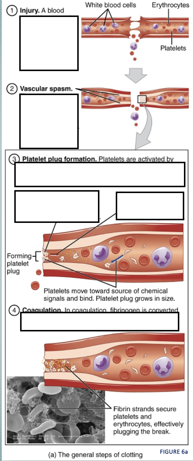
describe the hemostasis process as best as you can using the steps
clotting factors & pathways
chemicals prompting reactions activating other clotting factors during coagulation stage of hemostasis
extrinsic pathway: triggered by tissue damage
intrinsic pathway: triggered by vessel wall damage
→ both merge into the Common Pathway
12 known clotting factors (I - XII)
intrinsic pathway
(hemostasis → coagulation → clotting factors)
triggered by vessel wall damage
clotting factors all present in bloodstream
clotting factors lead to activation of factor X → activates Common Pathway
extrinsic pathway
(hemostasis → coagulation → clotting factors)
triggered by tissue damage
tissue factor / Factor III begins clotting factors
clotting factors lead to activation of Factor X → activates Common Pathway
common pathway
(hemostasis →