Week 3: Supporting the brain
1/42
There's no tags or description
Looks like no tags are added yet.
Name | Mastery | Learn | Test | Matching | Spaced | Call with Kai |
|---|
No analytics yet
Send a link to your students to track their progress
43 Terms
What are the three layers of meninges that cover the CNS?
Dura mater, arachnoid membrane, pia mater
What are meninges?
Tough connective tissue which covers the CNS and some of the PNS: protective layer dissipates as it moves to the PNS
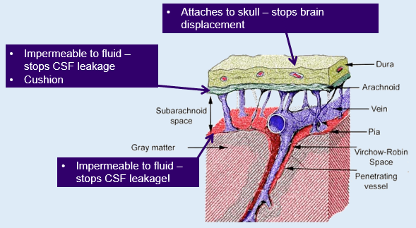
What is the function and structure of the dura mater?
It is the outer layer which is thick, tough and flexible but not flexible that provides protection and stops brain displacement.
What is the role of the arachnoid membrane?
It is the middle layer that is soft and spongy, located between the dura mater and pia mater.
What does the pia mater do?
It clings to the surface of the brain and spinal cord, being thin and delicate. Smaller surface blood vessels are located here.
What is the subarachnoid space?
A fluid-filled space between the arachnoid membrane and pia mater that cushions the brain, due to containing CSF.
Outline the differences between the CNS and PNS in terms of meninges
CNS - covered by 3 layers of meninges
Dura mater
Arachnoid membrane
Pia mater
PNS - 2 layers fuse
Dura and pia mater fuse
Sheath protects spinal and cranial nerves and the autonomic ganglia (large clusters of neurons)
Arachnoid membrane (CSF) not present
What is cerebrospinal fluid (CSF)?
A clear fluid that fills the ventricular system of the brain and the subarachnoid space.
What are ventricles?
Hollow spaces within the brain, filled with cerebrospinal fluid
How many ventricles are there in the brain?
Four ventricles: two lateral ventricles, one third ventricle, and one fourth ventricle.
→ and cerebral aqueduct
Outline the role of the each of the 4 ventricles
Lateral ventricles 1 and 2: sited in centre of telencephalon - largest, connected to 3rd ventricle
3rd ventricle: sited at midline in centre of diencephalon
Cerebral aqueduct: long tube in mesencephalon which connects 3rd and 4th ventricles
4th ventricle: found between cerebellum and pons - mesencephalon
Outline the main factors associated with cerebrospinal fluid (CSF)
Extracted from blood
Consists of ions, water, protein, glucose
Produced constantly from choroid plexus
Total volume of ~125 ml
6 hours for full replacement of CSF
Fills ventricular system of brain and subarachnoid space surrounding brain and spinal cord
Constantly being produced and flowing around our brain and spinal cord
Where is cerebrospinal fluid produced?
Produced by the choroid plexus of lateral ventricles - epithelium and ependymal cells
What are the 4 vital functions of cerebrospinal fluid?
Protection, buoyancy, waste reduction, and transport of nutrients and hormones.
What is the choroid plexus?
Tissue with a very rich blood supply and protrudes into the ventricles
Outline the flow of cerebrospinal fluid
CSF is produced in the choroid plexus (a tissue with a very rich blood supply and protrudes into the ventricles)
CSF flows to 3rd ventricle where more is produced
Flows through cerebral aqueduct to 4th ventricle through small openings - connect to subarachnoid space
Leaves ventricles to flow into subarachnoid space around CNS
It is reabsorbed back into blood stream through arachnoid granulations
What is hydrocephalus?
An accumulation of CSF within the cerebral ventricles causing ventricular dilation due to there being too much CSF fluid.
What are the two types of hydrocephalus?
Obstructive hydrocephalus and communicating hydrocephalus.
What causes obstructive hydrocephalus?
It is caused by a blockage to the natural ventricular drainage system and CSF flow
What is communicating hydrocephalus?
Reduced absorbance of CSF by the arachnoid villi
What are the three types of glial cells?
Astrocytes, oligodendrocytes, and microglia.
Outline the main roles of astrocytes
Provide physical support - ‘neuron glue’: attach to capillaries
Nourish neurons - wrap blood vessels to receive, store and release nutrients to neurons
Wrap multiple neurons or dendrites for CNS neurons
Helps control chemical composition of extracellular fluid
Surround and isolate synapse - limits the dispersion of neurotransmitters that are released by the terminal buttons
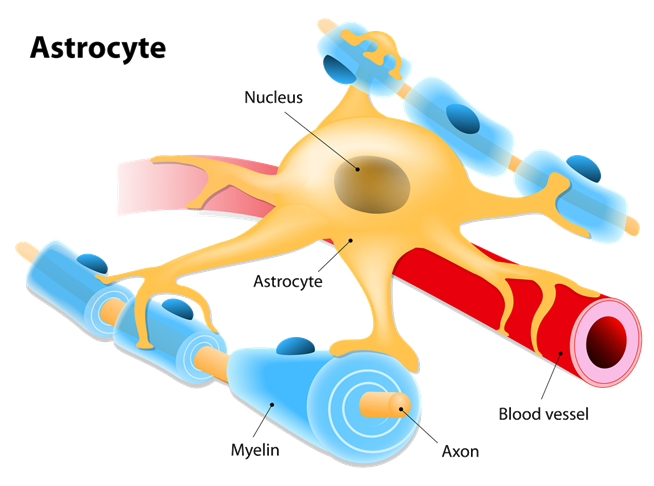
What is the function of astrocytes?
They provide physical support, nourish neurons, clean debris in the brain and help control the chemical composition of extracellular fluid.
What is phagocytosis in relation to astrocytes?
The process where astrocytes bind to and engulf dead cells in the CNS, cleaning up debris in the brain.
How to astrocytes form scar tissue?
Once astrocytes perform phagocytosis and the dead tissue has been broken down, scar tissue will be formed in place of dead tissue as astrocytes will be left to fill the vacant area.
What is amyotrophic lateral sclerosis (ALS)?
Most common form of motor neuron disease
Motor neurons are lost - can cause symptoms related to our muscles, e.g. twitching
Attacks nerve cells through astrocytes - toxin formed: inhibits action potentials
Rapidly aggressive, leading to fatality
Lose all voluntary muscle control
What has current research into ALS suggested about the role of astrocytes?
Neurodegeneration in ALS is partly mediated by non-cell autonomous mechanism
Astrocytes in ALS lose neuroprotective property and acquire toxic phenotypes
Astrocytes are a viable therapeutic target for ALS
What do microglia do?
They act as phagocytes, moving around the nervous system to engulf and break down dead or dying neurons.
Primarily responsible for inflammatory reaction in response to brain damage
What are microglia thought to have a role in? (Medical)
Neurodegenerative disorders (Alzheimers and Parkinsons disease)
Viral infections (HIV)
What is the role of oligodendrocytes?
They perform myelination in the CNS by wrapping around multiple axons.
How do Schwann cells differ from oligodendrocytes?
Schwann cells wrap individual axons in the PNS, while oligodendrocytes wrap several axons in the CNS.
What percentage of the body's blood supply does the brain receive?
15-20% of the body's blood supply.
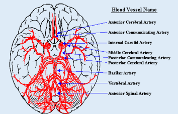
Name the blood vessels in the brain
Alien Attacks In My Parrot’s Pizza Box Very Aggressively
Anterior cerebral artery
Anterior communicating artery
Internal carotid artery
Middle cerebral artery
Posterior communicating artery
Posterior cerebral artery
Basilar artery
Vertebral artery
Anterior spinal artery
Alien Attacks In My Parrot’s Pizza Box Very Aggressively
What are the 2 basic functions of the blood?
Blood brings materials necessary for functioning: oxygen, hormones, nutrients (carbohydrates, amino acids, fats, vitamins)
Blood removes materials from the brain - toxic if left there, maintains homeostasis: carbon dioxide, lactate, hormones, ammonia
What is the blood-brain barrier (BBB)?
A semi-permeable barrier that protects the brain from foreign substances in the blood.
In the body, small spaces exist between endothelial cells so substances can move across capillary walls
→ In the brain, no spaces exist between the endothelial cells (composed of smaller subunits of transmembrane proteins) and substances cannot pass over capillary wall - BBB
What substances can cross the blood-brain barrier?
Lipid-soluble molecules can penetrate, while water-soluble molecules require specialised transport mechanisms.
Where is the blood-brain barrier weaker?
In areas like the area postrema (controls vomiting), which detects toxins in the blood.
What are the main functions of the blood-brain barrier?
Protects brain from 'foreign substances' in blood that may injure brain
Protects brain from hormones and neurotransmitters in the rest of body
Maintains a constant environment for brain
Makes transmission of messages from place to place in the brain more achievable
What is the total volume of cerebrospinal fluid in the body?
Approximately 125 ml.
How long does it take for half of the cerebrospinal fluid to be replaced?
About 3 hours.
What happens to cerebrospinal fluid after it leaves the ventricles?
It flows into the subarachnoid space and is reabsorbed into the bloodstream.
What is the function of the glial cells in the nervous system?
They insulate neurons, provide support, and maintain homeostasis.
Which substances are able to penetrate through the blood-brain barrier (BBB)?
Lipid soluble molecules via lipid membranes of cells
Water-soluble molecules can only use specialised carrier-mediated transport mechanisms
Active transport allows some substances to move across capillary walls, e.g. glucose transporters