Rad Qual II - Final Exam
1/69
There's no tags or description
Looks like no tags are added yet.
Name | Mastery | Learn | Test | Matching | Spaced | Call with Kai |
|---|
No analytics yet
Send a link to your students to track their progress
70 Terms
3v Cspine views (MC)
APOM, AP (APLC), Lateral
5v Cspine views
3v plus obliques OR flexion/extension
7v Cspine views (Davis Series)
3v plus obliques plus flexion/extension
**trauma cases
Which elbow projection is best for determining an intra-articular effusion as evidenced by anterior/posterior displacement of the distal humeral fat pad (Sail sign)
lateral
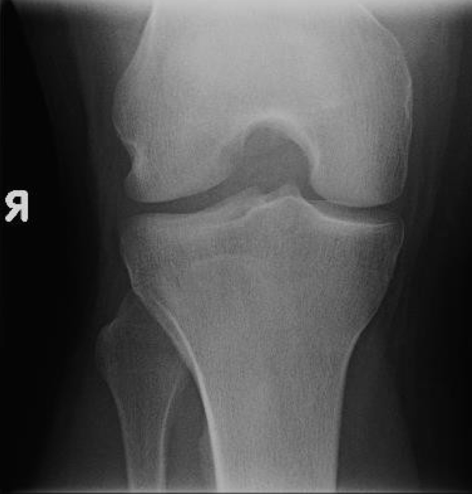
What projection of teh knee is this
intercondylar (holmblad)
What is the correct tube direction and angulation for a Scapular Y view projection
15 caudal
Which elbow view would be the best to assess for a potential avulsion fracture of the coronoid process
internal oblique
When performing a long bone study of the femur or humerus, if the entire bone does not fit on the image receptor, what modification should you make
include the joint closest to the pain
Which views are taken with a routine Knee series in the palmer clinics to perform a complete series
frontal and lateral
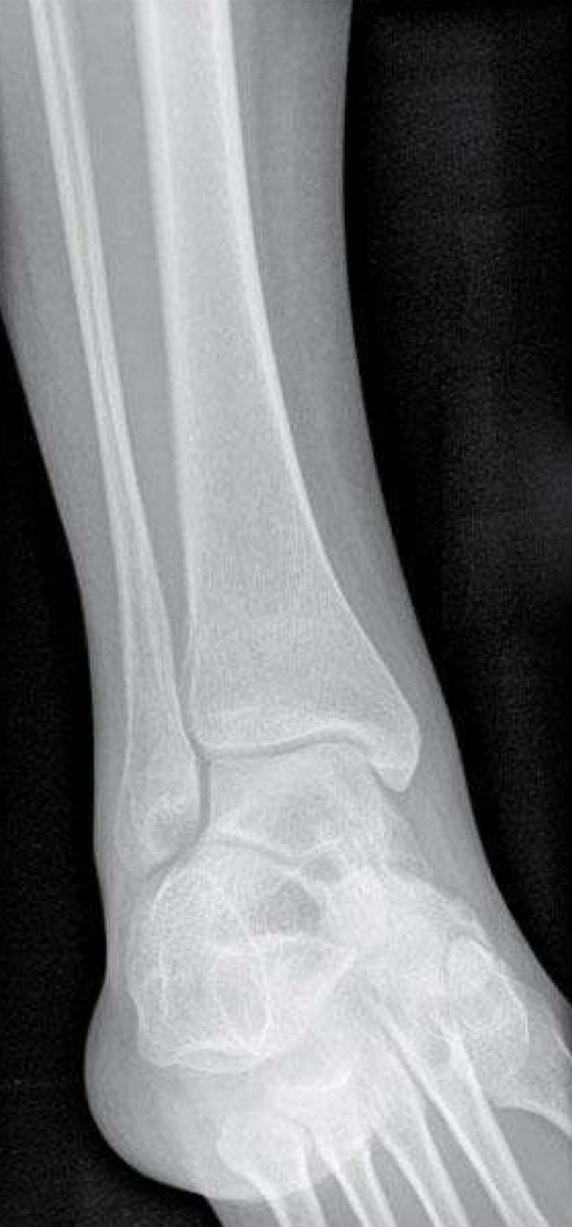
Name the projection demonstrated in this image
medial oblique ankle
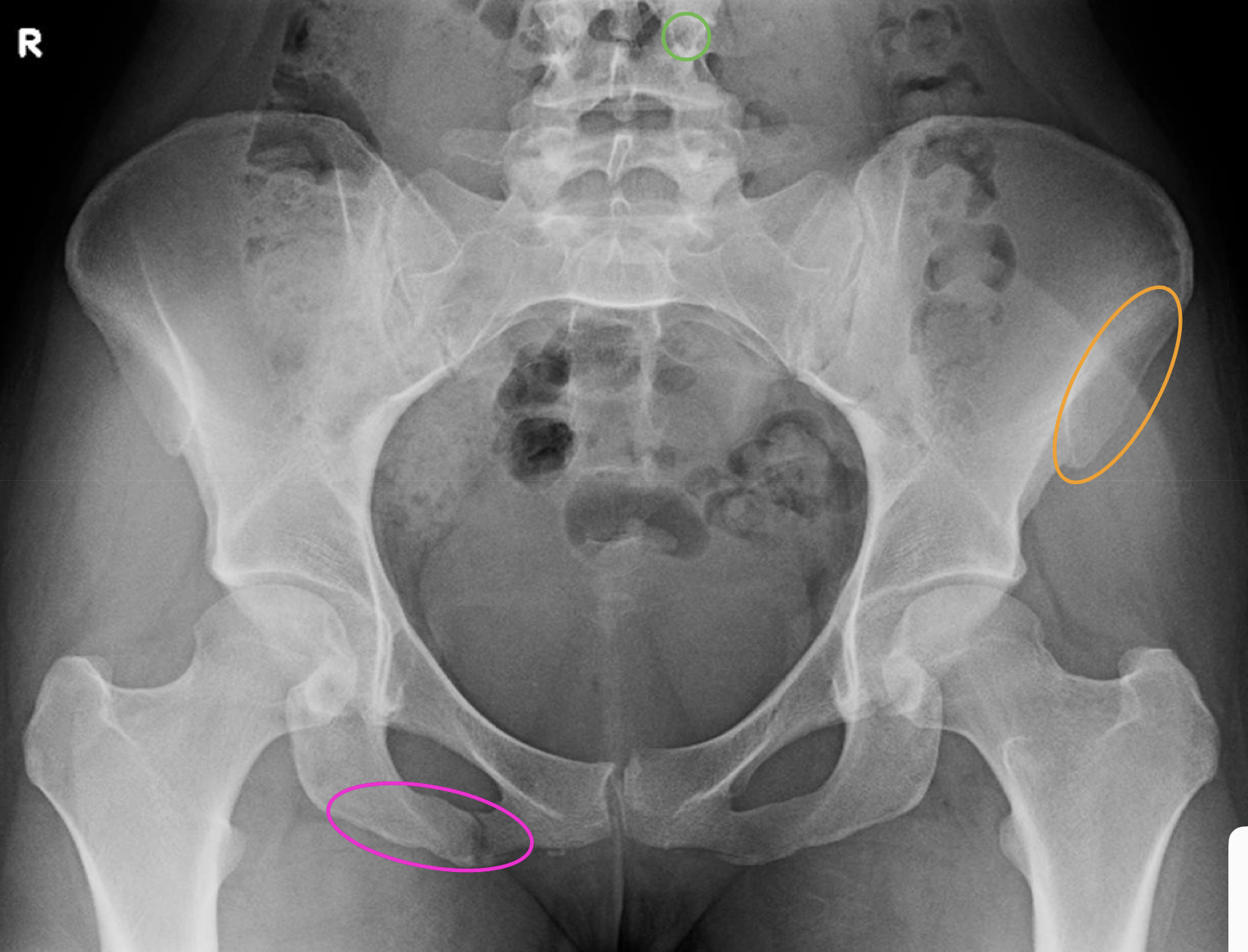
What are within each circle
green = left L4 pedicle
orange = anterior superior iliac spine
pink = inferior pubic ramus
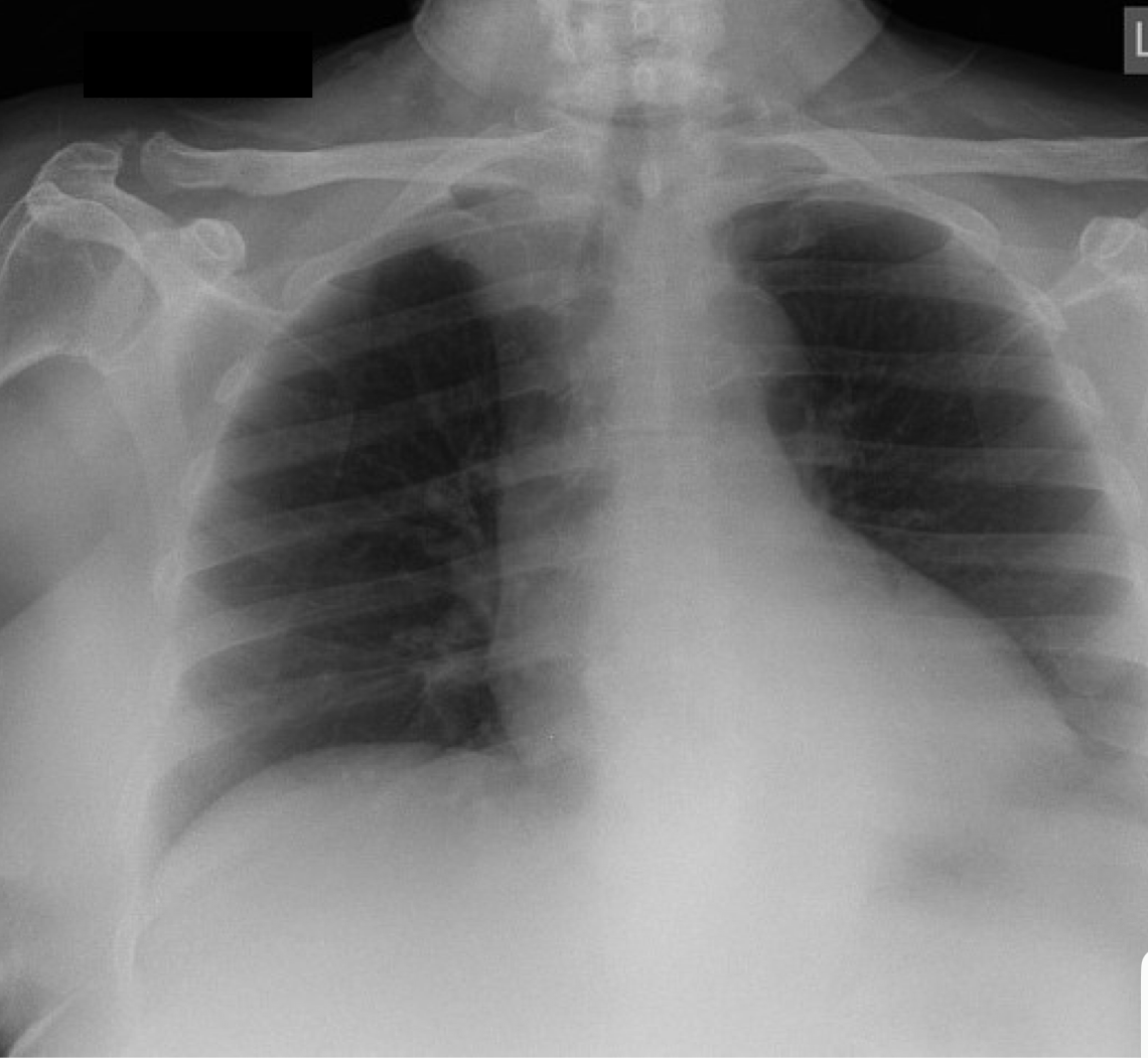
What projection is shown in the image
AP apical lordotic (Look for lung tumors)
Identify the projection and what would be the correct tube angulation and direction
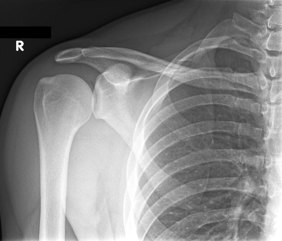
AP shoulder
No tube angulation
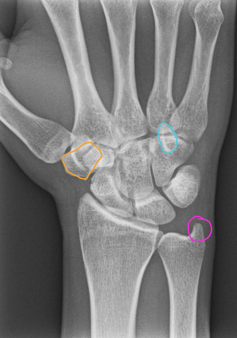
What are in each of the circles in this PA wrist image
orange = trapezium
blue = hamate
pink = ulnar styloid
What are the only views that are taken at 72” (everything else 40”)
Lateral cervical (Neutral, Flexion, Extension)
Cervical obliques (Anterior and Posterior)
Chest (PA and Lateral)
Standard projections of Chest
PA + Left Lateral
PA Chest
PP - standing w/chest touching bucky, shoulders rolled forward
Collimation - 14 × 17
CR - top of IR 1” above VP
SID - 72”
Breathing - Inhale and Hold
PA Chest demonstrates
lung fields, heart, greater vessels, ribs, shoulders, thoracic spine, upper abdomen

Supplementary Chest view = AP Lordotic view
evaluates suspicious areas within the lung apices that appeared obscured by overlying soft tissue
can tell it is this view b/c heart is too large (magnified) and clavicles are above apices
The __ hilum of the lung is normally 1-2cm higher
left
How many posterior/anterior ribs should be noted with a PA chest image
10 posterior
7 anterior
Lateral Chest
PP - standing w/left side touching bucky + arms above head
Collimation - 14 × 17
CR - top of IR 1” above VP
SID - 72”
Breathing - Inhale and Hold
Lateral chest view demonstrates
lung fields, heart, great vessels, ribs, sternum, thoracic spine
Standard projections of Ribs
AP, Oblique, PA Chest (included if complications)
AP Ribs (AD)
PP - standing w/back touching bucky
Collimation - 14 × 17
CR - top of IR 1 1/2” above VP
SID - 40”
Breathing - Inhale and Hold
AP Ribs view demonstrates
ribs 1-12 on affected side and some lung tissue
Posterior Oblique Ribs (AD)
PP - standing facing away from bucky
Collimation - 14 × 17
CR - top of IR 1 1/2” above VP
SID - 40”
Breathing - Inhale and Hold
Posterior Oblique Ribs view demonstrates
ribs 1-12 on affected side of pt w/increased clarity of costal angles
Supplementary projections - AP Ribs BD (lower ribs)
PP - standing w/back touching bucky
Collimation - 14 × 17
CR - bottom of IR at iliac crest
SID - 40”
Breathing - Exhale and Hold
Supplementary projections - Posterior Oblique Ribs BD (lower ribs)
PP - standing facing away from bucky
Collimation - 14 × 17
CR - bottom of IR at iliac crest
SID - 40”
Breathing - Exhale and Hold
Posterior Oblique Rib (BD) views demonstrate
inferior aspect of ribs and isolates curvature of the axillary rib aspect
Standard projections of Abdomen
AP supine
Acute Abdomen series = AP supine abdomen + PA Erect
Abdomen KUB
AP Abdomen
PP - lying supine or standing w/arms at side
Collimation - 14 × 17
CR - iliac crests
SID - 40” Table top or Bucky
Breathing - Exhale and Hold
AP Abdomen views demonstrate
spleen, liver, kidneys, psoas shadow, bowel, bladder
**ureters are not visible unless intravenous contrast has been administered
Bowel gas
Small bowel = up to 3cm
Large bowel = up to 5cm
Cecum = up to 9cm
Standard Projections of Shoulder
AP Internal Rotation
AP External Rotation (Grashey view)
Scapular Y
AP Internal Rotation
PP - back touching bucky w/dorsum of hand touching thigh
Collimation - 12 × 10
CR - 1” inferior to coracoid process
SID - 40”
Breathing - DBDM
AP Internal Rotation views demonstrate
lesser tuberosity, greater tuberosity, bicipital groove, distal clavicle, AC joint, scapula
AP External Rotation (Grashey view)
PP - scapula flat on bucky w/arm ext. rotated and extended
Collimation - 12 × 10
CR - 1” inferior to coracoid process
SID - 40”
Breathing - DBDM
AP External Rotation views demonstrate
greater tuberosity, lesser tuberosity, bicipital groove, distal clavicle, AC joint, scapula
What is BEST viewed with a AP External rotation shoulder image
GH joint space
Scapular Y Shoulder (PA)
PP - standing facing bucky obliquely, scapula perpendicular to film
Collimation - 10 × 12
CR - medial border of scapula + scapular spine intersection
Tube Tilt - 15 Caudal
SID - 40”
Breathing - DBDM
Scapular Y Shoulder (PA) views demonstrates
subacromial outlet and acromion process
shoulder dislocations and direction
What structures makes up the “Y” in the Scapular Y view
coracoid, acromion, humeral head
Standard Projections of Acromioclavicular joint
Bilateral without weights
Bilateral with weights (stress view)
**Unilateral views are supplementary
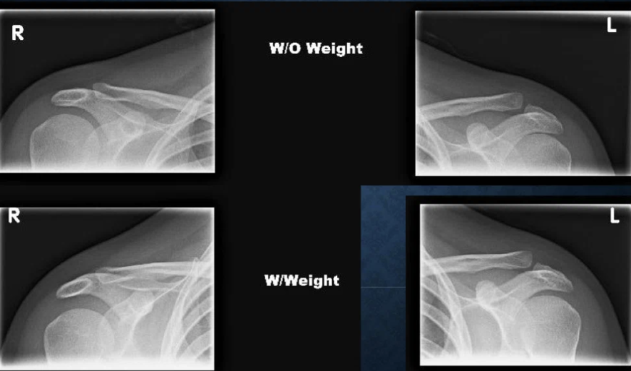
AP Acromioclavicular
PP - standing back touching bucky w/arms at side (w/weight hold in both hands)
Collimation - part size
CR - AC joint
Tube Tilt - 5 Cephalic
SID - 40”
Breathing - DBDM
**Use additional marker to indicate it is weighted
AP acromioclavicular views demonstrate
AC joint space and alignment
**used for AC separations
Standard projections of Clavicle
PA and Axial
PA Clavicle
PP - standing facing bucky w/head turned away from clavicle
Collimation - 12 × 10
CR - mid-clavicle
SID - 40”
Breathing - DBDM
PA Clavicle views demonstrate
medial to lateral dimension of clavicle
both AC and sternoclavicular joints
AP Axial Clavicle
PP - standing w/back touching bucky
Collimation - 12 × 10
CR - mid-clavicle
Tube Tilt - 15 Cephalic
SID - 40”
Breathing - DBDM
Standard projections of Humerus
AP and Lateral
AP Humerus
PP - standing w/back touching bucky leaning towards arm
Collimation - 7 × 17
CR - centered so jt closest to injury is in view
SID - 40”
Breathing - DBDM
Lateral Humerus
PP - standing w/back touching bucky arm supinated, elbow flexed in front
Collimation - 7 × 17
CR - centered so jt closest to injury is in view
SID - 40”
Breathing - DBDM
Standard projections of Elbow
AP, Lateral, Internal Oblique, External Oblique
AP Elbow
PP - hand supinated, elbow and shoulder in same plane
Collimation - part size
CR - center of cubital fossa
SID - 40” Table Top
Breathing - DBDM
AP Elbow views demonstrate
distal humeral diaphysis → bicipital/radial tuberosity of diaphysis
Internal Oblique Elbow
PP - arm and shoulder on same plane, hand pronated
Collimation - part size
CR - lateral to center of cubital fossa
SID - 40” Table top
Breathing - DBDM
External Oblique Elbow
PP - arm and shoulder in same plane, epicondyles 45 deg
Collimation - part size
CR - medial to center of cubital fossa
SID - 40” Table top
Breathing - DBDM
External oblique elbow is great to view
radial head and neck
Lateral Elbow
PP - elbow flexed 90, thumb up (karate chop)
Collimation - part size
CR - radial head
SID - 40” Table top
Breathing - DBDM
Supplementary projections for Elbow
Coyle (Radial head)
Jones (Olecranon)
What is the most important of the elbow views
lateral → identify fx (sail sign)
Elbow ossification centers appear (CRITOE) **NO Odd #**
Capitellum - 1
Radial head - 3
Internal/medial epicondyle - 5
Trochlea - 7
Olecranon process - 9
External/lateral epicondyle - 11
Knowing the age of the pt is important to determine fx/avulsion vs normal anatomy - if you see 5 ossification centers in a 5yo, think
avulsion fracture
If you see 5 ossification centers in a 10yo, think
most likely normal
Standard projections of the Forearm
AP and Lateral
AP Forearm
PP - arm and shoulder on same plane, hand supinated
Collimation - part size
CR - middle of anatomy
SID - 40” Table top
Breathing - DBDM
AP Forearm views demonstrate
above epicondyles → metacarpal bases of wrist
Lateral Forearm
PP - elbow flexed 90deg, thumb up (karate chop)
Collimation - part size
CR - middle of anatomy
SID - 40” table top
Breathing - DBDM