ANAT 5010 - unit 5
1/70
There's no tags or description
Looks like no tags are added yet.
Name | Mastery | Learn | Test | Matching | Spaced | Call with Kai |
|---|
No analytics yet
Send a link to your students to track their progress
71 Terms
- medial brachial cutaneous nerve joined by the intercostobrachial nerve which is the lateral cutaneous branch of the second intercostal nerve
- medial antebrachial cutaneous nerve
what are the cutaneous nerves on the medial side of the upper limb?
- upper lateral cutaneous nerve of arm (branch of axillary nerve)
- lower lateral cutaneous nerve of arm (branch of radial nerve)
- lateral antebrachial cutaneous nerve (continuation of the musculocutaneous nerve)
what are the cutaneous nerves on the lateral side of the upper limb?
- posterior cutaneous nerve of arm (radial nerve)
- dorsal cutaneous nerve of forearm (radial nerve)
what are the cutaneous nerves on the dorsal side of the upper limb?
cephalic vein
goes from the dorsal venous arch on the dorsum of the hand at the radial side; ascends the forearm to the anterolateral aspect at the elbow, then goes to the deltopectoral triangle; pierces clavipectoral fascia to join the axillary vein
basilic vein
goes from the dorsal venous arch on the ulnar side of the dorsum of the hand; ascends to the antero-medial side at the elbow; shortly above the elbow, it pierces the deep fascia to join the brachial vein and from the axillary vein
median cubital vein
formed at the front of the elbow; connects the basilic and cephalic veins; separated from the brachial artery and median nerve by the bicipital aponeurosis
cephalic, basilic, and median cubital veins
what are the superficial veins?
deep fascia
tubular connective tissue investment of deeper structures of arm
brachial fascia
covers muscles of the arm
lateral and medial intermuscular septa
separates arm muscles into anterior and posterior compartments
origin: lateral 1/3 of clavicle, acromion process, and spine of the scapula
insertion: deltoid tuberosity
what is the origin and insertion of the deltoid muscle?
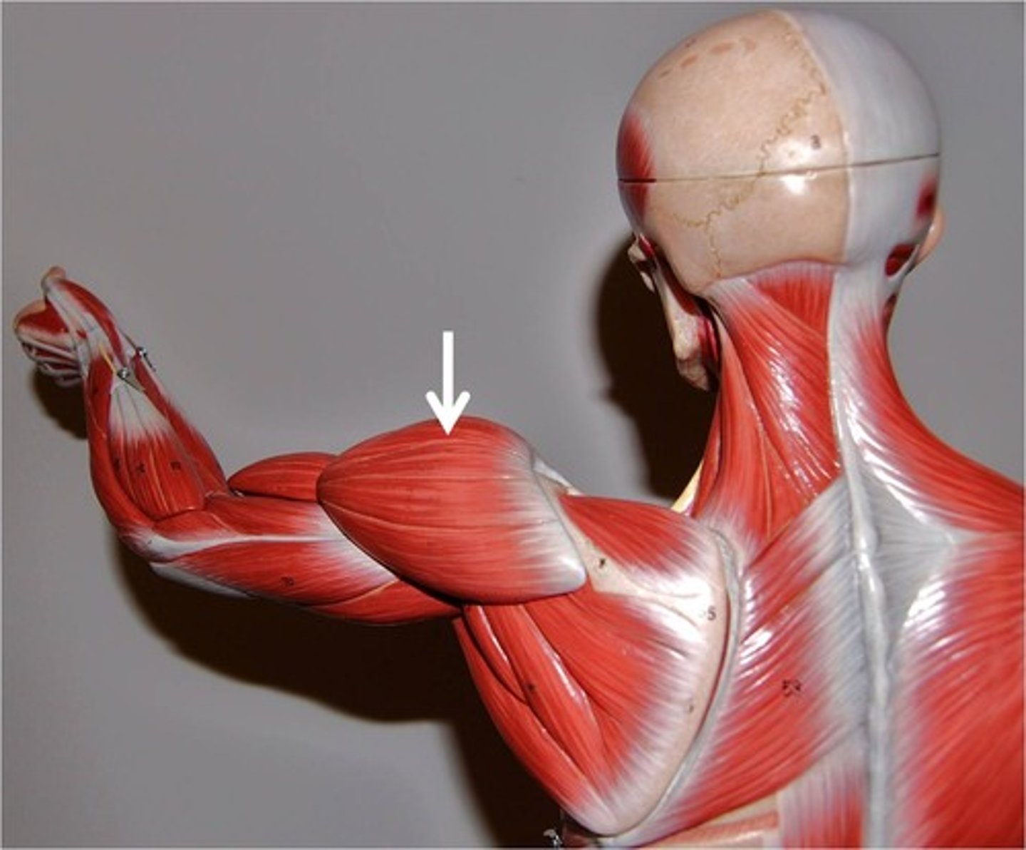
action: extends, adducts, and medially rotates the arm
innervation: thoracodorsal nerve (middle subscapular nerve)
what is the action and innervation of the deltoid muscle?

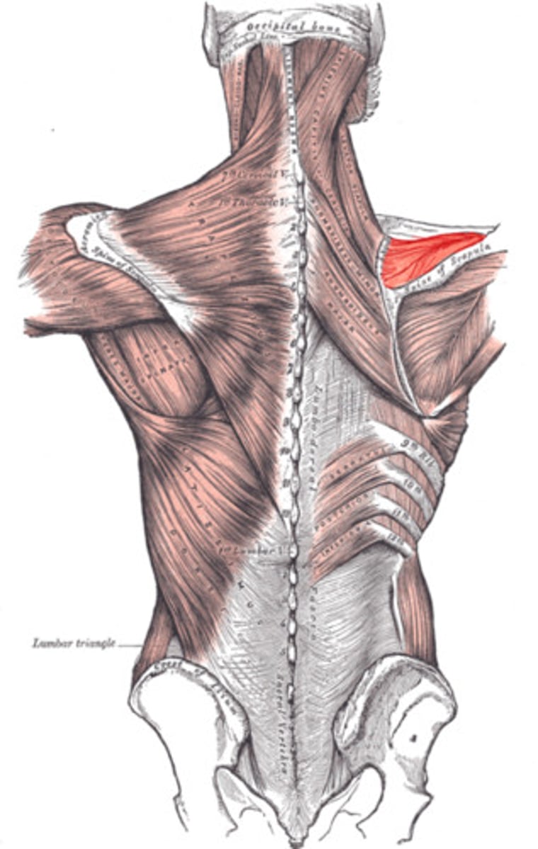
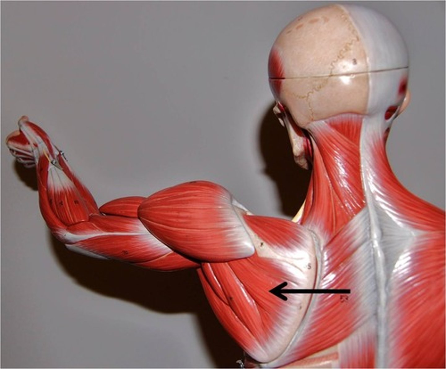
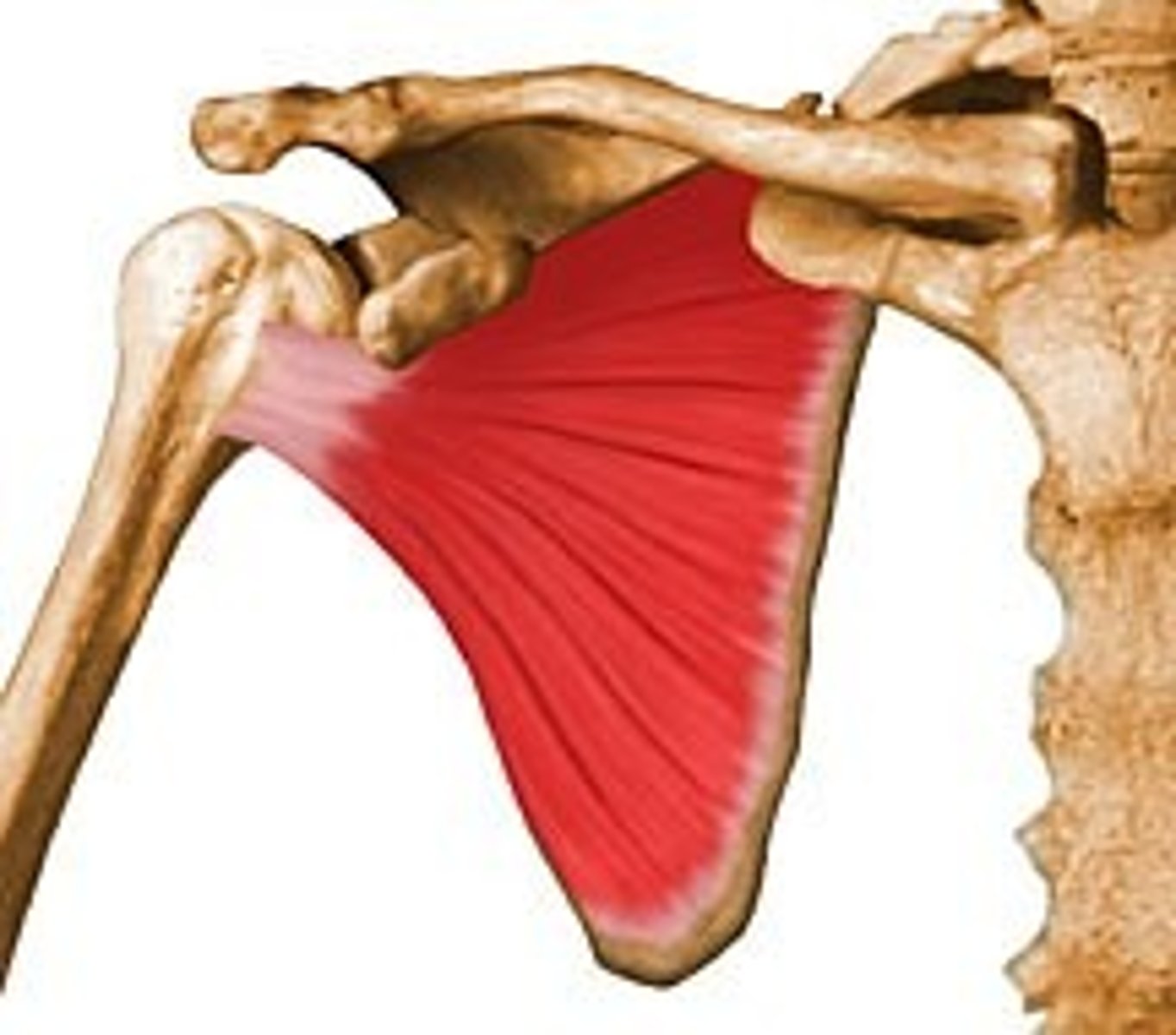
origin: inferior angle and axillary border of the scapula
insertion: crest of lesser tubercle of the humerus
what is the origin and insertion of the teres major muscle?

action: adducts and medially rotates the arm at the shoulder joint
innervation: inferior subscapular nerve
what is the action and innervation of the teres major muscle?

- supraspinatus
- infraspinatus
- teres minor
- subscapularis
what muscles make up the rotator cuff?
origin: supraspinatus fossa of the scapula
insertion: greater tubercle of the humerus
what is the origin and insertion of the supraspinatus?
origin: infraspinatus fossa of the scapula
insertion: greater tubercle of the humerus
what is the origin and insertion of the infraspinatus?
action: abducts the arm and assists the deltoid muscle in its full movement
innervation: suprascapular nerve
what is the action and innervation of the supraspinatus?
action: laterally rotates the arm
innervation: suprascapular nerve
what is the action and innervation of the infraspinatus?
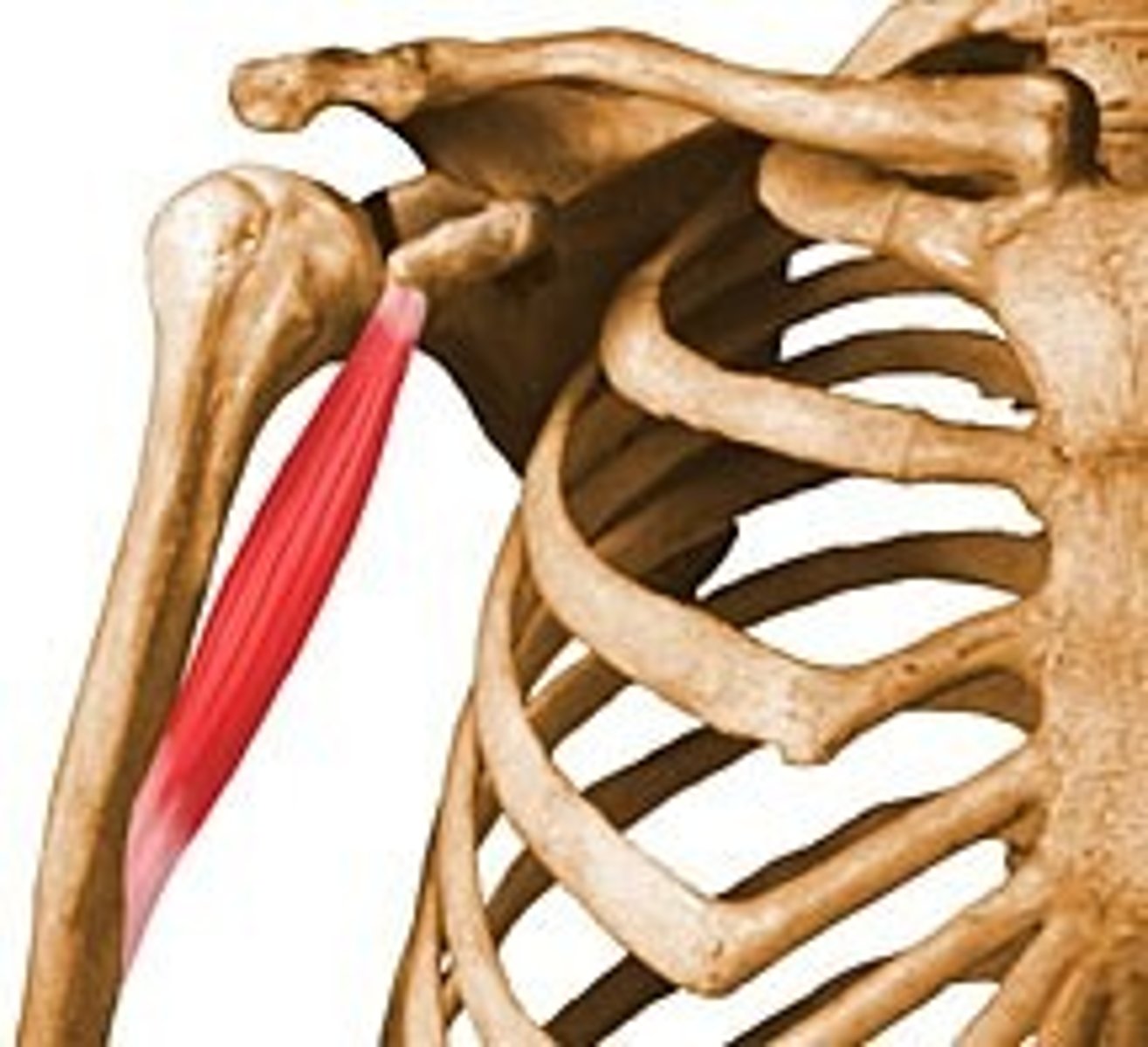
origin: lateral border of the scapula
insertion: greater tubercle of the humerus
what is the origin and insertion of the teres minor?
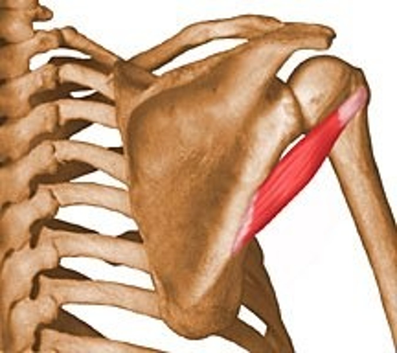
action: laterally rotates the arm
innervation: axillary nerve
what is the action and innervation of the teres minor?
origin: costal surface of scapula
insertion: lesser tubercle of the humerus
what is the origin and insertion of the subscapularis?
action: medially rotates and adducts the arm
innervation: superior and inferior subscapular nerves
what is the action and innervation of the subscapularis?
- humerus
- long head of the triceps muscle
- teres major
- teres minor
what forms the quadrangular space?
posterior humeral circumflex vessels and axillary nerve
what passes through the quadrangular space?
- teres minor
- teres major
- long head of the triceps muscle
what forms the triangular space?
branches of the scapular circumflex vessels
what can be seen through the triangular space?
- long head of triceps muscle
- lateral head of triceps muscle
- teres major
what makes up the triangular interval?
radial nerve and deep brachial vessels
what can be seen through the triangular interval?
- coracobrachialis
- biceps brachii
- brachialis
what muscles make up the anterior compartment of the arm?
origin: coracoid process
insertion: half way down the medial aspect of the humerus
what is the origin and insertion of the coracobrachialis?
action: adducts and flexes the arm
innervation: musculocutaneous nerve
what is the action and innervation of the coracobrachialis?
origin: supraglenoid tubercule (long head) and coracoid process (short head)
insertion: radial tuberosity
what is the origin and insertion of the biceps brachii?
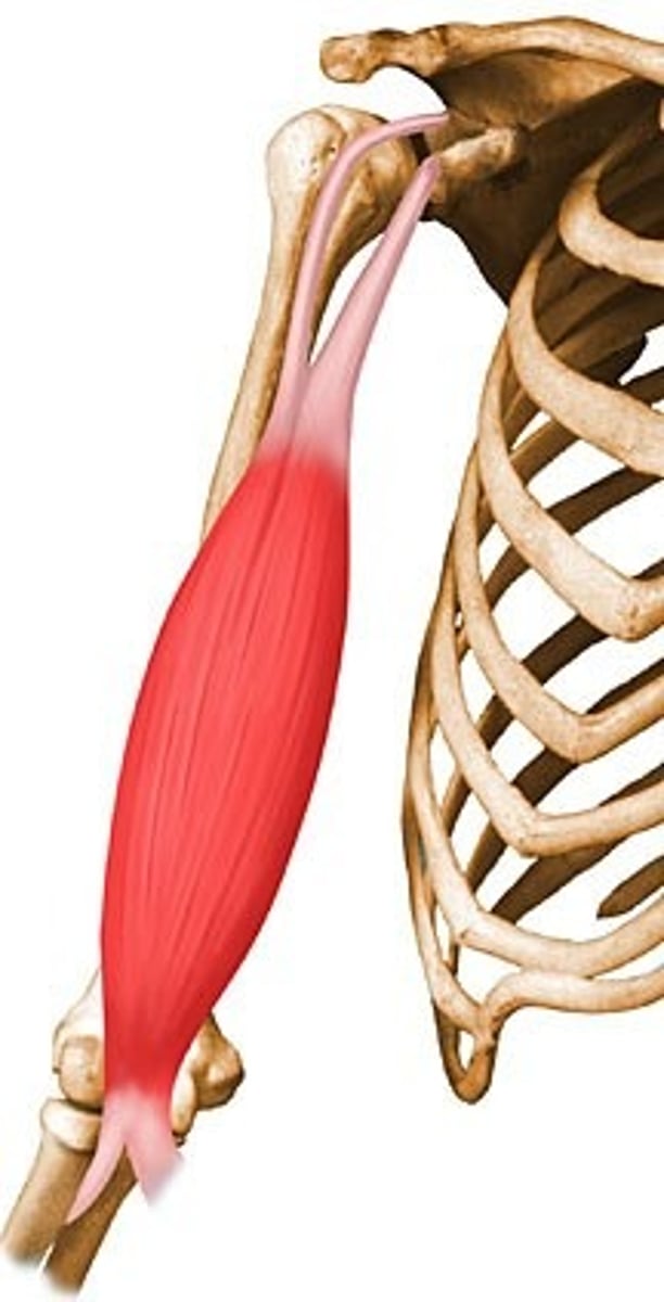
action: flexes shoulder, flexes forearm at elbow, supinates forearm
innervation: musculocutaneous nerve
what is the action and innervation of the biceps brachii?
origin: lower anterior half of humerus
insertion: ulnar tuberosity
what is the origin and insertion of the brachialis?
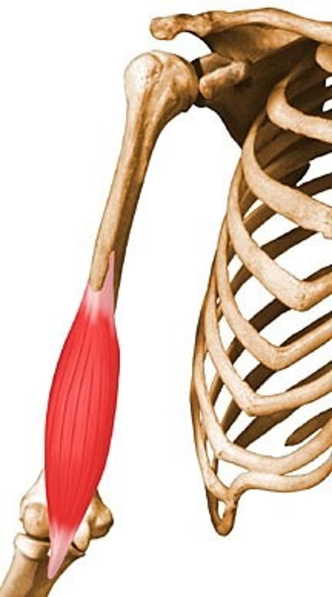
action: flexes forearm at elbow
innervation: musculocutaneous nerve
what is the action and innervation of the brachialis?
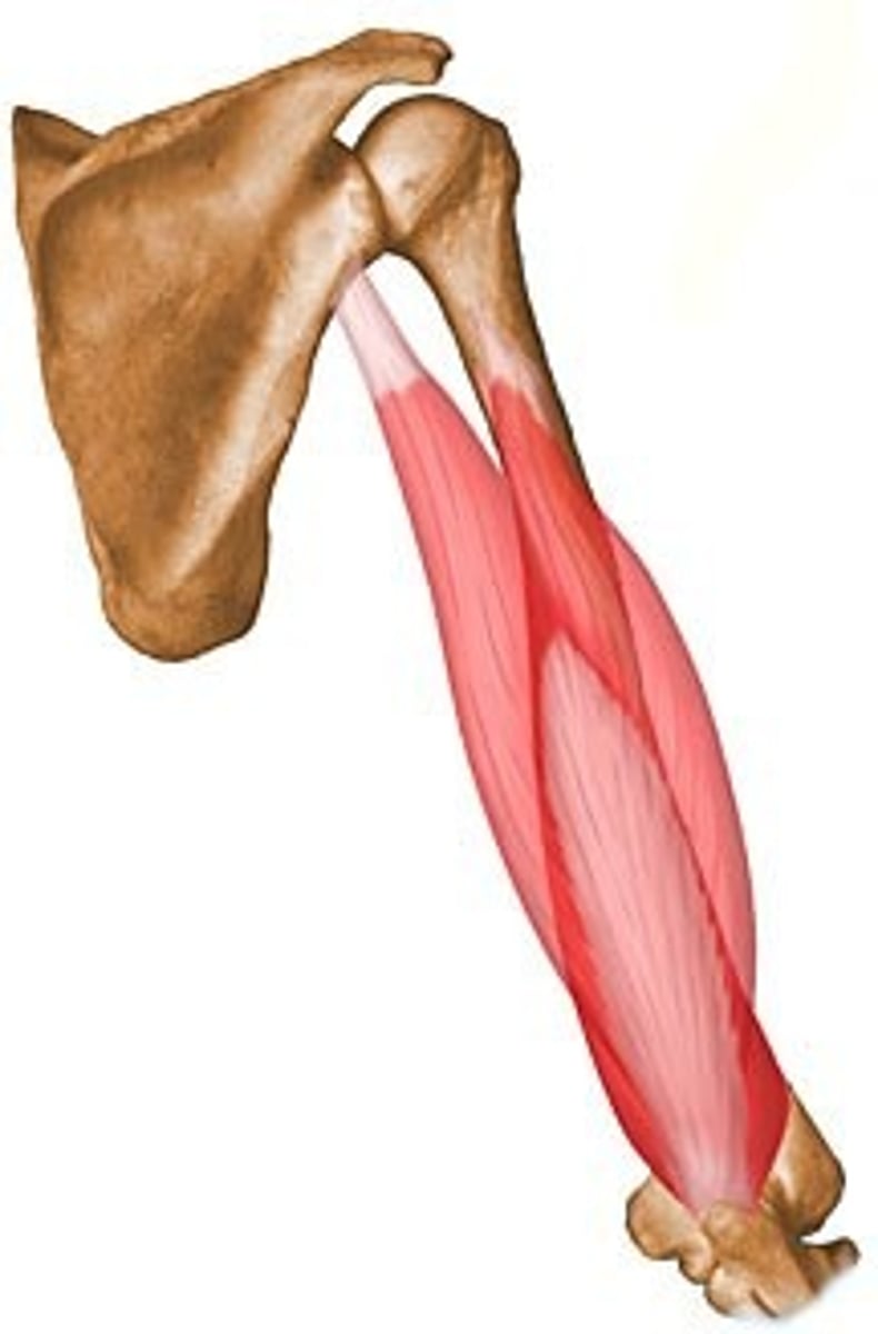
triceps brachii
what muscle is in the posterior compartment of the arm?
origin: long head -> infraglenoid tubercule of scapula, medial head -> lower half of humerus, upper head -> upper half of the humerus
insertion: olecranon process of ulna
what is the origin and insertion of the triceps brachii?
action: extends forearm at elbow and shoulder
innervation: radial nerve
what is the action and innervation of the triceps brachii?
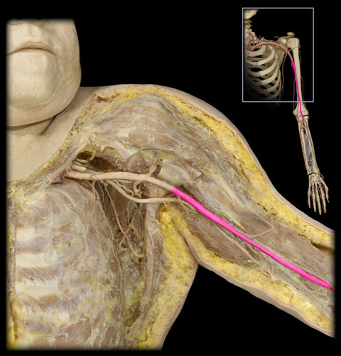
brachial artery
direction continuation of the axillary artery; begins at the lower border of the teres major muscle; ends by dividing into radial and ulnar arteries
- deep brachial artery
- superior ulnar collateral artery
- inferior ulnar collateral artery
what are the branches of the brachial artery?
deep brachial artery
travels with the radial nerve in the arm and ends by dividing into the radial collateral artery and the middle collateral artery
radial collateral artery
runs with the radial nerve; anastomoses with the radial recurrent artery; anterior to lateral epicondyle between brachioradialis and brachialis muscles
middle collateral artery
runs with the radial nerve to the anconeus muscle; anastomoses with the interosseous recurrent artery behind the elbow joint
superior ulnar collateral artery
travels with the ulnar nerve posterior to the medial epicondyle
inferior ulnar collateral artery
runs anterior to the medial epicondyle
brachial vein and axillary vein
what are the deep veins?
brachial veins
formed at the elbow by the union of the radial and ulnar veins; paired set of veins that accompany the brachial artery; contain valves and make frequent cross anastomoses with each other
axillary vein
formed at the lower border of either the teres major or subscapularis muscles; lateral brachial vein crosses the axillary artery to join the medial brachial vein to form this
sternoclavicular joint
a synovial joint between the medial end of the clavicle and the manubrium; represents the bony articulation between the upper limb and the axial skeleton
fibrocartilaginous disk
separates the cavity of the sternoclavicular joint into two
articular capsule
surrounds the medial end of the clavicle, disk, and other articular surfaces of the manubrium
interclavicular ligament
extends from one clavicle to the other; strengthens the superior surface of the joint capsule
anterior and posterior sternoclavicular ligaments
found on the anterior and posterior surfaces of the capsule of the sternoclavicular joint to strengthen it
costoclavicular ligament
a strong accessory ligament which joins the clavicle to the first rib
permits movement in anteroposterior and vertical planes and some rotation about the long axis of the clavicle
what does the sternoclavicular joint allow?
acromioclavicular joint
a plane synovial join between the lateral end of the clavicle and the medial surface of the acromion process of the scapula
- acromioclavicular ligament
- coracoclavicular ligament
what reinforces the capsular ligament of the acromioclavicular joint?
coracoclavicular ligament
an accessory ligament joining the coracoid process of the scapula to the undersurface of the clavicle and consists of the conoid ligament (medial) and the trapezoid ligament (lateral)
superior transverse scapular ligament
converts the superior scapular notch into a foramen for passage of the suprascapular nerve; suprascapular vessels pass over the ligament
coracoacromial ligament
passes from lateral margin of the coracoid process to the acromion process
glenohumeral joint
articulation between the glenoid fossa of the scapula and the head of the humerus; a ball and socket synovial joint exhibiting the greatest motility and least stability of the major joints of the body
glenoid labrum
a fibrocartilaginous ridge at the margins of the glenoid fossa which slightly deepens the fossa
transverse humeral ligament
attaches to the greater and lesser tubercles of the humerus, bridging over the intertubercular sulcus; crosses anterior to the tendon of the long head of the biceps brachii
coracohumeral ligament
from coracoid process to greater tubercle of the humerus
- superior glenohumeral ligament
- middle glenohumeral ligament
- inferior glenohumeral ligament
what are the three anterior thickenings of the shoulder joint capsule?
superior glenohumeral ligament
located above the communication between the joint cavity and the subscapular bursa
middle glenohumeral ligament
located immediately below the communication between the joint cavity and the subscapular bursa
inferior glenohumeral ligament
located inferior to the middle glenohumeral ligament
tendon of the long head of the biceps brachii
- passes under the transverse humeral ligament and enters the shoulder joint cavity
rotator cuff
major strength of the glenohumeral joint comes from the -