Imaging Quiz 1
1/61
There's no tags or description
Looks like no tags are added yet.
Name | Mastery | Learn | Test | Matching | Spaced | Call with Kai |
|---|
No analytics yet
Send a link to your students to track their progress
62 Terms
Who discovered the x-ray?
Wilhelm Roentgen
What are the types of ionizing radiation?
x-rays and gamma rays, some UV
What type of radiation are gamma rays and x-rays
wave type radiation
What type of materials stop wave-type radiation?
thick lead
what are the two types of radiation?
particle radiation (alpha and beta), wave-type radiation (gamma and x-rays)
what type of material stops neutron rays
water or concrete
what are direct and indirect effects of ionizing radiaton?
direct: x-rays hit DNA and rip off electron causing damage
indirect: x-ray creates free radical that interacts w DNA and causes damage
What are three possible outcomes of dna damage due to radiation?
1. successful repair - cell survives
2. damage is too great - cell dies
3. unsuccessful repair of damage - cell survives/mutated leads to cancer
what does stochastic mean?
so there is NO safe dose of ionizing radiation
What are the parts of the ALARA principle?
ALARA: as low as reasonably achievable
1. choose the right imaging study (dont get CT if xray works)
2. minimize the amount of exposure with correct technique and shielding
what are the 4 ways to acquire an image?
radiography (x-ray), CT, MRI, ultra-sound
What causes different shades of black,white,grey on an x-ray
differences in attenuation
what is an x-ray?
using ionizing radiation, projection of 3D anatomy on 2D film
What do darker images mean on an x-ray?
more x-rays made it to detector, things like the lung that are air filled
What do lighter images mean on an x-ray?
less x-rays made it to the detector, things like bone (DENSE)
what are the two elements that impact x-ray quality?
contrast and definition
what are things that impact x-ray definition?
source to detector distance, object to detector distance, abruptness of patient thickness changes, movement artifact
what are things that impact contrast?
absorption diffeerences within patient (tissue type, patient/tissue thickness), scatter from secondary radiation, wavelength/energy of primary radiation
what is the sagittal plane?
divides body into left and right
what is the coronal plane?
divides body into front and back
what is an oblique plane?
what does projection refer to?
the path of the x-ray beam as it passes through a patient
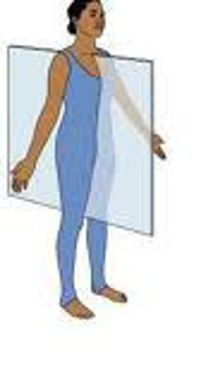
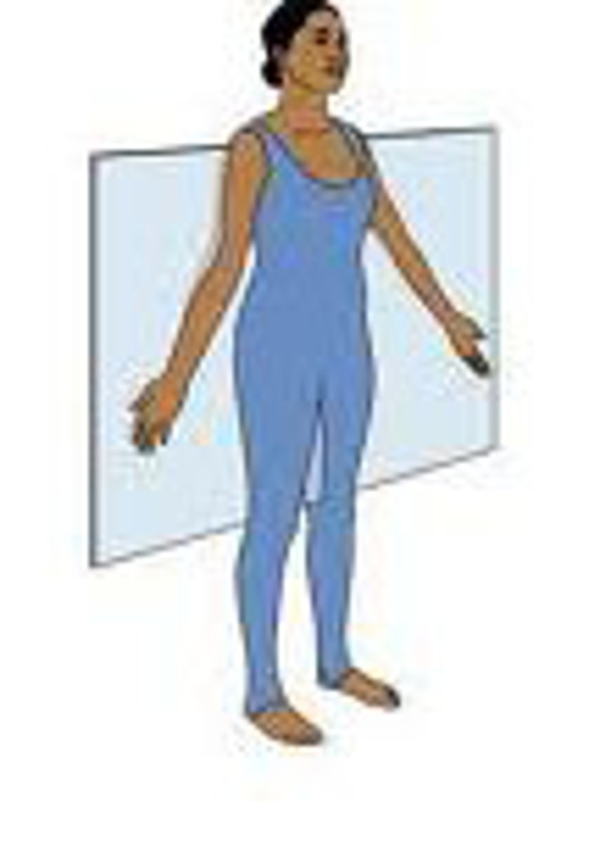
what is a PA/AP x-ray?
PA: x-ray enters posterior to anterior
AP: x-ray enters anterior to posterior
What x-ray plane is similar to a sagittal CT/MRI?
lateral
what x-ray plane is similar to a coronal CT/MRI?
AP/PA x-ray
what is fluoroscopy?
a live, real-time x-ray that is used during procedures and surgeries for guidance (on for periods of time not just a photo)
what is a CT scan?
computed tomography, transmission using ionizing radiation, 2D images reconstructed from 2D x-ray images taked 360
how does a CT work?
1. patient placed in scanner scout film obtained (original AP x-ray)
2. rotating x-ray tube revolves around patient with simultaneous revolving detector on other side
3. images reconstructed to make 3D rendering of anatomy with axial, sagittal, and cornal plane images
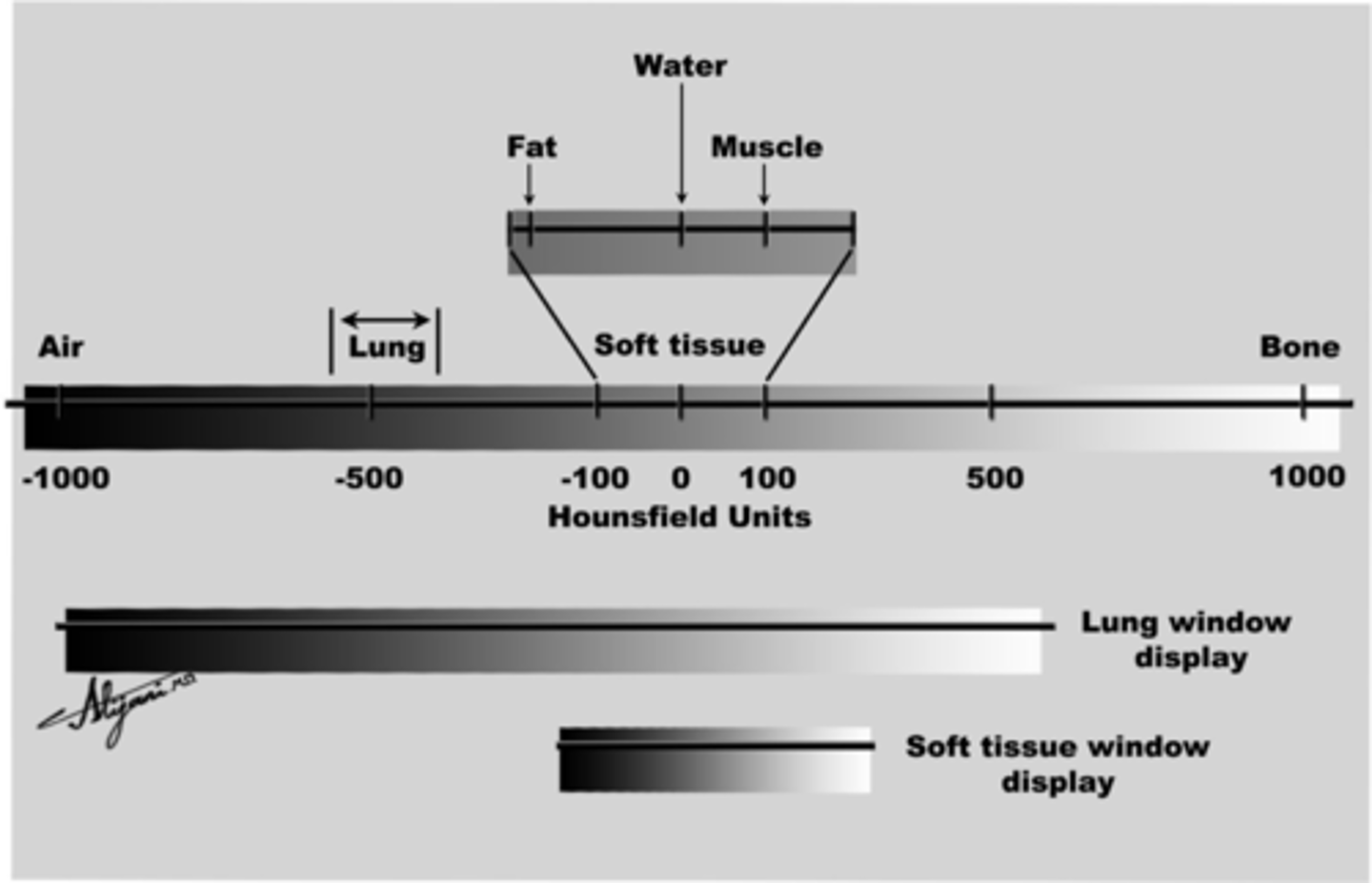
what does windowing mean?
choosing the gray scale range you want, you can choose upper and lower limit
what is the scale used for CT shades of gray?
Hounsfield Scale
what is contrast?
substances used to further enhance the visibility of structures being images in x-ray, CT, and MRI
when do you use iodinated IV contrast?
when you want to visualize organs, vessels, and abnormalities (perfussion differences)
when do you use barium contrast?
when you want to visualize GI mucousa (antegrade is oral, retrograde is rectally given)
what things are contrast not so helpful for?
brain (lots of blood in brain, will show up white)
bones/ortho (already white)
kidney stones (already white)
what is an MRI?
magnetic resonance imaging, NOT transmission imaging does NOT use ionizing radiation.
how does an MRI work?
magnetic field aligns hydrogen in the body and adding magnetic pulse changes changes orientation of these to create an image
what is the contrast for MRI?
Gadolinium
T1 MRI image
T1 you are seeing how long it takes for hydrogen nuclei to realign upwards with B0 field, instead of comparing signal fading from other tissues
fatty tissues are bright
T2 MRI image
based on differences in signal fading in the hydrogen in different tissues FROM EACH OTHER
-once B1 pulse turns hydrogens over how long does it take to fade energy? how does it compare to other tissues around it?
-hydrogens in smaller molecules fade faster = brighter (water)
-hydrogens in large molecules fade slower = darker (fatty)
do you give contrast with T1 or T2 MRI images
T1, NEVER T2
how can you tell the difference between a CT and MRI image?
-MRI has more detail in soft tissues
- on CT bones are bright white, they are darker on MRIs
what is an ultrasound?
imaging uses sound waves
how does an ultrasound work?
1. transducer placed on area of interest and sound waves propogate
2. sound waves variably bounce of different structures
3. time it takes to signal to return the transducer relates to depth of structure
What is an A-mode ultrasound?
amplitude mode, simplest type, single transducer
What is a B-mode US?
brightness mode, most often used, also known as 2-D, each echo depicted as a dot (baby ultrasounds)
what is a M-mode US?
motion mode, evaluate moving structures (lungs, heart valves)
what is a doppler US?
determines flow and velocity of blood, appears as blue/red, clogged vessels have higher velocity
what is a duplex US?
simultaneously using gray-scale and color doppler to visualize flow within a vessel
what is a PET scan?
positron emission tomography, emission imaging, does use ionizing radiation
how does a PET scan work?
1. FDG-18 is IV injected into patient
2. FDG-18 is taken up by metabolically active tissues like tumors
3.. FDG-18 begins beta decay, emit positrons, which create gamma rays for imaging
what does brigher area on pet scan mean?
higher uptake of FDG-18, lots of glucose use
5 things to determine xray adequacy
(PRIMA) penetration, rotation, inspiration, maginfication, angulation
penetration
powerful enough to show differences between bone and soft tissue (should be able to see thoracic spine through heart shadow)
rotation
spinous processes should fall equidistant between medial ends of clavicles (patient not rotated)
inspiration
should see at least 8-9 posterior ribs (tell patient to take a deep breath)
magnification
AP (mostly portable) magnify heart slightly
angulation
clavicle has normal "S" shape, medial end superimposes on 3/4th rib
patient not hunched over
other important xray things
Most superior portion of image should start just above apices of lung fields;
Most inferior portion of image should include AT LEAST both costophrenic angles;
Lateral margins of rib cage should be seen bilaterally in entirety
steps to reading a chest xray
1. confirm patient details (name, DOB, date and time of image)
2. assess image quality using PRIMA
3. ABCDE interpret
ABCDE method
Airway: trachea, carina, bronchi, hilar
Breathing: lungs and pleura
Cardiac: heart sizes and borders
Diaphragm: diaphragm and costovertebral angle
Everything else: mediastinal contours, bones, soft tissues, tubes, etc
reasons to get CT with contrast
malignancy workup/followup, vascular disease eval, eval of possible sternal/mediastinal infections,
TEE vs TTE echocardiogram
TTE: transducer on chest
TEE: transducer placed in esophagus