Cell Types
1/18
There's no tags or description
Looks like no tags are added yet.
Name | Mastery | Learn | Test | Matching | Spaced | Call with Kai |
|---|
No analytics yet
Send a link to your students to track their progress
19 Terms
Compared to other cell types, what makes neurons different
Describe the histology
Neurons Difference:
high rate of protein synthesis and metabolism
large amounts of Golgi apparatus + organized stacks of RER (Nissl substance)
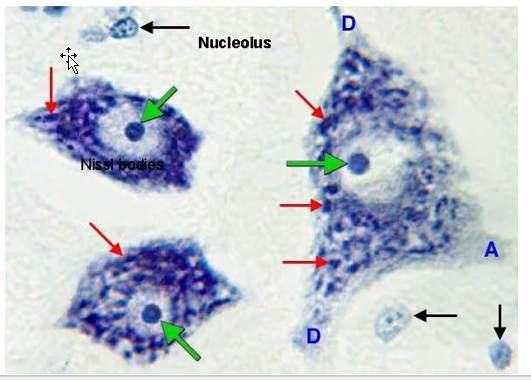
Histology
nucleic acids associated with the dense collection of ribosomal RNA can be stained with dyes that bind nucleic acids.
typical appearance of neurons histologically is a prominent, basophilic cell body with a clear nucleus and single nucleolus.
Describe the four types of neuron structure
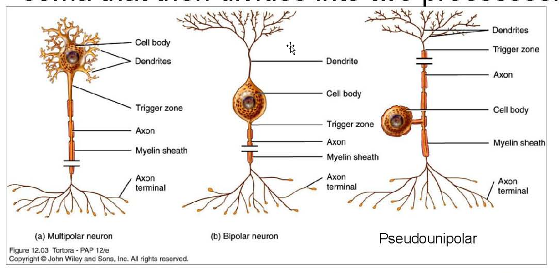
Types:
Multipolar:
multiple dendrites attached to the cell body and usually a single axon.
EX: Motor Neurons in SC
Bipolar:
single process from each end of the soma in opposite directions
EX: olfactory epithelial cells.
Unipolar:
Only in Dev
single process emerging from the cell body with no dendrites
Pseudounipolar:
single process that emerges from the soma that then divides into two processes
EX: Sensory Ganglion
Describe axonal transport
Fast vs Slow:
Both?
Consists of?
Rate?
Retrograde:
Importance?
Clinical Application:
Fast vs Slow:
Both:
Definition: process of components moving Soma → Axon terminals (anterograde transport)
Requires Energy:
Slow:
Consists of:
soluble substances
cytoskeletal proteins
EX: neurofilaments and components of microtubules
Rate: few millimeters a day.
Fast:
Consists of:
synaptic vesicles
neurotransmitter components
mitochondria
Rate: up to 400 mm/day
Clinical:
Transport tends to slow with age and in neurodegenerative diseases.
Retrograde Transport:
less well understood
different rates for different substances
Importance:
for feedback from axon terminals that can modify neuronal metabolism and responsiveness
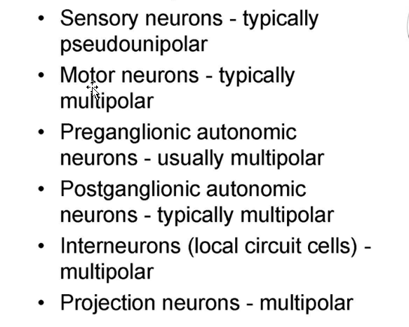
List the functional categories of neurons and what type they are
Draw out the functional zones of neurons
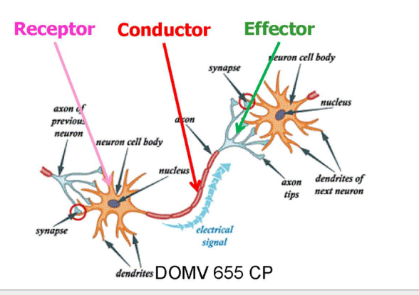
STATs: difference between dendrites and cell body
The dendrites typically receive most synapses (8,000 on a spinal motor neuron) while the cell body receives fewer (2,000).
Describe Axons:
STATs: Length/ Diameter
Relationship to Speed of AP?
STATs:
Length: can be more than a meter in length
Diameter: less than 1 micron to more than 20 microns.
AP Speed (conduction velocity):
depends upon the thickness of the axons.
Which Neurons are myelinated
Describe the characteristics of neurons that are likely to be myelinated
Myelination of Neurons
White matter in the CNS
most peripheral nerves in the PNS
Characteristics?
Larger axons are more likely to be myelinated
thickness of the "wrap" varies.
NOTE: even axons that don't have myelin are usually surrounded by either a Schwann cell or oligodendrocyte
Describe the Gray/White matter Difference in Cortext vs other places
Cerebral cortex (and brain nuclei): rich in neuronal cell bodies → gray matter
Areas under the cortex: rich in myelinated axons → White matter
Define laminae
Laminae - (layer, stratum) a flat slender sheet-like layer of functionally and sometimes anatomically similar neurons.
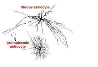
Describe Astrocytes
Two Types and Differences
Astrocytes:
protoplasmic
located in gray matter
puffy, protoplasmic appearance
fibrous astrocytes
in white matter
more fibrous appearance
greater abundance of intermediate filaments
NOTE:
These differences in morphology probably reflect the functional specialization related to the support of the cellular environment in synapse-rich regions and axons.
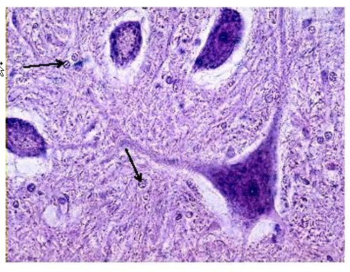
Describe the histological differences between Neurons and Neuroglials
Neuroglias:
less Nissl substance (nucleic acids)
smaller and generally do not have a prominent nucleolus.
nuclei have a speckled appearance
List out the function of the astrocytes
Function:
physical isolation and regulation of the neural environment of the CNS
scavenge K in extracellular environment
regulate extracellular ion concentrations
participate in neurotransmission (particularly glutamate and GABA)
participate in neurovascular coupling
supply nutrients to neurons and synapses from the circulation
Contribute to BBB (astrocyte end feet)
NOTE: most restriction is @ level of capillary endothelials
Repair/Response to injury/stress
important in the migration of neurons to their appropriate location in the CNS
Describe the location of astrocytes in relation to other neural structures
B/c of the above answer, what was believed?
Location: surround everything of CNS:
form a continuous layer on
surface of the CNS → glial-pial membrane
all blood vessesl of CNS
believed that astrocytes were primarily a scaffold element of the CNS much the same as fibroblasts are in the structure of organs peripherally.
Describe how astrocytes respond to injury
astrocytes respond to an area of tissue damage by walling off the injured area with astrocytic process, and re- establishing the isolated CNS environment.
Describe Myelination:
% lipid?
Function?
Large vs small axons?
Describe the Formation
myelination,
(80% lipid) neuroglial membranes.
Function:
insulates axons from one another
controls ionic environment of axon,
increases conduction velocity of axons.
Large vs small:
Large axons have thicker myelin sheaths and faster conduction velocities
Formation:
Glial cells respond will usually provide one segment to an axon (internodal segment), but may myelinate up to 40 axons
NOTE: segments vary between 200 and 1000 microns in length
Describe the other functions of oligodendrocytes
also observed in the vicinity of the cell bodies of neurons (perineuronal oligodendrocytes; satellite cells).
role of these perineuronal cells in both locations is not clear, but they appear to regulate the environment around the neuronal cell body.
Describe Microglia
Function?
Existence?
Establishes?
Describe when other immune cells will help the microglial cells
Microglial cells
Function
immune cells of the CNS
primary mediators of inflammation
major phagocytic cells (dead/injured) in the CNS
Existence?
1% of cells in the CNS
not derived from neuroectoderm (like astrocytes/oligodendrocytes)
exist in a quiescent state
establish interconnected territories, but are not coupled by gap junctions
Outside Help:
In areas of direct CNS injury,
peripheral macrophages "gitter cells" may enter to help
NOTE: Peripheral macrophages and immune cells (e.g. lymphocytes, neutrophils) do not normally enter the CNS in large numbers, but may do so with injury or infection
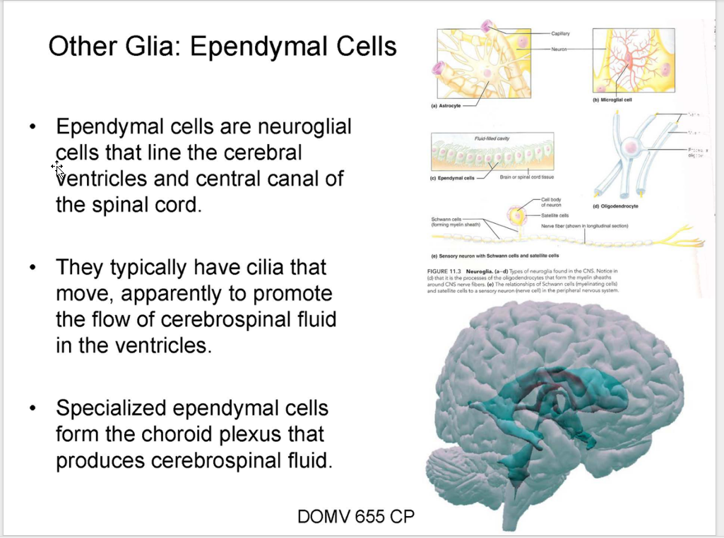
[REVIEW] Ependymal Cells