MUSCULAR SYSTEM
1/62
There's no tags or description
Looks like no tags are added yet.
Name | Mastery | Learn | Test | Matching | Spaced | Call with Kai |
|---|
No analytics yet
Send a link to your students to track their progress
63 Terms
is responsible for all types of body movement. Additional functions of this system include providing support, stabilizing joints, and generating heat for the body
muscular system
what do all muscles consist of?
muscle fibers
these contract to facilitate body movement
muscle fibers
what are muscle attached to?
attached to bones and to internal organs and blood vessels
how many muscles are there in the body?
over 600
3 types of muscle
cardiac, smooth, skeletal
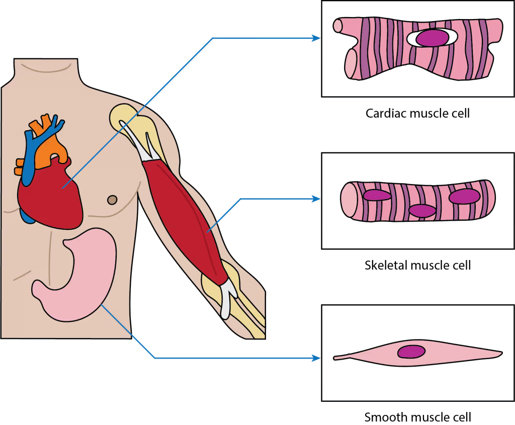
muscle is striated, short, and branched. Contains one nucleus, are branched, and rectangular.
Involuntary process and under the control of the autonomic nervous system.
Found in the walls of the heart
cardiac muscle
muscle is striated, long and cylindrical. Many nuclei in a cell.
Attached to bones in the body, contracts voluntarily - under conscious control
skeletal muscle
muscle consists of non-striated muscle cells that are spindle-shaped. Contain one nucleus.
Found in the walls of internal organs like the bladder and stomach.
Contraction is involuntary and controlled by the autonomic nervous system
smooth muscle
what do all the muscles share in common
excitability, contractility (muscle shortening), extensibility (muscle stretching), and elasticity
what muscle is excited by the nervous system
skeletal
what muscle are stimulated by the nervous system and by circulating hormones (dont got to think about it !)
cardiac and smooth
what muscles do skeletal muscles have?
endomysium, perimysium, epimysium
endomysium
encases individual skeletal muscle fibers
perimysium
muscle fibers are bundled together by a connective tissue
fasciculi
Bundles of skeletal muscle fibers
epimysium
Each fascicle is bundled together by a strong connective tissue
sarcolemma
cell membrane that surrounds a skeletal muscle fiber
sarcoplasm
cytoplasm of the skeletal muscle fiber
myofibrils
One muscle fiber is filled with several long, cylindrical protein, are the contractile units of the fiber
sarcomere
smallest contractile unit in a myofibril
myofilaments
they make up a myofibril
2 types of myofilaments
thick bands and thin bands
thick bands
made of several protein molecules called myosin
thin bands
several protein molecules, called actin, link together to form these bands
are attached to a Z-disk (or Z-line).
what color are the I-bands
light-colored bands
what color are the A-bands
dark-colored bands
where is the Z line found?
middle of the I-bands
where is the H zone found
middle of the A-bands
what is in the middle of the H zone
M line, which is the center of the sarcomere
which band is thick and thin filaments
A band
what band is thin filament only
I band
what band is actin filament attachment site
Z line
which band is thick filament only
H band
what does slide filament theory explain?
muscle contraction
actin filaments slide past myosin filaments, pulling the actin filaments closer to the center of the sarcomere, or M line
what attachments do head of myosin form with the actin myofilaments?
crossbridges
what is the head of actin
a round protein shaped like a ball. Several of these round proteins link together to form a long chain, or thin myofilament
with the help of ATP, what do myosin heads do?
attach to binding sites in actin and form a crossbridge. After energy in the myosin head is released, the myosin pulls actin myofilaments closer to the M line. This head can only form another crossbridge when another molecule of ATP attaches to the head, reenergizing it
how does calcium play a role?
determining when contraction happens
where is calcium found in?
sarcoplasmic reticulum
ligaments
attach bones to bones, they form a joint
3 type of joints
immovable, partly movable, synovial
immovable joint
known as fibrous joints, these consist of bones held together by connective tissues. The bones are in very close contact.
ex: intersection of cranial bones in the skull.
partly moveable joint
known as cartilaginous joints, these consist of bones held together by cartilage. These joints allow some degree of movement.
ex: vertebral discs in the spine.
synovial joint
allow the largest freedom of movement because the bones are separated by a joint cavity.
ex: hip and shoulder
6 types of synovial joints
pivot, hinge, saddle, condyloid, plane, ball/socket
pivot joint
a type of freely moveable, uniaxial synovial joint that allows only rotational movement around a single axis
Allow turning, twisting, pronation, and supination
ex of pivot joint
Atlanto-axial joint: Located between the first (atlas) and second (axis) cervical vertebrae, this joint allows the head to turn from side to side.
Proximal Radioulnar Joint: Located near the elbow, where the head of the radius rotates against the ulna, enabling the forearm to twist.
Distal Radioulnar Joint: Located near the wrist, working in conjunction with the proximal joint for forearm rotation
hinge joint
a type of synovial joint allowing movement in only one plane, enabling flexion and extension like a door hinge
ex of hinge joint
Elbow: Articulation between the humerus and ulna.
Knee: Between the femur and tibia (note: allows slight rotation when flexed).
Ankle: Between the tibia/fibula and the talus.
Interphalangeal Joints: Fingers and toes.
saddle joint
a highly flexible, biaxial synovial joint where one bone is concave and the other convex, fitting together like a rider on a saddle
ex of saddle joint
Trapeziometacarpal Joint (Thumb): Connects the trapezium and the metacarpal of the thumb, allowing complex, opposable movements.
Sternoclavicular Joint (Shoulder): Connects the sternum (breastbone) and clavicle (collarbone).
Incudomalleolar Joint (Ear): Connects the incus and malleus, part of the ossicle chain
condyloid joint
a biaxial synovial joint featuring an oval-shaped condyle that fits into an elliptical cavity, allowing movement in two planes: flexion/extension and abduction/adduction
ex of condyloid joint
Wrist (Radiocarpal joint): Between the radius and carpal bones.
Knuckles (Metacarpophalangeal joints): Between the metacarpals and phalanges.
Foot (Metatarsophalangeal joints): Between metatarsals and phalanges.
Jaw (Temporomandibular joint): Although often considered a complex hinge, it functions as a condyloid joint.
Head/Spine (Atlanto-occipital joint): Allows nodding,.
plane joint
a type of synovial joint with flat or slightly curved bone surfaces that permit limited, non-axial gliding movements
ex plane joint
Acromioclavicular Joint: Connects the scapula to the clavicle.
Intercarpal/Intertarsal Joints: Joints between the small bones of the wrist and ankle.
Zygapophyseal Joints: The facet joints connecting vertebrae in the spine.
Sternocostal Joints: Joints connecting the ribs and sternum.
Sacroiliac Joint: Connects the sacrum to the pelvis.
ball socket joint
a type of synovial joint where a spherical bone end fits into a cup-like cavity, allowing multi-axial movement, including rotation, flexion, extension, abduction, and adduction
ball socket joint ex
hip and shoulder joints
tendons
attach muscle to bone
consist of tough connective tissue that is found on either side of the joint where two bones are connected
work with skeletal muscles to move bones
flexor muscle
muscle that causes a joint to bend
extension muscle
muscle that contracts and causes a joint to straighten
what happens when one of the flexor or extension muscles contracts
the other remains elongated.
ex of flexor and extension muscle
Biceps and triceps muscles in the arm work together to bend and lengthen the elbow. As a biceps muscle contracts, the triceps muscle remains elongated, or relaxed. Thus, the biceps is the flexor and the triceps is the extensor of the elbow joint.