Exchange
1/69
Earn XP
Description and Tags
Name | Mastery | Learn | Test | Matching | Spaced | Call with Kai |
|---|
No analytics yet
Send a link to your students to track their progress
70 Terms
True or false? The larger the organism, the larger it’s surface area to volume ratio.
False. THE LARGER THE ORGANISM, THE SMALLER IT’S SURFACE AREA TO VOLUME RATIO/ THE SMALLER THE ORGANISM, THE LARGER IT’S SURFACE AREA TO VOLUME RATIO.
Why can very small organisms simply exchange substances across their surface?
They have a very large surface area to volume ratio.
The distance from their surface to their centre is very short (short diffusion distance).
Overall this means that they can receive a sufficient supply of substances solely via simple diffusion.
Why do smaller mammals have a higher rate of metabolism and respiration?
They have a larger surface area to volume ratio meaning they lose heat easily, having a higher rate of metabolism and respiration allows them to release more energy needed to release more heat to maintain a stable body temperature.
Give five examples of adaptations in specialised exchange surfaces to increase surface area to volume ratio in different organisms.
Alveoli and bronchioles (mammals).
Villi and microvilli.
Spiracles and tracheoles (insects).
Gill filaments and lamellae (fish).
Thin, wide leaves (plants).
Define ventilation.
The movement of air into and out of the lungs (inhalation and exhalation).
Give three general adaptations of gas exchange surfaces.
Large surface area to volume ratio.
Short diffusion distance.
Ability to maintain steep concentration gradient.
Draw and label a simple diagram of the human gas exchange system.
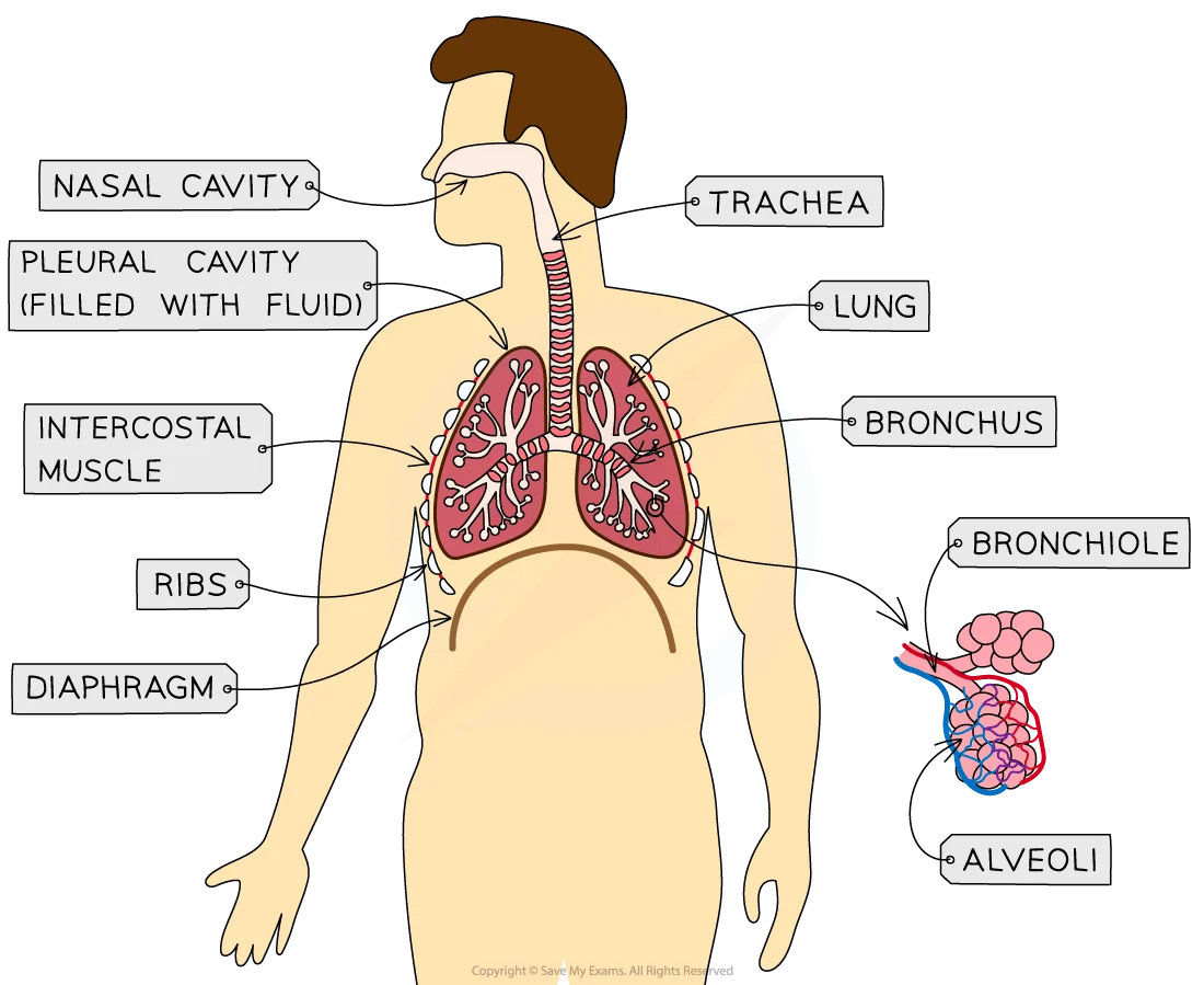
What is the name of the interaction between the internal and external intercostal muscles?
Antagonistic.
What happens to:
a) the external intercostal muscles
b) the internal intercostal muscles
c) the ribs
d) the diaphragm
e) the air pressure in the thoracic cavity
f) volume of the lungs
upon inhalation?
a) contract
b) relax
c) move up and out
d) contracts to move down and flatten
e) decreases
f) increases
What happens to:
a) the external intercostal muscles
b) the internal intercostal muscles
c) the ribs
d) the diaphragm
e) the air pressure in the lungs
f) volume of the lungs
upon exhalation?
a) relax
b) contract
c) move down and in
d) relaxes to move up and dome
e) increases
d) decreases
What is pulmonary ventilation?
The total volume of air that moves into the lungs in one minute (dm3 min-1).
What is the tidal volume?
The volume of air inhaled in one breath (dm3).
What is the ventilation rate?
The number of breaths per minute (min-1).
Give an equation for calculating pulmonary ventilation with tidal volume and ventilation rate.
Pulmonary ventilation = tidal volume x ventilation rate
Where exactly in the lungs does gas exchange take place?
In the alveoli.
How do alveoli increase the surface area to volume ratio of the lungs?
Because there are so many alveoli in the lungs.
How do alveoli provide a short diffusion distance for gas exchange?
Walls of the alveoli are only one cell thick with thin epithelium cells.
How is a concentration gradient maintained in the alveoli?
Each alveolus is surrounded by a network of capillaries to remove exchange gases, therefore maintaining a steep concentration gradient allowing for more gas exchange to take place.
Where is the site of gas exchange in fish?
The gills.
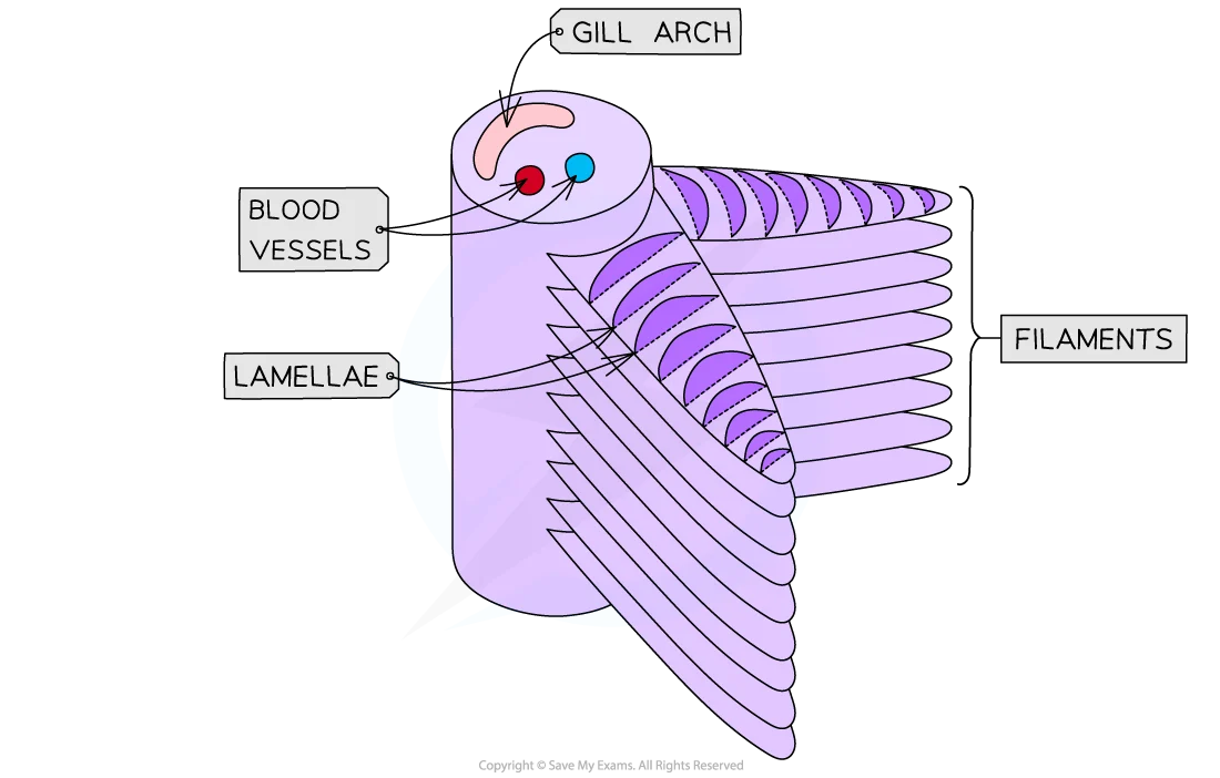
Describe how the structure of the gills provides a large surface area for gas exchange.
On the gill filaments there are further extensions called lamellae which increase the surface area of the gill filaments.
How is a short diffusion distance maintained in the gills?
Through every lamella having a capillary network and being very thin.
Draw a simple, labelled diagram of fish gills.
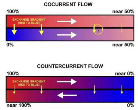
What is the general idea of countercurrent flow?
Water flowing over the gills in the opposite direction to the flow of blood in the gills to ensure that a concentration gradient is maintain across the entire length of the gill lamellae.
Talk me through how countercurrent flow works.
1) Water goes into the gill filament with a high concentration of oxygen as it has not yet given any to the blood.
2) As water moves down the gill filament, it’s oxygen concentration decreases as oxygen diffuses into the blood.
3) Blood goes into the gill filament (from the opposite end) with a low concentration of oxygen as it has not yet received any from the water.
4) The low oxygen concentration water meets with the low oxygen concentration blood at the ‘start’ of the gill filament, since water still has a slightly high oxygen concentration oxygen still diffuses into the blood.
5) The new high oxygen concentration water meets with the now high oxygen concentration blood at the ‘end’ of the gill filament, since water still has a slightly higher oxygen concentration oxygen still diffuses into the blood.
This maximises oxygen diffusion into the blood.
Give two advantages of insects having an exoskeleton.
Provides protection.
Prevents water loss.
What do insects have instead of lungs for gas exchange?
A tracheal system (spiracles → trachea → tracheoles).
What are spiracles?
Round, valve-like openings on the exoskeleton of an insect that can open and close.
How does oxygen enter/carbon dioxide leave the insect?
Through the spiracles.
Where are spiracles found on an insect?
Along the insect’s abdomen.
What are the trachea in an insect? What do they branch off into?
A network of internal tubes. They branch off into tracheoles.
Where do the tracheoles go in an insect?
The tracheoles extend into all internal tissues.
How is the collapsing of the trachea prevented in insects?
The trachea are strengthened with rings (of chitin) within them.
Give three ways in which the tracheoles are adapted to provide a short diffusion distance.
They have thin walls.
They extend throughout all tissues.
They are highly branched so provide a large surface area.
Talk me through how a concentration gradient is maintained between the tracheoles and the atmosphere when an insect is at rest.
1) Cells respire which uses up oxygen and produces carbon dioxide.
2) This creates a steep concentration gradient for oxygen and carbon dioxide between the atmosphere and the tracheoles.
3) This means oxygen simply diffuses into the cells while carbon dioxide simply diffuses out of cells.
Talk me through how a concentration gradient is maintained between the tracheoles and the atmosphere when the insect is in HIGHER activity.
1) Insects contract and relax their abdominal muscles.
2) This causes air to be actively moved in and out of the tracheal system.
3) This helps maintain an oxygen and carbon dioxide concentration gradient.
Talk me through how a concentration gradient is maintained between the tracheoles and the atmosphere when the insect is in VERY HIGH activity.
1) In very high activity, the insect anaerobically respires more.
2) Increased anaerobic respiration produces more lactate in muscle cells.
3) More lactate in muscle cells means that the water potential of the muscle cells is lowered.
4) This causes water in the ends of the tracheoles to move into the muscle cells via osmosis.
5) This movement of water out of the tracheoles ends decreases the volume of fluid in the tracheoles.
6) Gases diffuse faster in air than in liquid, so diffusion takes place faster.
Give and explain three adaptations insects have for limiting water loss.
Water proof exoskeleton (reduces evaporation from the surface of the insect).
Spiracles can open and close (stops water evaporating out of the spiracles all the time).
Spiracles are surrounded by hairs (they trap water vapour (humid air) which reduces the water potential gradient from the atmosphere to the trachea meaning less water evaporates).
Label this cross section of a leaf.
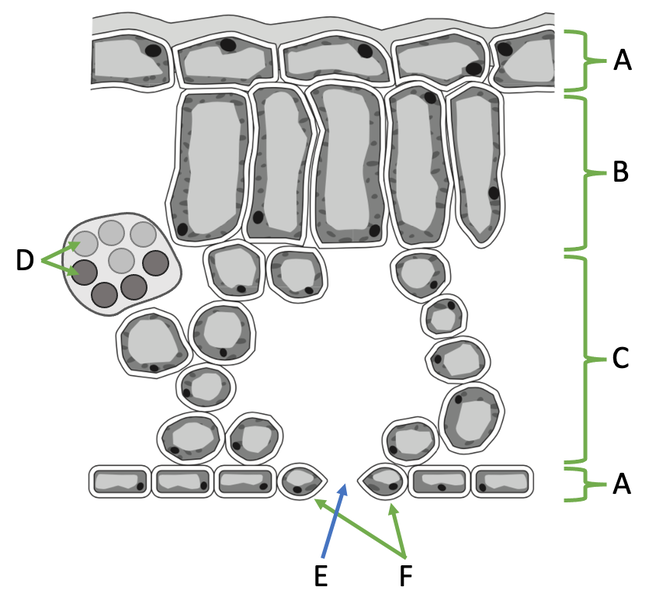
A) Upper epidermis/lower epidermis.
B) Palisade mesophyll.
C) Spongy mesophyll.
D) Vascular bundle (made up of xylem and phloem).
E) Stoma.
F) Guard cells.
Where in a leaf is the site of photosynthesis?
The palisade mesophyll.
Talk me through how gas exchange takes place in a leaf.
1) Photosynthesis takes place in the palisade mesophyll, this decreases the concentration of carbon dioxide and increases the concentration of oxygen in the palisade mesophyll.
2) Oxygen moves down it’s concentration gradient, through the spongy mesophyll and out of the leaf via the open stomata.
3) Carbon dioxide moves down it’s concentration gradient, in through the open stomata and up the spongy mesophyll to the palisade mesophyll where it is needed.
How do the stomata limit water loss when the plant is not photosynthesising?
The stomata close when the plant is not photosynthesising meaning water cannot evaporate out of them.
How are leaves adapted to provide a short diffusion distance for gas exchange?
Leaves are very thin.
What are xerophytic plants?
Plants that are adapted to survive in environments with little water.
Give and explain five ways in which xerophytes are adapted to their environment.
Leaves curl inwards (this means that all stomata are facing inwards so any water that evaporates out of them is trapped which reduces the water potential between the atmosphere and the leaf meaning less water evaporates out of the leaf).
Hair-like structures (trap moisture, same idea as the curled leaves).
Sunken stomata (trap moisture, same idea as the curled leaves).
Thicker waxy cuticle (reduces evaporation).
Longer root network (reach more water).
What is digestion?
The process in which larger molecules are hydrolysed into smaller molecules that can cross cell-surface membranes.
Where is amylase produced?
Pancreas
Salivary glands
Where are membrane-bound disaccharidases produced?
Ileum.
Talk me through the process of the digestion of a carbohydrate.
1) Amylase from the salivary glands hydrolyses some of the polysaccharides into maltose (a disaccharide) by hydrolysing glycosidic bonds within the polysaccharide.
2) Hydrolysis of the rest of the polysaccharides with amylase continues in the duodenum.
3) Maltose reaches the ileum and is hydrolysed (breaking a glycosidic bond) by membrane-bound disaccharidases into glucose (a monosaccharide).
Where does protein digestion start and end?
Starts in the stomach, (continues in the duodenum), ends in the ileum.
Which three types of enzymes digest proteins?
Endopeptidases.
Exopeptidases.
Membrane-bound disaccharidases.
How do endopeptidases work when digesting a protein?
Endopeptidases hydrolyse peptide bonds between amino acids that are within the polypeptide chain, forming multiple shorter polypeptides.
How do exopeptidases work when digesting a protein?
Exopeptidases hydrolyse peptide bonds between amino acids on the ends of the polypeptide, forming dipeptides.
How do membrane-bound dipeptidases work in digesting proteins?
Membrane-bound dipeptidases hydrolyse peptide bonds between amino acids in dipeptides, forming amino acids.
Where are endopeptidases and exopeptidases found?
In the stomach and duodenum.
Where are membrane-bound dipeptidases found?
In the ileum.
What physically digests lipids?
Bile salts.
What chemically digests lipids?
Lipase.
Where is lipase produced?
In the pancreas.
How does lipase work?
Lipase hydrolyses ester bonds between fatty acids and glycerol in triglycerides, forming monoglycerides and fatty acids.
How do bile salts work?
Bile salts emulsify lipids (forming micelles) so that smaller lipid droplets are formed, this increases the surface area of the lipid meaning lipase action to digest the lipid is faster.
Where are bile salts produced and stored?
Produced in the liver, stored in the gallbladder.
Where does lipase action take place?
In the small intestine.
Where are the products of digestion absorbed in mammals?
In the ileum.
Give and explain seven ways in which the ileum is adapted to maximise absorption.
Villi (increase surface area).
Microvilli on epithelial cells (increase surface area).
Epithelium is one cell thick (short diffusion distance).
Surrounded by capillaries (short diffusion distance, maintain concentration gradient).
Many carrier/channel proteins in epithelial cell-surface membranes (more active transport and facilitated diffusion into the blood at a time).
Many mitochondria in epithelial cells (more ATP provided which is needed for active transport).
Many ribosomes in epithelial cells (produce more membrane proteins for active transport and facilitated diffusion).
What are micelles?
Water-soluble vesicles formed from fatty acids, glycerol, monoglycerides, bile salts.
How do micelles help with lipid absorption?
They make lipids more water-soluble, allowing them to stay suspended in the watery content on the small intestine.
They bring fatty acids to the cell-surface membrane of epithelial cells which helps maintain a steep concentration gradient of fatty acids near the epithelial cells.
Talk me through the process of lipid absorption (from micelles to lymph).
1) Fatty acids and monoglycerides leave micelles at the cell-surface membrane of the epithelial cell and diffuse into the epithelial cell.
2) In the golgi apparatus, the fatty acids and monoglycerides are reformed (ester bonds reformed) into triglycerides.
3) Triglycerides are combined with proteins to form chylomicrons which are packaged into vesicles.
4) Vesicles move towards to other side of the cell-surface membrane of the epithelial cell.
5) Vesicles fuse with the cell-surface membrane of the epithelial cell and release the chylomicrons via exocytosis.
6) Chylomicrons diffuse into lymphatic lacteal.
True or false? There is usually a higher concentration of glucose in the lumen of the ileum than in the epithelial cells of the ileum.
False. THERE IS USUALLY A HIGHER CONCENTRATION OF GLUCOSE IN THE EPITHELIAL CELLS OF THE ILEUM THAN IN THE LUMEN OF THE ILEUM.
What is the name of the process in which glucose/amino acids are absorbed?
Co-transport.
Talk me through the process of the absorption of glucose in the ileum via co-transport.
1) A sodium ion pump actively transports sodium ions out of the epithelial cell and into the blood, creating a sodium ion concentration gradient between the lumen of the ileum and the epithelial cell (higher in lumen).
2) Sodium ions diffuse into the epithelial cell down their concentration gradient.
3) The co-transporter protein that transports sodium ions back into the epithelial cells also has glucose attach to it meaning glucose is transported into the epithelial cell with sodium ions(against it’s concentration gradient).
4) Glucose moves via facilitated diffusion into the blood.