UNIT 1 SEMESTER 1 EXAM- Biological, Lifespan and Science Inquiry Psychology Flashcards
1/47
Earn XP
Description and Tags
Covers all ATAR Psychology Syllabus content from Unit 1
Name | Mastery | Learn | Test | Matching | Spaced | Call with Kai |
|---|
No analytics yet
Send a link to your students to track their progress
48 Terms
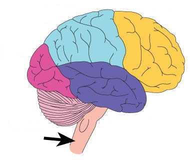
Label brain structure and functions- frontal lobe, pariteal lobe, occipital lobe, temporal lobe, cerebellum, hippocampus, amygdala, broca's area, wernickes area, corpus callosum.
Frontal lobe - higher‑order thinking, planning, decision‑making, problem‑solving; voluntary movement via the primary motor cortex; speech production (Broca's area).
Parietal lobe - processes sensory information such as touch, pressure, temperature via the primary somatosensory cortex; spatial awareness.
Occipital lobe - visual processing via the primary visual cortex.
Temporal lobe - auditory processing via the primary auditory cortex; memory; language comprehension (Wernicke's area).
Cerebellum - coordination of balance, posture and fine motor movements. Hippocampus - formation of new long‑term memories.
Amygdala - processing of emotions, especially fear and threat detection.
Broca's area - speech production (left frontal lobe). Wernicke's area - language comprehension (left temporal lobe).
Corpus callosum - bundle of nerve fibres connecting the two hemispheres, enabling communication between them.
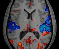
What are fMRI scans?
Uses magnetic fields to measure changes in blood flow.
Shows which brain areas are active during tasks.
Used to study brain function and identify abnormalities.
Strengths and Limitations of fMRI scans
Strengths: Non‑invasive, no radiation, produces detailed images of brain activity, good for studying function in real time.
Limitations: Expensive, sensitive to movement, not suitable for people with metal implants or severe claustrophobia.
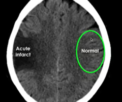
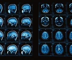
What are CT scans?
Uses X‑ray images taken from multiple angles to create a 3D structural image of the brain. Shows physical abnormalities such as tumours, bleeding or injury.
Strengths and Limitations of CT scans
Strengths: Quick and non‑invasive. Useful for detecting structural damage.
Limitations: Uses radiation. Less detailed than MRI.
Define: Neuron
A specialised cell that transmits information through electrochemical signals.
Label a neuron
A: Dendrite. B: Cell Body. C: Axon. D: Axon Terminal. F: Myelin Sheath.
How to neurons work?
Dendrites receive information. An electrical impulse (action potential) travels down the axon. Neurotransmitters are released from the axon terminals into the synapse.
Define: Neurotransmitters
Chemical messengers that transmit signals across the synapse to the next neuron.
Define: Synapse
The gap between two neurons where neurotransmission occurs
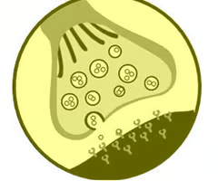
Label a synapse.
Axon terminal
Vesicles containing neurotransmitters
Synaptic cleft
Neurotransmitter
Receptor
Dendrite
How to synapses work?
Action potential reaches axon terminal.
Neurotransmitters are released into the synaptic cleft.
They bind to receptors on the next neuron.
Excess neurotransmitter is reabsorbed
What are the 4 main brain scanning techniques?
EEG, CT, MRI, fMRI
What is the role of the CNS?
Central processing and control point for all human behaviour. Processes all information from the senses and is responsible for controlling behaviour.
What is the role of the spinal cord in the CNS?
Connects the brain to the rest of the body, and allows messages to be passed from body to brain and from brain to other parts of the body
Synaptic transmission
Process of communication between neurons using neurotransmitters.
How is a synaptic transmission generated?
1. Electrical impulse down the axon triggers release of neurotransmitters from axon terminals.
2. Released into synaptic gap to be picked up by the receptors on dendrites or to be taken up again for re-use
Neurotransmitters in synaptic transmission
- Released from vesicles when action potential arrives.
- Bind to receptors on the next neuron.
Receptors in synaptic transmission
- Bind to specific neurotransmitters.
- Trigger response in the post‑synaptic neuron.
3 common neurotransmitters
Noradrenaline
Dopamine
Serotonin
Noradrenaline
Arousal, alertness, attention.
Dopamine
Movement, motivation, reward.
Serotonin
Mood regulation, sleep, appetite.
The brain is in two halves, called...
Hemispheres, which is lateralisation.
Pre-frontal cortex
The front part of the frontal lobe, located behind the forehead, and is responsible for executive functions like decision-making, planning, personality expression, emotional regulation, and social behavior.
Peripheral Nervous System (PNS)
All nerves outside the CNS that carry sensory information to the brain and motor commands back to the body, allowing communication between the CNS and organs, muscles and glands.
Somatic Nervous System
Controls voluntary movement by sending sensory messages to the CNS and motor signals to skeletal muscles, enabling actions like walking, writing and speaking
Autonomic Nervous System
Controls involuntary functions such as heart rate, breathing and digestion, automatically regulating internal processes to maintain balance and survival.
Sympathetic Nervous System
Activates the fight‑or‑flight response by increasing heart rate, widening pupils and slowing digestion, preparing the body for immediate action during stress.
Parasympathetic Nervous System
Returns the body to a calm resting state by slowing heart rate, increasing digestion and conserving energy once danger or stress has passed.
Sensory Neuron
Carries information from sensory receptors to the CNS, allowing the brain to detect environmental changes like touch, temperature and pain.
Motor Neuron
Carries commands from the CNS to muscles and glands, producing movement and physical responses based on brain and spinal cord signals.
Interneuron
Connects sensory and motor neurons within the CNS and processes information, playing a key role in reflexes, decision‑making and complex thinking.
Medulla
Controls vital survival functions such as breathing, heart rate and swallowing, operating automatically to keep the body alive without conscious effort.
Reticular Formation
Regulates alertness, arousal and the sleep-wake cycle, filtering incoming sensory information to help maintain attention and consciousness
Thalamus
Relay station that directs all sensory information except smell to the correct cortical areas, helping the brain organise and interpret incoming signals.
Primary Motor Cortex
Controls voluntary movement of skeletal muscles and is organised by body part, allowing precise, coordinated and intentional physical actions.
Primary Somatosensory Cortex
Processes touch, pressure, temperature and pain signals from the body, creating a detailed sensory map that helps us interpret physical sensations.
Primary Auditory Cortex
Processes sound information such as pitch, volume and speech patterns, allowing us to recognise and understand auditory signals.
Primary Visual Cortex
Processes visual information including colour, shape and motion, enabling recognition, interpretation and understanding of what we see.
Phineas Gage
- Survived an 1848 accident where an iron rod blasted through his left frontal lobe.
- Before the accident he was responsible and polite; afterwards he became impulsive, aggressive and unreliable.
- Memory, intelligence and language stayed intact, showing the damage was specific to personality and executive functioning.
- Demonstrates the frontal lobe's role in planning, impulse control, emotional regulation and socially appropriate behaviour.
Roger Sperry Split‑Brain
- Studied patients whose corpus callosum was severed to treat severe epilepsy.
- Found the left hemisphere specialises in language, logic and analytical tasks, while the right hemisphere specialises in spatial, visual and creative processing.
- When information was presented to the left visual field (right hemisphere), patients could not verbally describe it but could draw or identify it.
- Demonstrated hemispheric specialisation and the importance of the corpus callosum for communication between the two hemispheres.
Walter Freeman Lobotomy
- Performed frontal lobotomies by inserting an ice‑pick‑like tool through the eye socket to sever connections in the prefrontal cortex.
- Patients often became emotionally flat, passive, childlike or unable to plan, make decisions or control impulses.
- Showed the prefrontal cortex is essential for personality, judgement, emotional regulation and executive functioning.
- Procedure was later abandoned due to severe side effects and ethical concerns, highlighting the dangers of early psychosurgery.
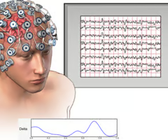
EEG (electroencephalogram)
Measures electrical activity in the brain to study sleep, consciousness and abnormal activity, excellent for tracking timing of brain activity but cannot pinpoint exact locations.
EEG Strengths and Limitations
Strengths: non‑invasive, inexpensive, excellent temporal resolution.
Limitations: poor spatial resolution, cannot locate exact brain areas, only measures surface activity
MRI (magnetic resonance imaging)
Produces detailed structural images of the brain without radiation; excellent for detecting tumours and injuries but expensive and unsuitable for people with metal implants.
MRI Strengths and Limitations
Strengths: highly detailed structural images, no radiation, good for detecting tumours and injury.
Limitations: expensive, slow, unsuitable for metal implants or claustrophobic patients.
Developmental Plasticity
Brain changes that occur naturally during growth, including synaptogenesis, pruning and myelination, most active in childhood when learning is rapid.