DH228 Oral Path M4 KC Ch 5 (ALL 3 Attempts) (",)
1/76
There's no tags or description
Looks like no tags are added yet.
Name | Mastery | Learn | Test | Matching | Spaced | Call with Kai |
|---|
No analytics yet
Send a link to your students to track their progress
77 Terms
*Which tumor frequently arises from a dentigerous cyst?
Sarcoma
Ameloblastoma
Odontoma
Dens in dente
Ameloblastoma
An ameloblastoma frequently arises from a dentigerous cyst.
*A small elevated mass of thyroid tissue located near the foramen cecum or posterior lateral borders of the tongue, which forms as a result of failure of the embryonic thyroid tissue to migrate to its proper position, is called a(n)
ameloblastic fibroma.
hemangioma.
lingual thyroid nodule.
thyroglossal duct cyst.
lingual thyroid nodule.
A small elevated mass of thyroid tissue located near the foramen cecum or posterior lateral borders of the tongue, which forms as a result of failure of the embryonic thyroid tissue to migrate to its proper position, is a lingual thyroid nodule. An ameloblastic fibroma is a mixed odontogenic tumor. A hemangioma is a benign proliferation of capillaries. A thyroglossal duct cyst is located below the hyoid bone.
*Clinically, the lingual thyroid nodule appears as a smooth nodular mass
at the base of the tongue posterior to the circumvallate papillae.
on the anterior ventral tongue.
on the lateral borders of the middle third of the tongue.
anterior to the circumvallate papillae.
at the base of the tongue posterior to the circumvallate papillae.
Clinically the lingual thyroid nodule appears as a smooth nodular mass at the base of the tongue posterior to the circumvallate papillae. The lingual thyroid nodule is not found on the anterior ventral tongue. The lingual thyroid nodule is not found on the lateral borders of the middle third of the tongue. The lingual thyroid nodule is not found anterior to the circumvallate papillae.
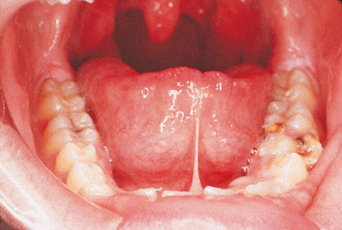
*This patient exhibits an extensive adhesion of the tongue to the floor of the mouth caused by the short lingual frenum. What condition is suspected?
Ankyloglossia
Frenectomy
Lingual thyroid
Total ankyloglossia
Ankyloglossia
floor of the mouth caused by a short lingual frenum.
A frenectomy is a surgical procedure performed to remove a portion of the lingual frenum in the treatment of ankyloglossia.
Lingual thyroid is a smooth nodular mass at the base of the tongue posterior to the circumvallate papillae and near the midline.
Total ankyloglossia rarely occurs.
What is a condition likely to reveal ankylosed teeth?
Presence of a dentigerous cyst
Existence of supernumerary teeth
Orthodontic appliances
Retained deciduous teeth
Retained deciduous teeth
Ankylosed teeth are fused to the alveolar bone, a condition especially common with deciduous teeth. A dentigerous cyst surrounds the crown of an unerupted or impacted tooth. Supernumerary teeth are not associated with ankylosed teeth. Orthodontic appliances are not associated with ankylosed teeth.
*Deciduous teeth in which bone has fused to cementum and dentin, preventing exfoliation of the deciduous tooth and eruption of the underlying permanent tooth are termed
embedded.
ankylosed.
impacted.
erupted.
ankylosed
A tooth is ankylosed if it is fused to bone. This condition is especially common with retained deciduous teeth. Embedded teeth do not erupt because of a lack of eruptive force. Impacted teeth cannot erupt because of physical obstruction. Erupted teeth are not fused to cementum and dentin.
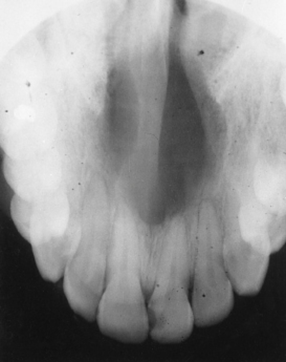
*Radiographically, this radiolucent cyst is often heart shaped, caused by the anatomic Y shape of the area. It is called the _____ cyst.
nasopalatine canal
median palatine
nasolabial
globulomaxillary
nasopalatine canal
The nasopalatine canal cyst is often heart shaped. The median palatine cyst appears as a well-defined unilocular radiolucency in the midline of the palate. The nasolabial cyst is a soft tissue cyst with no alveolar bone involvement. The globulomaxillary cyst is a well-defined pear-shaped radiolucency found between the roots of the maxillary lateral and cuspid.
*The presence of Hutchinson incisors and mulberry molars would indicate the presence of which condition?
Hepatitis B
HIV disease
Syphilis
Chickenpox
Syphilis
Congenital syphilis is transmitted from an infected mother to her fetus; teeth affected in the child include the incisors and molars. Hepatitis B does not cause any changes in the sizes of permanent teeth. HIV disease does not cause any changes in the sizes of permanent teeth. Chickenpox that occurs during the time of tooth formation may result in the pitting of enamel.
*Multiple supernumerary teeth may be a component of which condition?
Cleidocranial dysplasia
Dermoid cyst
Syphilis
Static bone cyst
Cleidocranial dysplasia
Multiple supernumerary teeth may be a component of cleidocranial dysplasia or Gardner syndrome, both described in Chapter 6. The dermoid cyst does not have teeth in the cyst wall. Children with congenital syphilis have mulberry molars and Hutchinson incisors but not supernumerary teeth. Static bone cyst has nothing to do with supernumerary teeth.
*During embryonic development of the face, the frontal process divides into three parts. These three parts include the median nasal process, the right lateral nasal process, and the left lateral nasal process.
Both statements are true.
Both statements are false.
The first statement is true; the second is false.
The first statement is false; the second is true.
Both statements are true.
During embryonic development of the face, the frontal process divides into three parts. These three parts include the median nasal process, the right lateral nasal process, and the left lateral nasal process. Both statements are true.
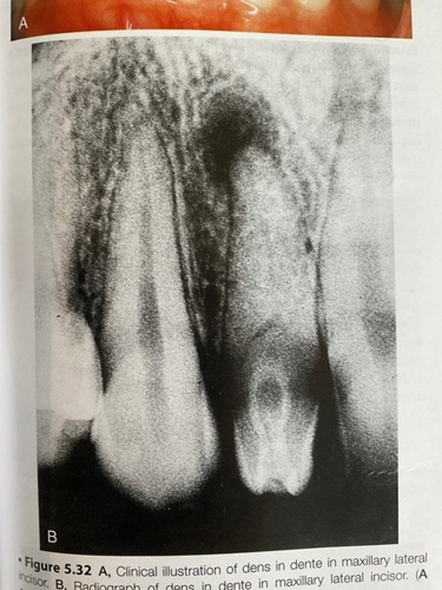
*Which tooth is most commonly affected by dens in dente?
Maxillary central
Mandibular lateral
Maxillary lateral
A supernumerary tooth
Maxillary lateral
The maxillary lateral is the tooth most commonly affected by dens in dente. The maxillary central is not the most common tooth seen with dens in dente. The mandibular lateral is not the most common tooth seen with dens in dente. A supernumerary tooth is not seen with dens in dente.
*Another name for dens invaginatus is
taurodontism.
dens in dente.
dens evaginatus.
enamel pearl.
dens in dente.
Dens in dente is another name for dens invaginatus. Taurodontism is a developmental anomaly in which teeth exhibit elongated large pulp chambers and short roots. Dens evaginatus is a rare developmental anomaly in which an enamel cusp is found on the occlusal surface of mandibular premolars. Enamel pearl or enameloma is a projection of enamel found on the furcation area of maxillary molars.
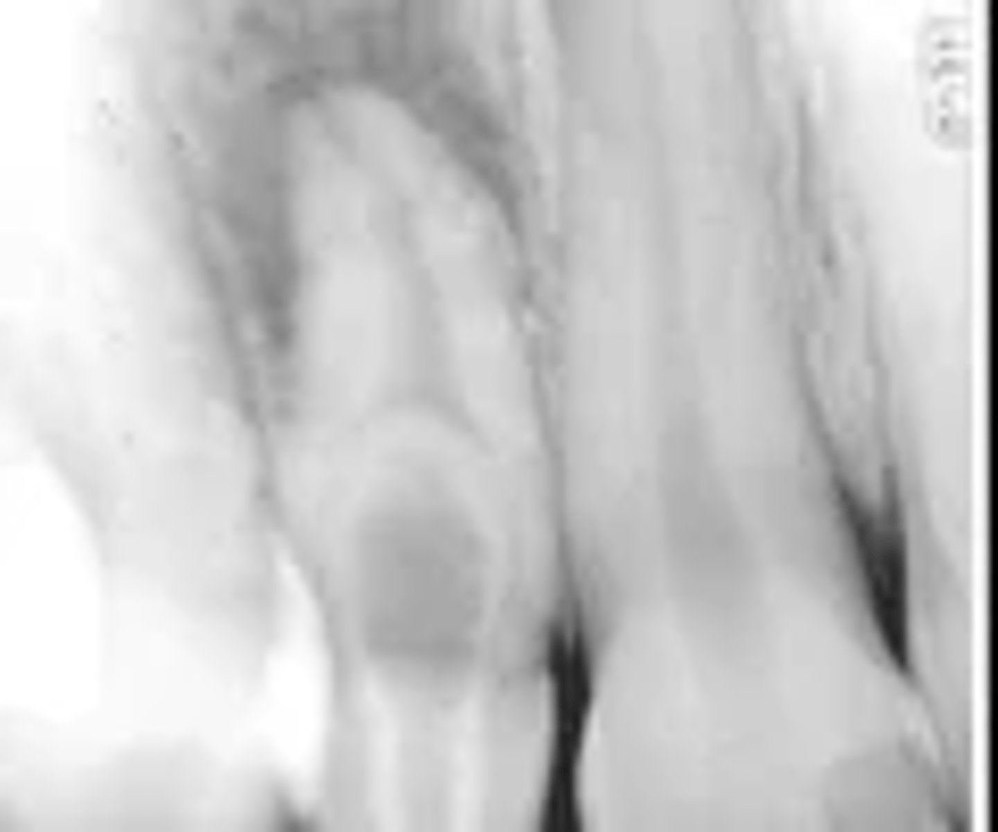
*Since the dens in dente is often a nonvital tooth, it may be seen in association with
an impacted tooth.
a periapical lesion.
swelling and displacement of surrounding teeth.
malocclusion.
a periapical lesion.
*Dens in dente is a developmental anomaly often seen with
extra cusps.
a periapical lesion.
tuberculated premolars.
supernumerary roots.
a periapical lesion.
Dens in dente is a developmental anomaly often seen with a periapical lesion. Dens evaginatus is an accessory occlusal cusp found on mandibular premolars. Tuberculated premolars occur when the mandibular premolars are affected with dens evaginatus. Dens in dente does not exhibit evidence of supernumerary roots.
*This radiographic image clearly shows which developmental anomaly?
Dens in dente
Periapical pathology (PAP)
Caries
Open contacts
Dens in dente
**This radiographic image clearly shows which developmental anomaly?
Dens in dente
Periapical pathology (PAP)
Caries
Open contacts
Dens in dente
*After the tooth erupts into the oral cavity, how long is it before the root length is complete?
6 months
1 year
1-4 years
2-6 years
1-4 years
The pseudocyst seen in this radiographic image is surrounded by salivary gland tissue. It is a(n) _____ bone cyst.
simple
Stafne
traumatic
aneurysmal
Stafne
*Trauma or change in the environment at the time of birth can eventually cause enamel hypoplasia in the child. Which cells are so sensitive and easily damaged to cause this defect?
Ameloblasts
Neutrophils
Macrophages
C-reactive proteins
Ameloblasts
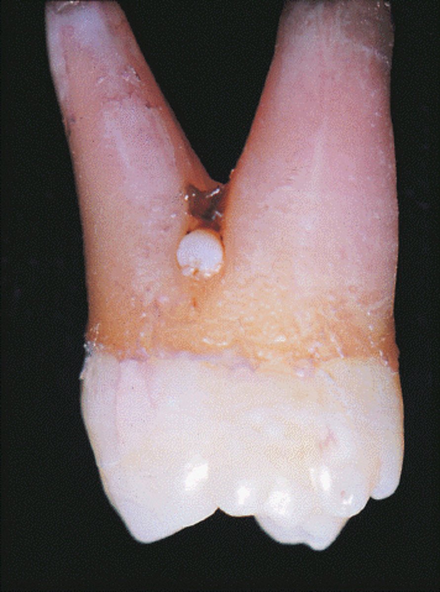
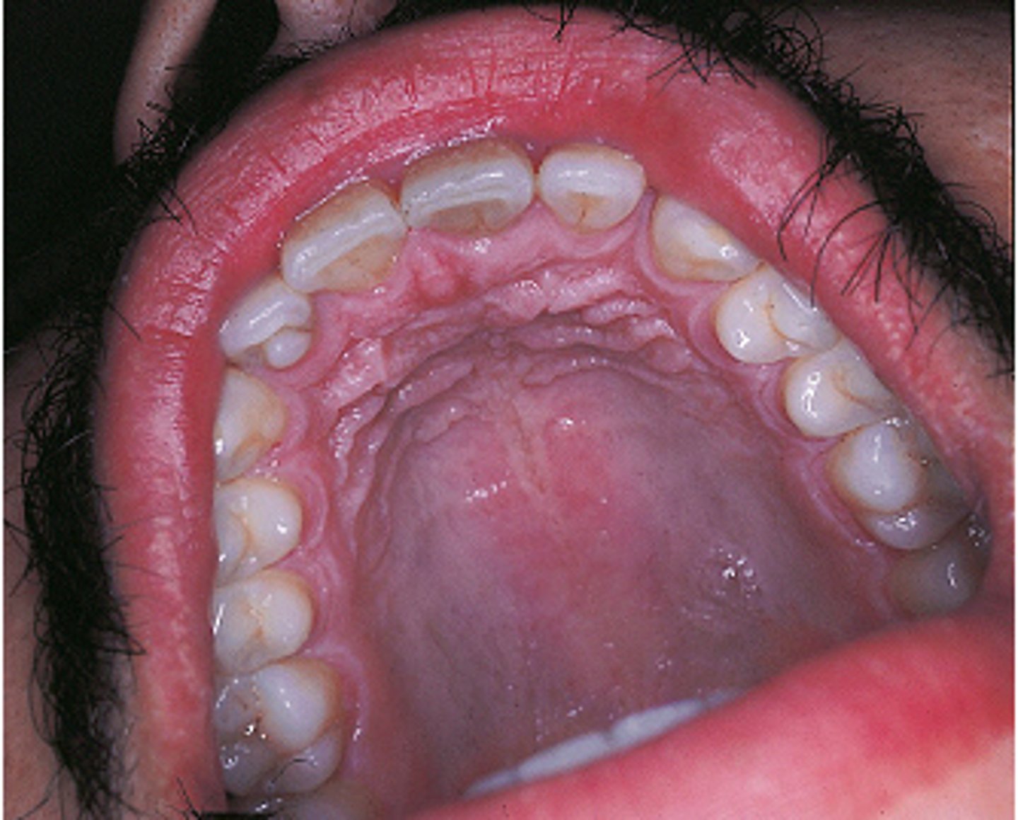
*The projection of white material seen at the furcation area in this maxillary molar is a developmental anomaly. Which condition is suspected?
Dens evaginatus
Enamel pearl
Supernumerary cusp
Calculus
Enamel pearl
The enamel pearl is a projection of enamel caused by abnormal displacement of ameloblasts during tooth formation. It is found near the furcation in maxillary molars. Dens evaginatus is an accessory enamel cusp found on the occlusal surfaces of mandibular premolars. A supernumerary cusp would be on or near the occlusal surface. Calculus is not a developmental anomaly.
*The cyst that appears in the bone in this radiographic image surrounds the fully formed crown of an unerupted premolar. The dental hygienist should refer to this as a(n) _____ cyst.
eruption
follicular
lateral periodontal
primordial
follicular
*Which is not considered a pseudocyst?
Thyroglossal tract cyst
Static bone cyst
Simple bone cyst
Aneurysmal bone cyst
Thyroglossal tract cyst
The thyroglossal tract cyst can be lined by various types of epithelia. The static bone cyst is not lined with epithelium. The simple bone cyst is not lined with epithelium. An aneurysmal bone cyst is a pseudocyst that contains blood-filled spaces surrounded by multinucleated giant cells and fibrous connective tissue.
*An asymptomatic, well-defined unilocular radiolucency was discovered in the region of tooth #32 on a panoramic image of a young adult patient. This tooth had never formed and therefore was never extracted. Identify this cyst:
Primordial
Odontogenic keratocyst
Periapical
Static bone cyst
Primordial
The primordial cyst develops in place of a tooth and is most commonly found in place of the third molar.
The odontogenic keratocyst is often seen in the mandibular third molar region, yet often appears as a multilocular radiolucency that can move teeth and resorb tooth structure.
The periapical cyst is always associated with a nonvital tooth.
The static bone cyst is a well-defined radiolucency seen in the posterior mandible; clinically, an anatomic depression may be felt in this area on the lingual side of the mandible.
What is the pseudocyst filled with salivary gland tissue that may be an extension of the sublingual gland?
Ranula
Static bone cyst
Lymphoepithelial cyst
Traumatic bone cyst
Static bone cyst
*What is the radiographic feature of a cyst found within soft tissue?
Unilocular
Multilocular
Diffuse
No radiographic features are evident.
No radiographic features are evident.
No radiographic features are seen when a cyst is found within soft tissue. Unilocular refers to a single rounded compartment. Multilocular describes multiple rounded compartments that may appear "soap bubble-like." Diffuse denotes a border that is not well defined, and the parameters of the lesion are unknown.
*The lateral periodontal cyst occurs most often on the lateral aspect of a tooth root, which is usually the
mandibular third molar.
maxillary premolars.
mandibular cuspid/premolars.
maxillary anteriors.
mandibular cuspid/premolars.
The mandibular cuspid/premolar area is the most common site for the lateral periodontal cyst. The mandibular third molar is not the site for a lateral periodontal cyst. Maxillary premolars are not the site for a lateral periodontal cyst. Maxillary anteriors are not the site for a lateral periodontal cyst.
*Which term describes a disorder present at and existing from the time of birth?
Anomaly
Inherited
Congenital
Developmental
Congenital
A congenital disorder is present at and existing from the time of birth. An anomaly is a marked deviation from normal that can be the result of congenital or hereditary defects. Inherited disorders are caused by abnormalities in the genetic makeup transmitted from parent to offspring. Developmental disorders occur when failure or disturbances occur during the complex series of cell division, multiplication, or differentiation.
*Which defines a disturbance of the maturation of the enamel matrix?
Turner tooth
Mulberry molar
Premature birth
Enamel hypocalcification
Enamel hypocalcification
Enamel hypocalcification is a disturbance of the maturation of the enamel matrix. Turner tooth results from enamel hypoplasia. Mulberry molar results from enamel hypoplasia associated with congenital syphilis. Premature birth can contribute to enamel hypoplasia.
*The _____ cyst has a strong predilection for females.
lateral periodontal
nasopalatine canal
nasolabial
gingival
nasolabial
The nasolabial cyst has a strong predilection for females. The lateral periodontal cyst is most often found in males. The nasopalatine canal cyst has a predilection for males. The gingival cyst has no sex predilection.
*Pitting is the most common type of enamel hypoplasia seen in patients who have which condition during tooth development?
Febrile illness
Drinking water with 2.4 ppm of fluoride during tooth development
Congenital syphilis
Herpes simplex
Febrile illness
Febrile illnesses such as measles and chickenpox cause enamel hypoplasia showing pitting of the enamel. Drinking water with twice the recommended fluoride content causes white flecks or chalky areas of the enamel. Congenital syphilis causes mulberry molars and Hutchinson incisors. Herpes simplex is characterized by oral ulcers involving the soft tissues and not enamel hypoplasia.
*Pitting of the enamel may be seen in these conditions except one. Which is the exception?
Measles
Vitamin A deficiency
Scarlet fever
Talon cusp
Talon cusp
The presence of a talon cusp does not cause pitting of the enamel. Febrile illnesses, such as measles, that occur during the time of tooth formation can result in pitting of the enamel. Vitamin deficiencies, such as vitamins A, C, and D, that occur during the time of tooth formation can result in pitting of the enamel. Febrile illnesses, such as scarlet fever, that occur during the time of tooth formation can result in pitting of the enamel.
*Which epithelium-lined tract is a developmental anomaly located in the corners of the mouth?
Commissural lip pit
Angular cheilitis
Fistula
Congenital lip pit
Commissural lip pit
Commissural lip pits are epithelium-lined blind tracts located in the corners of the mouth. Angular cheilitis is often caused by Candida organisms. It appears as erythema or fissuring at the labial commissures. A fistula is a drainage tract from an area of infection. A congenital lip pit occurs near the midline of the vermilion border of the lip, and it appears as a depression.
*Regional odontodysplasia is
(*****unerupted teeth in a quadrant.)
a decrease in radiodensity seen on one or more unerupted teeth in a quadrant.
a genetic condition.
caused by systemic illness.
most often seen in the mandible.
a decrease in radiodensity seen on one or more unerupted teeth in a quadrant.
*Which term best describes a disorder caused by abnormalities in the genetic makeup transmitted from parent to offspring?
Anomaly
Inherited
Congenital
Developmental
Inherited
*The most common cyst observed in the oral cavity is caused by pulpal inflammation and is called a(n) _____ cyst.
dentigerous
eruption
radicular
primordial
radicular
*For which condition would pulp vitality be nonvital?
Radicular cyst
Median mandibular cyst
Median palatal cyst
Periapical cemento-osseous dysplasia
Radicular cyst
*During tooth development, ectoderm and ectomesenchymal cells give rise to each of the following except one. Which one is the exception?
Periodontal ligament
Ameloblasts
Odontoblasts
Cementoblasts
Periodontal ligament
The dental sac that surrounds the developing tooth germ provides cells that form the periodontal ligament. Ectoderm and ectomesenchymal cells give rise to ameloblasts. Ectoderm and ectomesenchymal cells give rise to odontoblasts. Ectoderm and ectomesenchymal cells give rise to cementoblasts.
*The most common supernumerary tooth is termed
distomolar.
mesiodens.
mulberry molar.
urner tooth.
mesiodens.
A mesiodens is a supernumerary tooth located between the maxillary central incisors at the midline.
*The supernumerary tooth in this illustration is
a mesiodens.
a dilaceration.
the result of twinning.
the result of gemination.
mesiodens
A mesiodens is a supernumerary tooth located between the maxillary central incisors at the midline.
*Nonerupted supernumerary teeth should be extracted because of which risk?
Malignant tumor development
Cysts around the crowns
Internal resorption
Condensing osteitis
Cysts around the crowns
*Regional odontodysplasia is also referred to as
hypodontia.
ghost teeth.
taurodontism.
supernumerary teeth.
ghost teeth.
*Which statement about macrodontia is true?
A common developmental anomaly.
Commonly affects a single tooth.
Seen in cases of pituitary gigantism.
Treatment involves extraction and prosthetic replacement.
Seen in cases of pituitary gigantism.
Macrodontia is seen occasionally in cases of pituitary gigantism. Macrodontia is an uncommon developmental anomaly in which one or more teeth are larger than normal. Macrodontia affecting a single tooth is uncommon. No treatment is indicated for macrodontia.
*The _____ is characterized by its unique histologic appearance and its frequent recurrence rate.
radicular cyst
residual cyst
dentigerous cyst
odontogenic keratocyst
odontogenic keratocyst
The odontogenic keratocyst is characterized by its unique histologic appearance and its frequent recurrence rate. The radicular cyst is caused by pulpal inflammation. The residual cyst remains after extraction of the tooth with the radicular cyst. The radicular cyst is left behind and not removed. The dentigerous cyst is treated by complete removal of the cyst and the tooth involved.
*This unilocular radiolucency around the crown of an unerupted second premolar is most likely a
normal developmental sac.
dentigerous cyst.
primordial cyst.
lateral periodontal cyst.
dentigerous cyst.
A dentigerous cyst is a well-defined unilocular radiolucency around the crown of an unerupted tooth. A normal developmental sac has a much smaller radiolucency around the crown. A primordial cyst develops in place of a tooth. The lateral periodontal cyst is most often seen in the mandibular cuspid and premolar region.

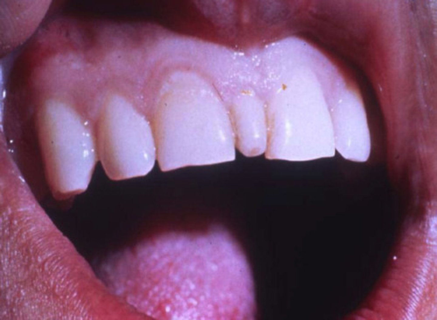
*A solitary hypoplastic defect in this dentition is located on the facial surface of a permanent maxillary central incisor. The most likely cause of this defect is
A dietary deficiency during tooth formation.
Absence of the primary mandibular central incisor.
Physical injury of the primary maxillary central incisor.
Neonatal hypoplasia of the primary anterior teeth.
physical injury of the primary maxillary central incisor.
Hypoplastic defects in a patient's dentition are indicative of physical injury of the primary teeth. A dietary deficiency during tooth formation would not typically appear in only one tooth. This description is not indicative of an absence of the primary mandibular central incisor. This description is not characteristic of neonatal hypoplasia of the primary anterior tooth.
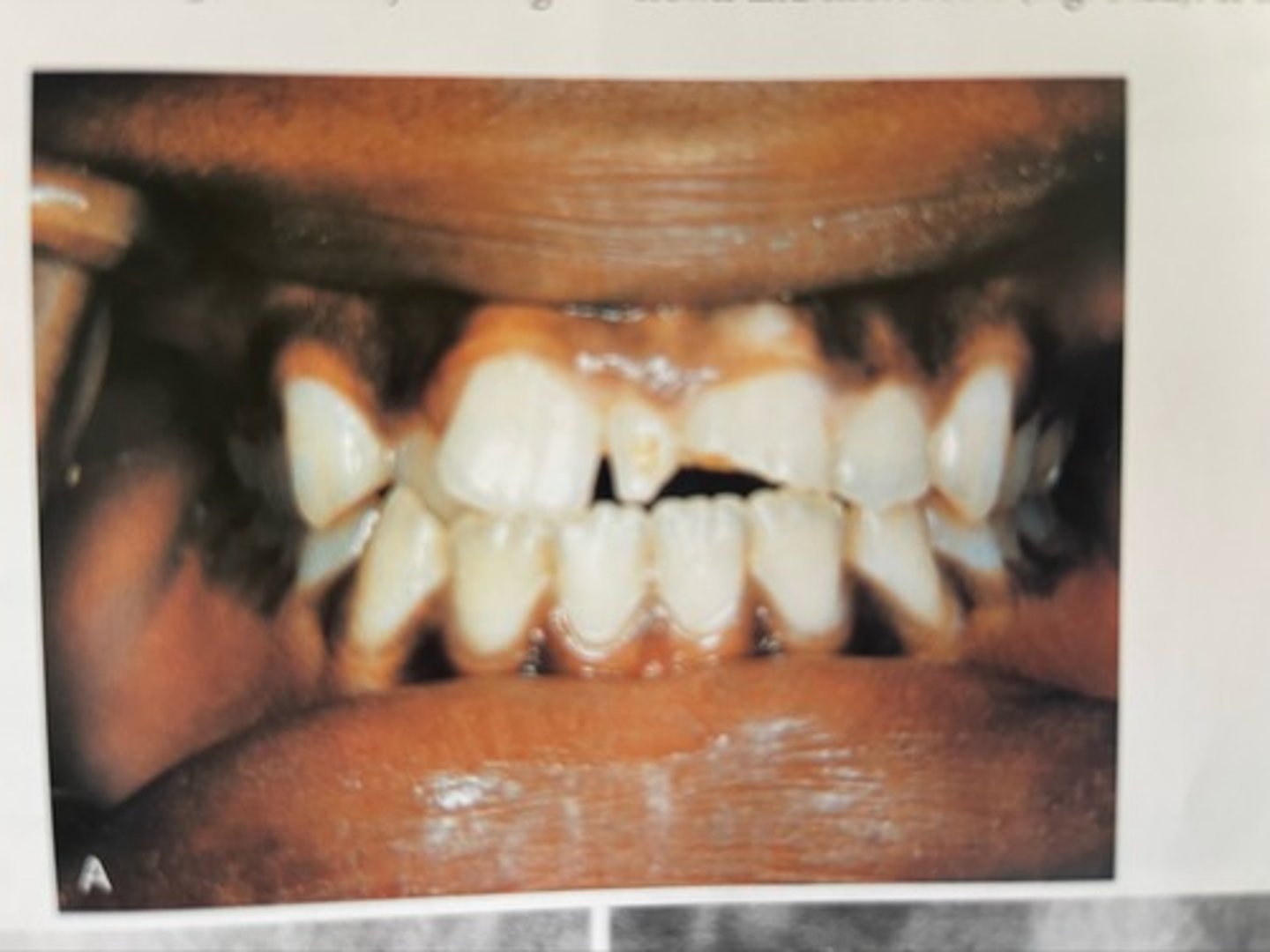
*When counting the maxillary anterior teeth of this adolescent patient, it appears that five are present clinically, if the large tooth is counted as one. A radiographic image reveals that this large central tooth has two roots. This tooth demonstrates
geminism.
concrescence.
dilaceration.
fusion.
fusion.
Fusion is the union of two normally separated adjacent tooth germs. Gemination is when a single enamel organ (tooth germ) divides partially. Concrescence occurs when two adjacent teeth are united by cementum. Dilaceration refers to an abnormal curve or angle in the root.
*The following groups of teeth are most often missing with hypodontia except one. Which is the exception?
Third molars
Mandibular canines
Maxillary lateral incisors
Mandibular second premolars
Mandibular canines
The mandibular canines are not typically missing in a case of hypodontia. The teeth most often missing with hypodontia include the maxillary and mandibular third molars. The teeth most often missing with hypodontia include the maxillary lateral incisors. The teeth most often missing with hypodontia include the mandibular second premolars.
*Odontogenesis in the human embryo occurs at
3 weeks.
5 weeks.
5 months.
1 month.
at birth.
5 weeks.
Odontogenesis in the human embryo takes place at approximately 5 weeks.
Odontogenesis in the human embryo takes place before 2 months.
Odontogenesis in the human embryo takes place before 3 months.
Odontogenesis in the human embryo takes place before birth.
*Which term describes partial anodontia or the lack of one or more teeth?
Anodontia
Ankylosed
Hypodontia
Gemination
Hypodontia
Hypodontia defines partial anodontia or the lack of one or more teeth. Anodontia is the congenital lack of teeth. Ankylosed teeth are those fused to alveolar bone, usually retained deciduous teeth. Gemination occurs when a single tooth germ attempts to divide, resulting in the incomplete formation of two teeth.
*Odontogenesis in the human embryo takes place at approximately
5 weeks.
2 months.
3 months.
at birth.
5 weeks.
Odontogenesis in the human embryo takes place at approximately 5 weeks. Odontogenesis in the human embryo takes place before 2 months. Odontogenesis in the human embryo takes place before 3 months. Odontogenesis in the human embryo takes place before birth.
*Which term is unlike the others?
Distomolar
Supernumerary
Hypodontia
Mesiodens
Hypodontia
Hypodontia refers to a lack of one or more teeth. A distomolar is an extra tooth, also known as a maxillary fourth molar. Supernumerary is a term to describe extra teeth found in the dentalarches. A mesiodens is a supernumerary tooth located between the maxillary central incisors at the midline.
*Which cyst develops from a preexisting periapical granuloma found at the apex of a nonvital tooth?
Radicular
Follicular
Eruption
Calcifying odontogenic
Radicular
The radicular (or periapical) cyst is always associated with a nonvital tooth; it develops from a preexisting periapical granuloma found at the apex of a nonvital tooth. The follicular (dentigerous) cyst forms around the crown of an unerupted or developing tooth. The eruption cyst is found in the soft tissue around the crown of an erupting tooth. The calcifying odontogenic cyst resembles the epithelium of the ameloblastoma.
*Impacted teeth cannot erupt because of
lack of eruptive force.
physical obstruction.
ankylosis.
bone pathology.
physical obstruction.
Impacted teeth cannot erupt because of physical obstruction. Lack of eruptive force does not play a role in eruption of impacted teeth. A tooth is ankylosed if it is fused to bone. This condition is especially common with retained deciduous teeth. Bone pathology can affect the eruption of teeth, but it is not the main reason that impacted teeth do not erupt.
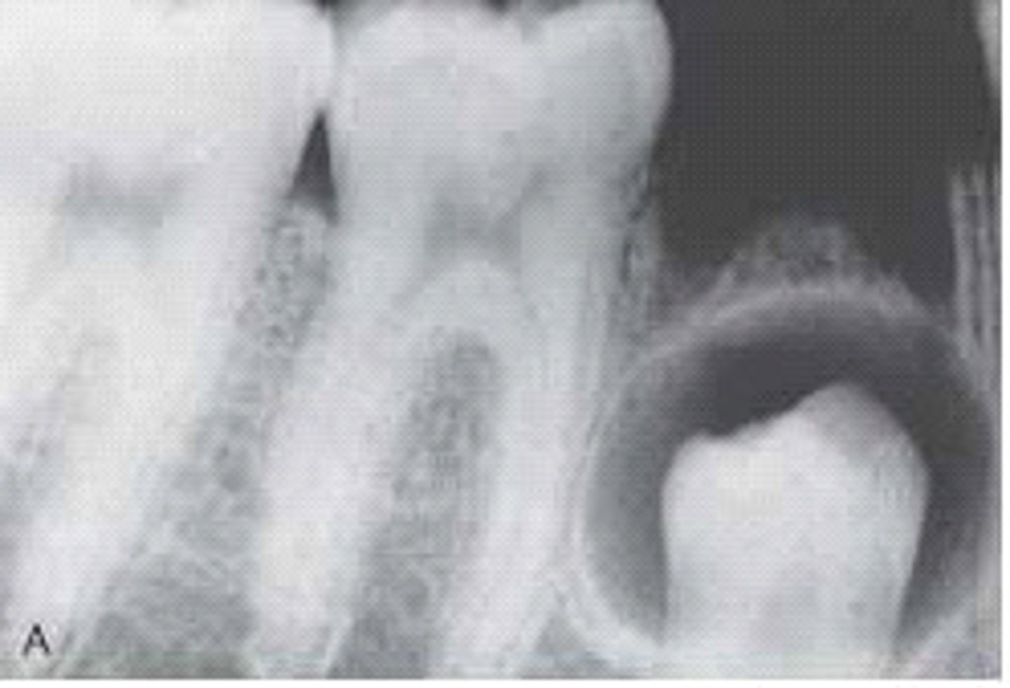
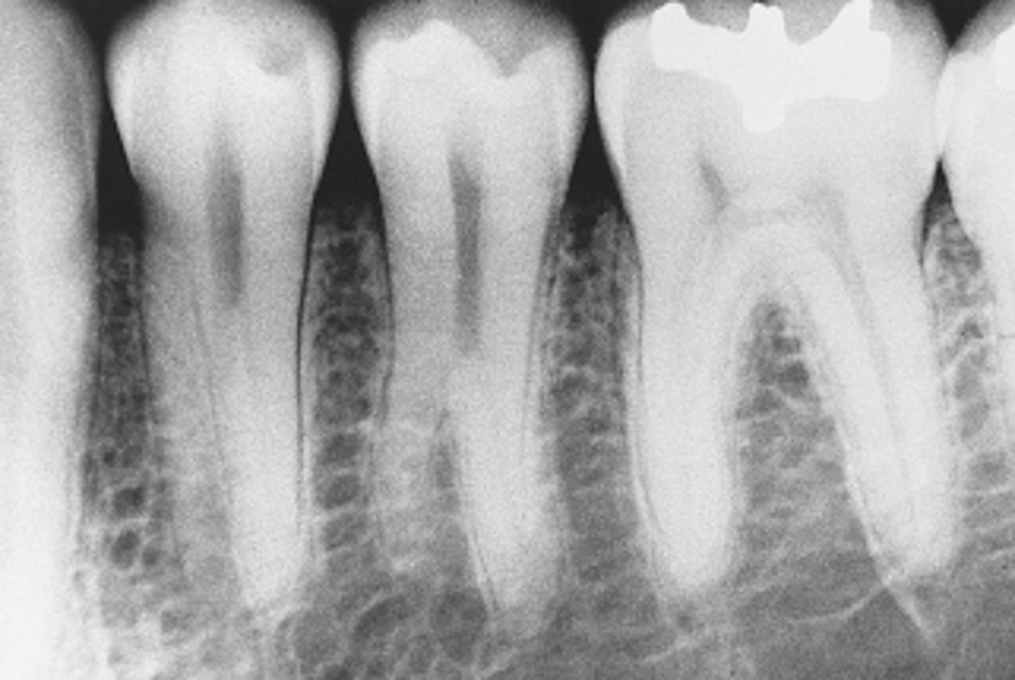
*The developmental anomaly seen in this radiographic image is
taurodontism.
mulberry molar.
supernumerary roots on the mandibular premolars.
dilaceration.
supernumerary roots on the mandibular premolars.
This radiographic image shows supernumerary roots on the mandibular premolars. Taurodontic teeth, or bull's teeth, show large pulp chambers and short roots, not seen in this radiograph. Mulberry molars result from congenital syphilis. Small globules of enamel make up the occlusal surface of the first molar. Dilaceration is a sharp bend or curve in the root.
*The body of the tongue develops from the
frontal process.
first branchial arch.
second branchial arch.
third branchial arch.
first branchial arch.
The body of the tongue develops from the first branchial arch. The frontal process is above the first branchial arch. The second and third branchial arches form the base of the tongue.
The first branchial arch divides into two maxillary processes and the _____ process.
.mandibular
frontal
median nasal
globular
mandibular
*Periapical radiographic examination reveals a well-defined unilocular radiolucency located in the midline of the hard palate. The diagnosis is _____ cyst.
nasolabial
globulomaxillary
branchial cleft
median palatine
median palatine
A median palatine cyst is a well-defined unilocular radiolucency located in the midline of the hard palate. A nasolabial cyst is a soft tissue cyst with no alveolar bone involvement. A globulomaxillary cyst is a well-defined, pear-shaped radiolucency found between the roots of the maxillary lateral incisor and cuspid. A branchial cleft cyst is located on the lateral neck at the anterior border of the sternocleidomastoid muscle.
*Total anodontia is often associated with a hereditary disturbance termed
taurodontism.
amelogenesis imperfecta.
ectodermal dysplasia.
cleidocranial dysplasia.
ectodermal dysplasia.
*Ingesting water with four times the amount of fluoride causes
brown-to-black staining.
cusp fractures.
white spots on the middle third of smooth crowns.
increased dental caries.
brown-to-black staining.
*The radiographic image of this patient exhibits a biloculated, well-defined radiolucency lateral to the tooth root. It is asymptomatic. This is a _____ cyst.
residual
follicular
lateral periodontal
primordial
lateral periodontal
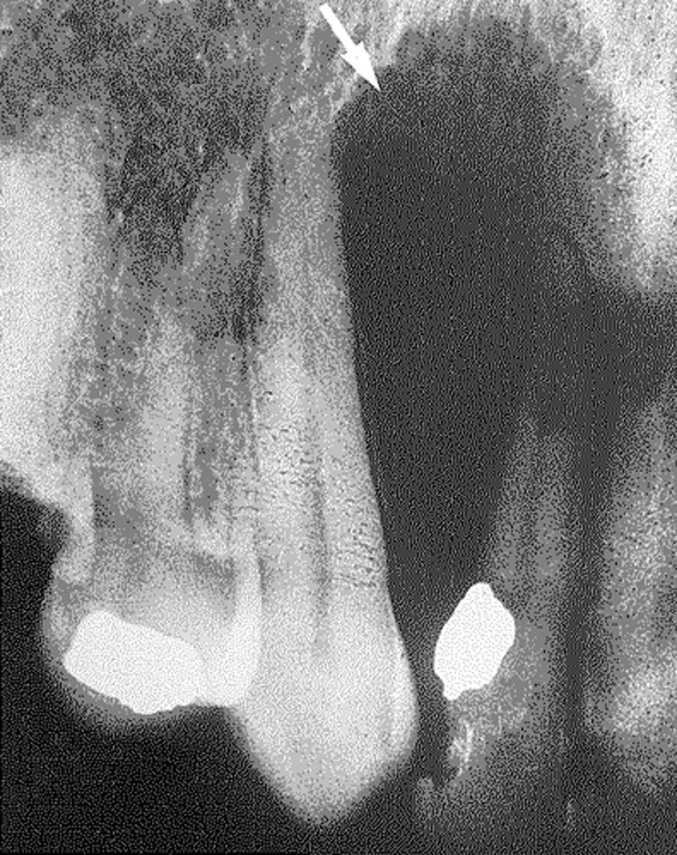
*The pear-shaped radiolucency observed in this radiographic image is most likely a _____ cyst.
radicular
globulomaxillary
lateral periodontal
nasopalatine canal
globulomaxillary
The globulomaxillary cyst is a pear-shaped radiolucency found between the roots of a maxillary lateral and cuspid. The radicular cyst is a root end cyst found at the apex of a tooth that is usually involved with caries. A lateral periodontal cyst is usually found between the roots of the mandibular cuspid and premolar. The nasopalatine canal cyst is usually heart shaped and found near the apices of the maxillary centrals, lingual aspect.
*Your patient presents with several horizontal rows of deep pits traversing the surfaces of the permanent central and lateral incisors, canines, and first molars. The pits are stained and unsightly. Which condition is suspected?
Dens in dente
Concrescence
Enamel hypoplasia
Oligodontia
Enamel hypoplasia
Enamel hypoplasia is the incomplete or defective formation of enamel, causing the alteration of tooth form or color. Dens in dente is a developmental anomaly that results when the enamel organ invaginates into the crown of a tooth before mineralization. Concrescence is a condition in which two adjacent teeth are united by cementum. Oligodontia is a term describing a type of hypodontia in which six or more teeth are congenitally missing.
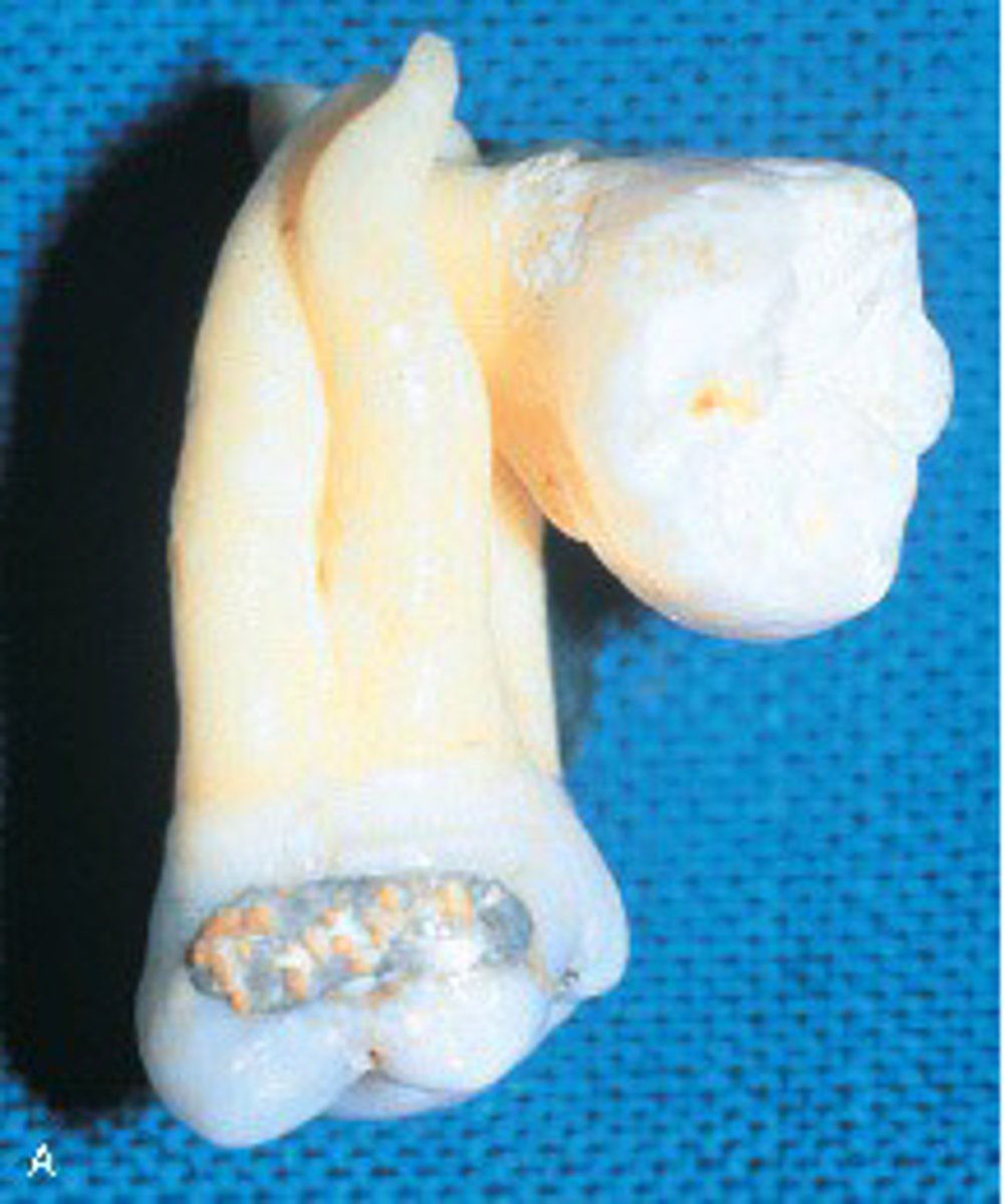
*When two or more teeth are joined by cementum, as shown in this picture, it is termed
concrescence.
dilaceration.
enamel pearl.
gemination.
concrescence.
Concrescence occurs when two adjacent teeth are united by cementum. Dilaceration refers to an abnormal curve or angle in the root. Enamel pearl is a small spherical enamel projection located on a root surface. Gemination occurs when a single enamel organ (tooth germ) divides partially.
Which term defines the joining of two adjacent teeth by cementum only?
Twinning
Concrescence
Cementogenesis
Fusion
Concrescence
The process by which parts of a whole join together (fuse) to make one (squamous cell carcinoma)
Coalescence
The deformity seen here with a bend in root apices is characteristic for
dilaceration
gemination
fusion
concrescence
Dilaceration
*With concrescence, which tissue unites two adjacent teeth?
Enamel
Dentin
Cementum
Pulp
Cementum
*Odontogenic keratocysts are a clinical component of
nevoid basal cell carcinoma syndrome.
neurofibromatosis of von Recklinghausen.
cherubism.
Gardner syndrome.
nevoid basal cell carcinoma syndrome is AKA
Odontogenic keratocysts are a clinical component of nevoid basal cell carcinoma syndrome.
*Enamel hypoplasia is the result of a disturbance of or damage to ameloblasts during enamel matrix formation. Which is not be a factor?
Genetics
Ingestion of high concentrations of fluoride during tooth development
Vitamin deficiency during tooth development
Shingles
Shingles
Shingles is caused by the herpes zoster virus and is seen in adults. Genetic problems do cause enamel hypoplasia. High fluoride intake during tooth development does cause enamel hypoplasia. Vitamin deficiency during tooth development does cause enamel hypoplasia.
*Which teeth are most commonly affected by microdontia
#7 and #16
#10 and #17
#17 and #24
#25 and #31
#7 and #16
maxillary laterals (#7, #10) and third molars (#1, #16).
*Microdontia most commonly occurs in
maxillary laterals and third molars.
maxillary canine.
mandibular molars.
mandibular incisors and molars.
maxillary laterals and third molars. (#7, #10 and #1, #16)
*This patient exhibits an accessory cusp located in the cingulum of the maxillary right lateral permanent incisor. This can be diagnosed as a
talon cusp.
dens in dente.
taurodontism.
dens evaginatus.
talon cusp.
A talon cusp is an accessory cusp located in the cingulum of a maxillary or mandibular permanent incisor. A dens in dente is a developmental anomaly that results when the enamel organ invaginates into the crown of a tooth before mineralization. Taurodontism is a term used to describe a developmental dental anomaly in which the teeth exhibit elongated, large pulp chambers and short roots. Dens evaginatus is an accessory enamel cusp found on the occlusal tooth surface.
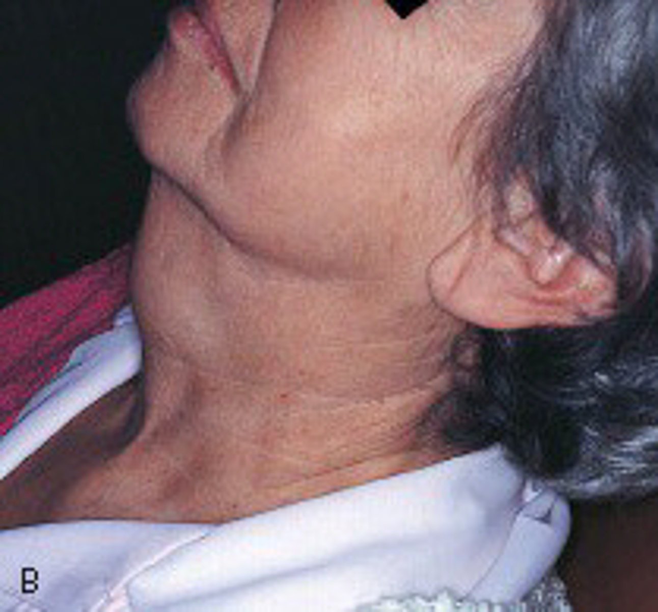
*This enlargement on the lateral neck of this patient has been present for months and is slowly increasing in size. It is painless and feels soft. Histologic examination shows this to be an epithelium-lined sac filled with clear, yellow fluid. It is a _____ cyst.
thyroglossal duct
branchial
median palatine
globulomaxillary
thyroglossal duct
A thyroglossal duct cyst appears on the lateral neck and slowly increases in size. It is painless and feels soft. Histologic examination shows an epithelium-lined sac filled with clear, yellow fluid. A branchial cleft cyst is located on the lateral neck at the anterior border of the sternocleidomastoid muscle. A median palatine cyst is a well-defined unilocular radiolucency located in the midline of the hard palate. A globulomaxillary cyst is a well-defined, pear-shaped radiolucency found between the roots of a maxillary lateral incisor and cuspid.
*Proliferation is defined as
congenital lack of teeth.
formation of dentin.
multiplication of cells.
disposition in favor of something.
multiplication of cells.
Proliferation is the multiplication of cells.
Anodontia is the congenital lack of teeth.
Dentinogenesis is the formation of dentin.
Predilection is a disposition in favor of something; preference.
*The formation of dentin is termed
amelogensis
Dentinogenesis
dens in dente
odontogensis
Dentinogenesis
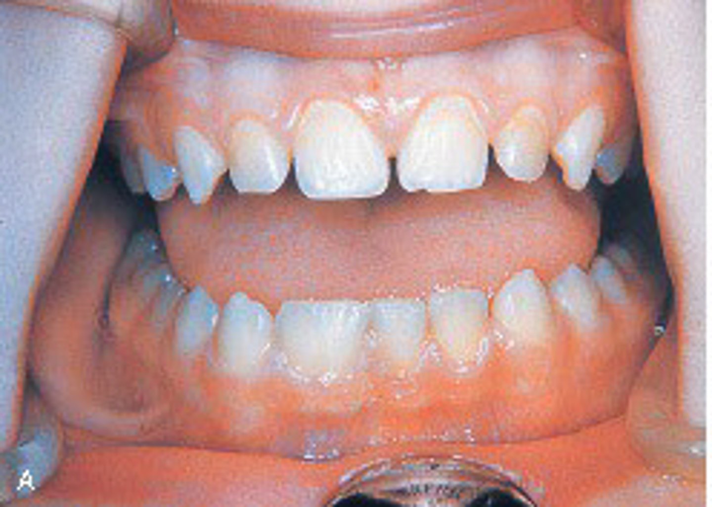
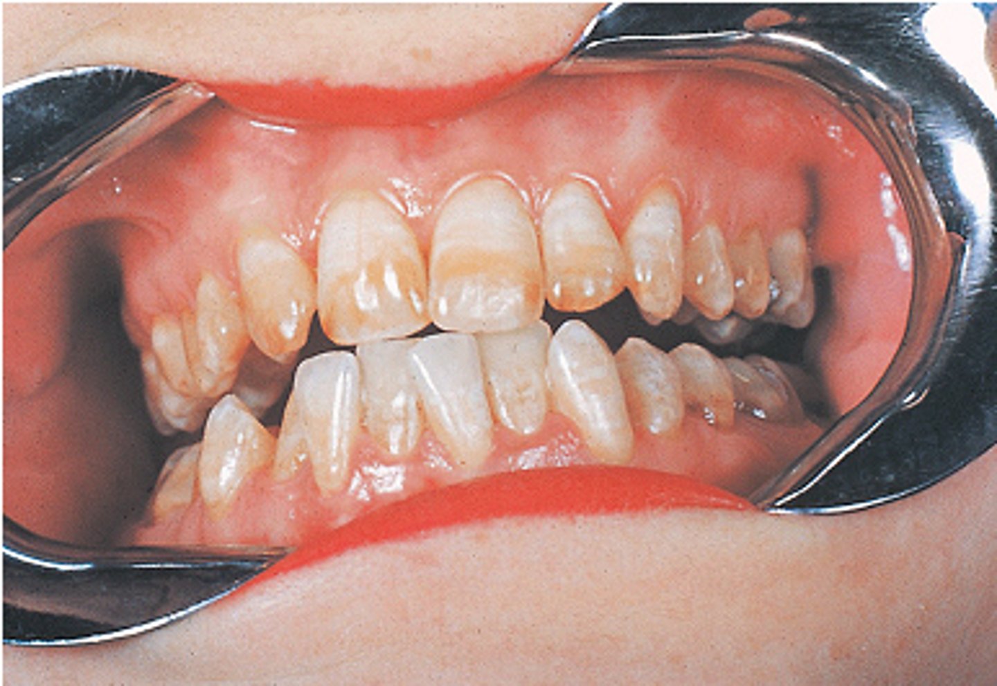
*This patient is healthy with no history of local or systemic infection or disease. The patient's teeth are caries free, as are all of the teeth of all of the patients who exhibit this defect. This is characteristic of
fluorosis.
Hutchinson incisors.
a Turner tooth.
attrition.
fluorosis.
Fluorosis occurs from ingestion of a high concentration of fluoride during tooth development. The teeth affected by fluorosis are generally decay resistant. Hutchinson incisors are a result of congenital syphilis. A Turner tooth is the result of infection from a deciduous tooth. Attrition is the result of the wearing away of tooth structure during mastication.
*Which is NOT true about the thyroglossal tract cyst?
It is found in individuals younger than 20 years.
No sex predilection exists.
Clinically, it is located below the hyoid bone.
Conservative nonsurgical treatment is sufficient.
Conservative nonsurgical treatment is sufficient.
Treatment of the thyroglossal tract cyst requires complete excision of the cyst and tract, usually including part of the hyoid bone and muscle within the tract. The thyroglossal tract cyst is found in individuals younger than 20 years. The thyroglossal tract cyst has no sex predilection. Clinically, the thyroglossal tract cyst is located below the hyoid bone.