Histology Lab Practical 2 - Reproductive System
1/136
There's no tags or description
Looks like no tags are added yet.
Name | Mastery | Learn | Test | Matching | Spaced | Call with Kai |
|---|
No analytics yet
Send a link to your students to track their progress
137 Terms
What is the job of the testes?
produce male gametes and hormones
What is the site of sperm production?
seminiferous tubules
What cells produce testosterone to promote the development of male sex characteristics and sperm and form the blood-testis barrier?
Interstitial/Leydig cells

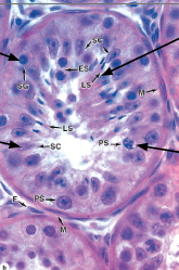
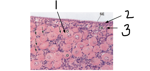
What structure is this?
seminiferous tubule
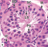
What is M?
myoid cells

What is SC?
sertoli cells

PS?
primary spermatocytes

What is IC?
Interstitial (Leydig) cells

SG?
spermatogonia

LS?
late spermatids
What is the tissue type of sertoli cells?
columnar support
What shape are spermatogonia that divide to form sperm?
round
What lines the periphery of each seminiferous tubule?
germinal/spermatogenic epithelium
What two things are contained in spermatogenic epithelium?
sertoli cells and spermatogonia
What structure is this overall?
seminiferous tubule

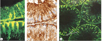
What cells are immunolabeled?
sertoli cells

1?
sertoli cells

2?
interstitial cells

IC?
interstitial cells
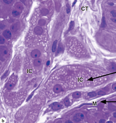
M?
myoid cells
What do myoid cells do?
contract to propel sperm
What are the 4 stages of spermatogenesis?
spermatogonia → primary spermatocytes → secondary spermatocytes → haploid spermatids
What is spermatogenesis?
the initial stage of sperm formation
What is spermiogenesis?
the final differentiation process where spermatids become sperm

SG?
spermatogonia

PS?
primary spermatocytes
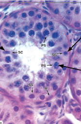
SC?
sertoli cells
What structure is this?
seminiferous tubule

PS?
primary spermatocyte

LS?
late spermatid

SC?
sertoli cells

SG?
spermatogonia
Describe spermatogonia in a histology slide:
located on the basement membrane; spherical nuclei
What do sertoli cells look like in a histology slide?
columnar cells with oval nuclei
What do primary spermatocytes look like on a slide?
spherical with euchromatic nuclei, abundant at all levels between basement membrane and lumen
What do late spermatids look like on a slide?
lose volume → develop into mature spermatozoa through spermiogenesis
What is the pathway of sperm from the seminiferous tubules to the urethra? (8 steps)
Seminiferous tubules
Tubuli recti
Rete testis
Efferent ductules
Epididymis
Ductus/Vas deferens
Ejaculatory duct
Urethra
Temporary storage and maturation of sperm
epididymis
transports sperm by peristalsis
ductus deferens
mix sperm and seminal fluid; transport to urethra
ejaculatory duct

1
epididymis duct

2
pseudostratified columnar epithelium with long stereocilia (principal cells)
3
principal cells

4
smooth muscle

5
sperm
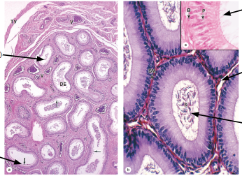
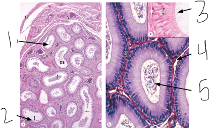
What overall structure is this?
epididymis
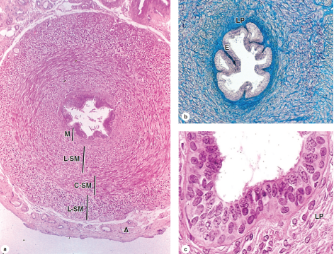
What is the structure?
ductus deferens
How many layers of smooth muscle in the ductus deferens?
3
What tissue lines the lumen of the ductus deferens?
pseudostratified columnar epithelium
What three sets of glands connect the ductus deferens to the urethra?
seminal vesicles
prostate
bulbourethral
What are the three tissue types of the seminal vesicles?
smooth muscle, thin lamina propria, simple columnar epithelium
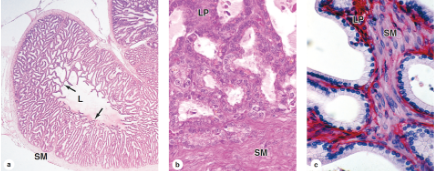
What structure is this?
seminal vesicles
What structure is this?
prostate gland
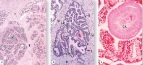
What is the histology of the prostate gland?
tubuloacinar glands (G) made up of pseudostratified columnar epithelia and surrounded by connective tissue stroma (S).
The prostate gland often have partially calcified concretions called _______ _________ that accumulate with age.
corpus amylacea

CA?
corpus amylacea
What are the three cylindircal masses of erectile tissue of the penis?
corpus cavernosa (x2) and corpus spongiosum (x1)
Tissue type of the penile urethra:
pseudostratified columnar epithelium
Pseudostratified columnar becomes stratified squamous at the ______ _______.
glans penis
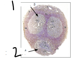
1
corpus cavernosa

2
corpus spongiosum

What overall structure is this?
penis

1
urethral glands

2
penile urethra

3
corpus spongiosum

4
corpus cavernosum
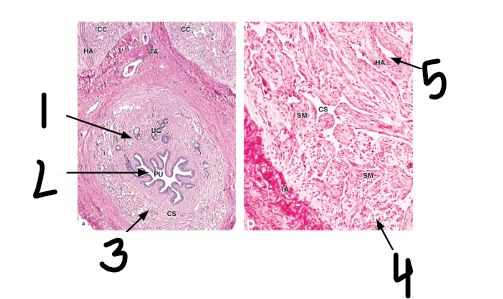
5
helicine arteries
produce oocytes
ovaries
transport oocyte and facilitate fertilization
uterine tubes
supports developing infant during gestation
uterus

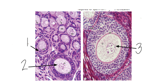
What structure of follicle development is this?
primordial follicles
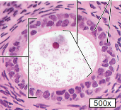
What structure of follicle development is this?
primary follicle
What structure of follicle development is this?
secondary or antral follicle
What structure of follicle development is this?
mature follicle
What structure of follicle development is this?
corpus luteum
What structure of follicle development is this?
corpus albicans
What are the stages of oogenesis?
oogonia → primary oocyte → secondary oocyte → zygote
What are the stages of follicle development?
primordial follicle → primary follicle → secondary follicle → graafian follicle → corpus luteum
At birth, primary oocytes are surrounded by __________ cells creating a __________ follicle.
granulosa; primordial
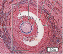
1
primary oocyte in primordial follicle

2
surface epithelium

3
tunica albuginea

What is the overall image?
primordial follicles

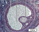
1
unilaminar primary follicle
What is the tissue type of the unilaminar primary follicle?
simple cuboidal epithelium
2
primary oocyte

3
primary oocyte

What is ZP?
zona pellucida

What is G?
granulosa cells

What is the overall structure?
primary follicle

ZP?
zona pellucida
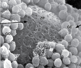
GC?
granulosa cells

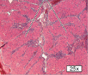
1
antrum

2
primary oocyte
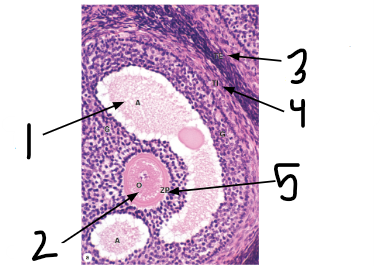
3
theca externa

4
theca interna

5
zona pellucida

What is the overall structure?
secondary follicle

1
corona radiata
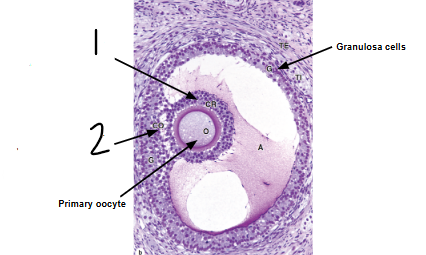
2
cumulus oophorus