Lab #4 Cerebellum
1/32
There's no tags or description
Looks like no tags are added yet.
Name | Mastery | Learn | Test | Matching | Spaced | Call with Kai |
|---|
No analytics yet
Send a link to your students to track their progress
33 Terms
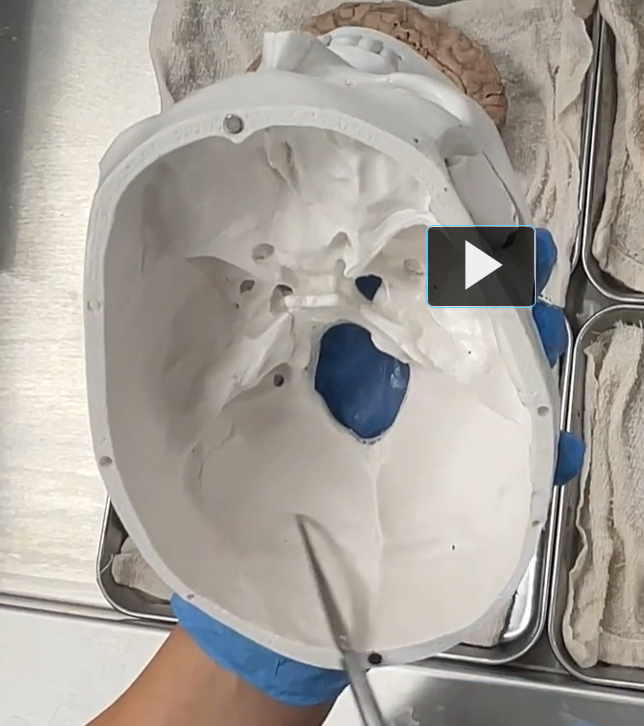
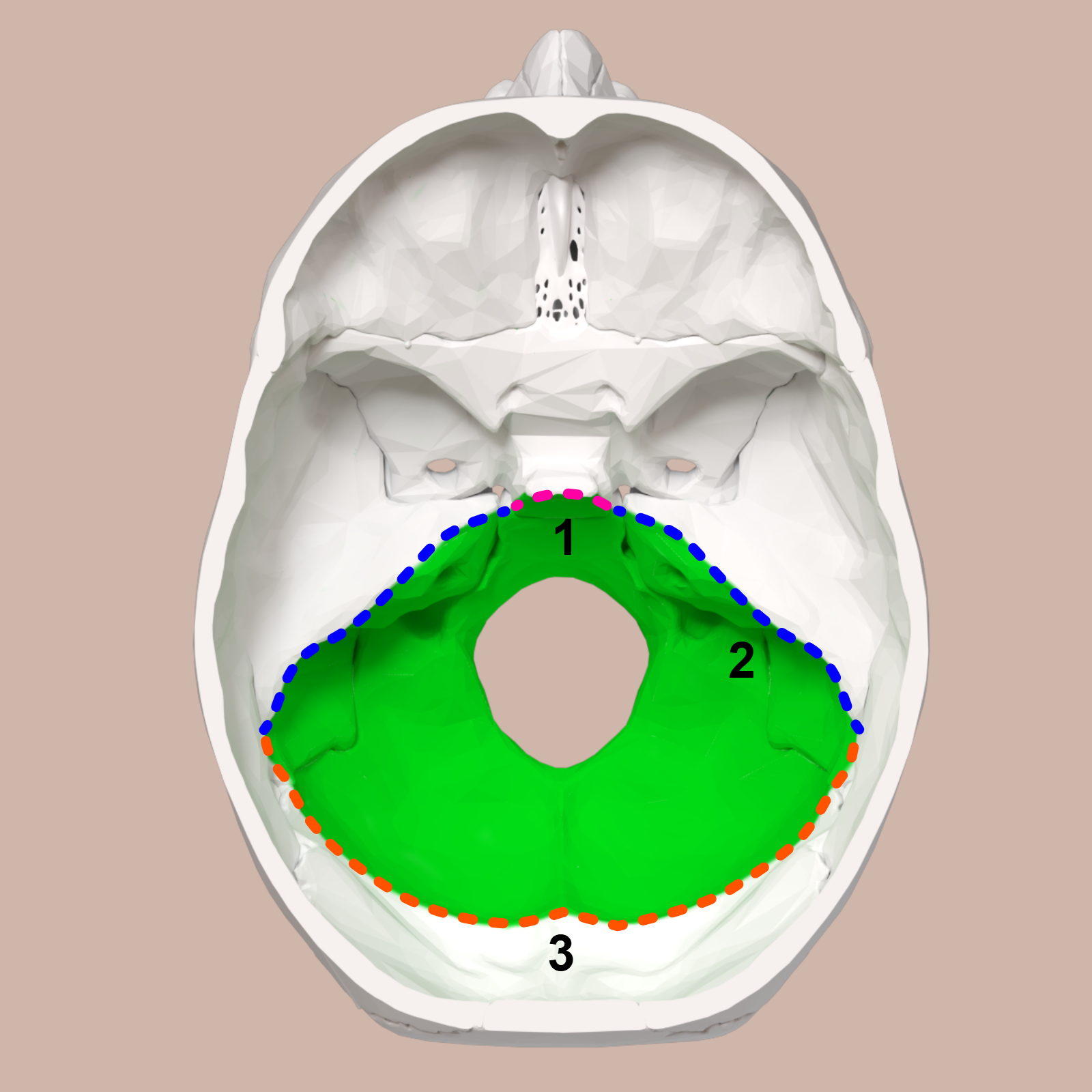
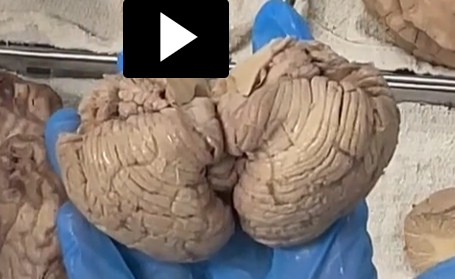
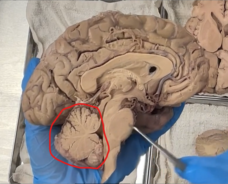
The cerebellum sits in a region called the posterior cranial fossa
The posterior cranial fossa is the deepest and most posterior depression on the floor of the skull. It houses the cerebellum, pons, and medulla oblongata. It is the part of the cranial cavity behind the petrous temporal bones and around the foramen magnum.
Posterior: behind
Cranial: skull
Fossa: ditch
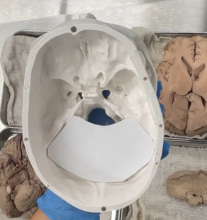
Tentorium Cerebelli
she cut out a piece of paper to show the tentorium Cerebelli.
The tentorium cerebelli is superior to the cerebellum, and inferior to the cerebrum.
The tentorium cerebelli is a fold of dura mater that forms a horizontal “tent-like” partition separating:
the cerebellum (below)
fromthe occipital lobes of the cerebrum (above)
It acts as a support structure for the brain and helps compartmentalize the cranial cavity.
Tentorium: from Latin tentorium = tent
Cerebelli: from Latin cerebellum = little brain (cerebellum)
So it literally means:
“the tent over the little brain.”
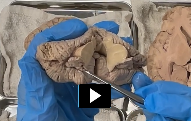
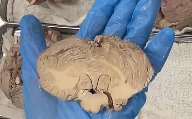
superior surface of the cerebellum
inferior surface of the cerebellum
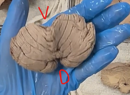
V: ventral (front)
D: dorsal (back)
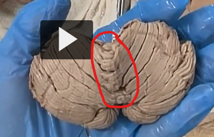
The cerebellum can be separated into left and right hemispheres.
The left and right hemisphers are separated by a midline structure called the vermis.
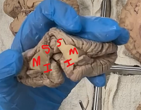
S: superior peduncle
M: middle peduncle
I: inferior peduncle
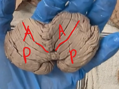
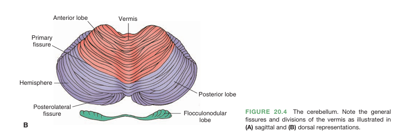
Anterior lobe
Posterior lobe
Primary fissure
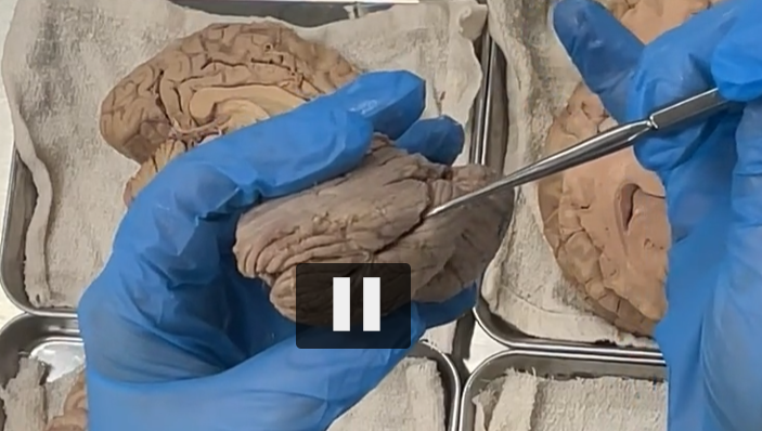
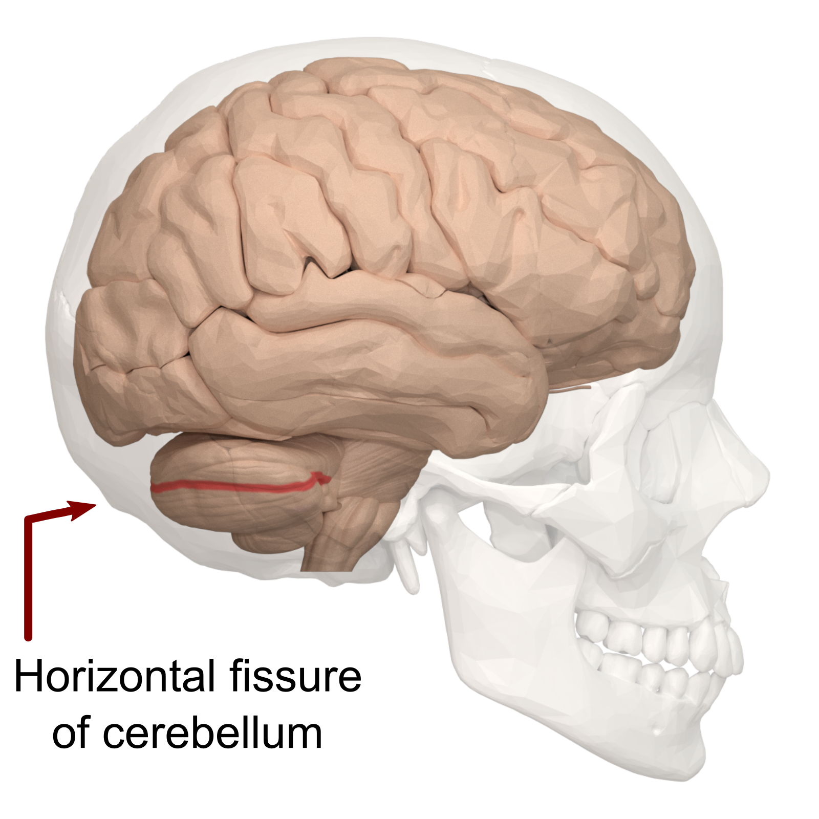
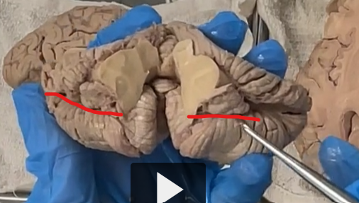
lateral view of the horizontal fissure: splits the cerebellum into a superior and inferior or upper and lower part of the cerebellum.
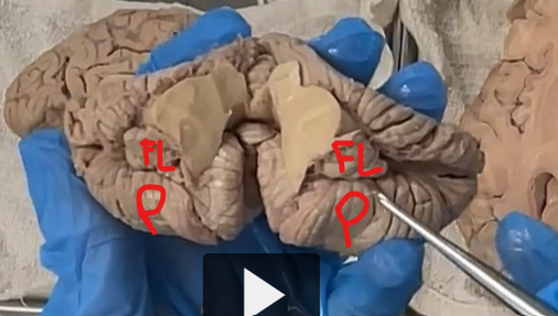
postero-lateral fissure
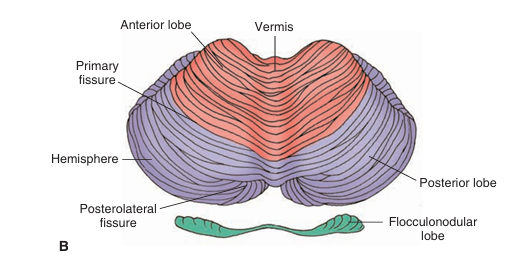
postero-lateral fissures
posterior-lateral fissures separates the Flocculus (which is a feature of the floccular-nodular lobe) from the posterior lobe.
The little bumps on the posterior lobes are the cerebellar tonsils.
Cerebellar: from Latin cerebellum = “little brain”
Tonsils: from Latin tonsillae = “almonds” (referring to their shape)
Literal meaning:
“almond-shaped parts of the little brain.”
The cerebellar tonsils are two rounded lobules on the inferior (bottom) surface of the cerebellum, located just above the foramen magnum.
They are the lowest part of the cerebellum, sitting close to where the brain transitions into the spinal cord.
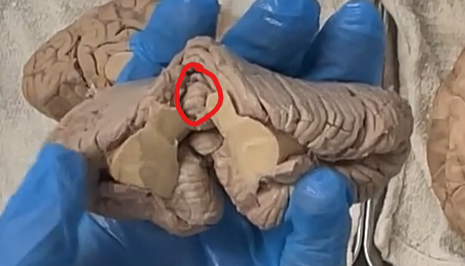
lingula, feature of the superior vermis.
In neuroanatomy, the lingula is a small, tongue-shaped lobule of the cerebellar vermis located on the superior surface of the cerebellum.
It is the most anterior (front-most) part of the vermis
Lies just above the superior medullary velum
Etymology
Lingula: from Latin lingula = “little tongue”
Literal meaning:
“small tongue-like structure.”
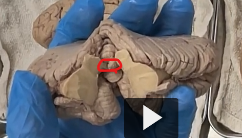
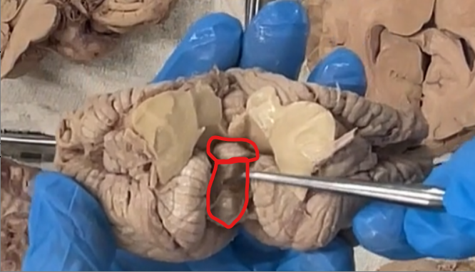
superior medullary velum
Definition
The superior medullary velum is a thin sheet of white matter that forms part of the roof of the fourth ventricle.
It connects the left and right superior cerebellar peduncles
Lies just beneath the lingula of the cerebellum
Forms the upper portion of the fourth ventricle roof
Etymology
Superior: Latin superior = above
Medullary: from Latin medulla = marrow / inner core (referring to brainstem/white matter)
Velum: Latin velum = veil or sail
Literal meaning:
“upper veil of the medulla (brainstem region)”
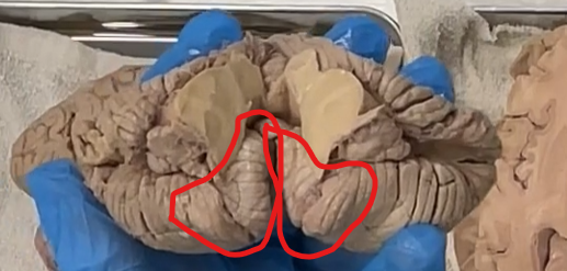
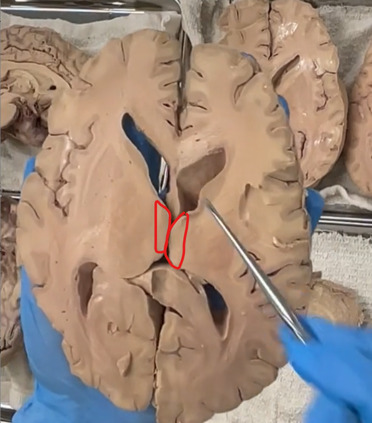
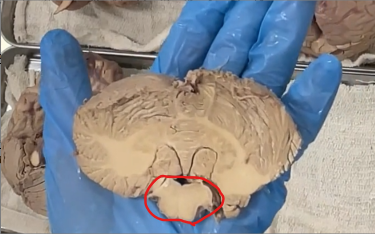
spread the tonsils
superior small circle: nodule
inferior large circle: uvula
These are features of the inferior vermis.
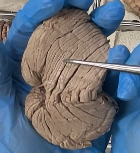
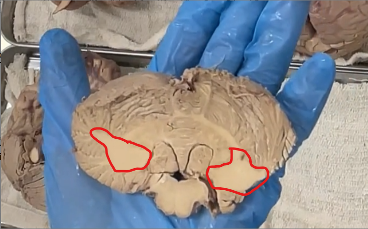
There are little folds called “folia”, which are separated by sulci.
This is a hemisection of the cerebellum.

We can see there is a white fiber tract, which is called the arbor vitae.
Arbor Vitae — Etymology & Definition
Etymology
Latin roots:
arbor = “tree” 🌳
vitae = “of life” (from vita = life)
So, arbor vitae literally means “tree of life.”
Definition (Neuroanatomy)
The arbor vitae refers to the tree-like pattern of white matter seen within the cerebellum.
It is formed by myelinated nerve fibers that branch extensively, resembling a tree.
Functional Meaning
The arbor vitae:
Carries signals between the cerebellar cortex and deeper brain regions
Helps coordinate movement, balance, and motor learning
Therefore, the folia look like little leaves that are part of the arbor vitae.
From Latin folium = “leaf” 🍃
folia = plural form → “leaves”
👉 So, folia literally means “leaves.”
Definition (Neuroanatomy)
Folia are the thin, leaf-like folds of the cerebellum.
They are the cerebellum’s version of cortical folds (like gyri in the cerebrum), but:
Much thinner
More tightly packed
Arranged in parallel, repeating layers
anterior lobe (top red)
Primary fissure circled in black.
posterior lobe (bottom red)
horizontal fissure


little red cirlce: floculonoddular
black circle: postero-lateral fissure
large red circle: posterior lobe
“T”: Tonsils (feature of the posterior lobe)
Cerebellar Tonsils (Posterior Lobe) — Etymology & Definition
Etymology
“Tonsil” comes from Latin tonsillae = “almonds”
👉 Named for their rounded, oval shape, similar to the palatine tonsils in the throat
Definition (Neuroanatomy)
The cerebellar tonsils are:
Paired, rounded lobules located on the inferior surface of the cerebellum
Part of the posterior lobe (the largest lobe of the cerebellum)
Positioned just above the foramen magnum
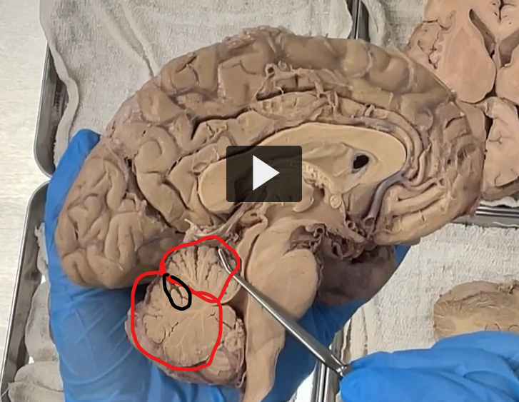
Window through the septum pellucidum to look into the lateral ventricle.
Septum Pellucidum — Etymology & Definition
Etymology
Latin roots:
septum = “partition” or “wall”
pellucidum = “transparent” (per = through + lucidus = light)
So, septum pellucidum literally means “transparent partition.”
Definition (Neuroanatomy)
The septum pellucidum is:
A thin, translucent membrane
Located in the midline of the brain
It separates the left and right anterior horns of the lateral ventricle
Lateral Ventricle — Etymology & Definition
Etymology
Latin roots:
lateralis = “side”
ventriculus = “little belly” or “small cavity”
So, lateral ventricle literally means “side cavity.”
Definition (Neuroanatomy)
The lateral ventricles are:
A pair of large, C-shaped fluid-filled cavities
Located within each cerebral hemisphere
Part of the brain’s ventricular system that contains cerebrospinal fluid (CSF)

Third ventricle
Third Ventricle — Etymology & Definition
Etymology
Latin:
ventriculus = “little cavity”
third = refers to its position in the sequence of brain ventricles
So, third ventricle = the “third cavity” of the ventricular system
Definition (Neuroanatomy)
The third ventricle is:
A narrow, midline, slit-like cavity
Located in the diencephalon
Lies between the right and left thalamus
The lateral ventricle and the third ventricle communicate through an interventricular foramen.
Interventricular Foramen — Etymology & Definition
Etymology
Latin roots:
inter- = “between”
ventriculus = “little cavity”
foramen = “opening”
So, interventricular foramen literally means “opening between the ventricles.”
Definition (Neuroanatomy)
The interventricular foramen (also called the foramen of Monro) is:
A small channel connecting each lateral ventricle to the third ventricle
Present on both sides (one per hemisphere)


Corpus Collosum (structure that connects the left and right sides of the brain).
Corpus Callosum — Etymology & Definition
Etymology
Latin roots:
corpus = “body”
callosum = “hard” or “tough”
So, corpus callosum literally means “tough body.”
(This reflects its dense bundle of myelinated fibers.)
Definition (Neuroanatomy)
The corpus callosum is:
The largest white matter structure in the brain
A commissural fiber bundle connecting the left and right cerebral hemispheres
Essential for interhemispheric communication
anterior commissure
Anterior Commissure — Etymology & Definition
Etymology
Latin roots:
anterior = “front”
commissura = “joining together” (com- = together + mittere = to send)
So, anterior commissure literally means “front connection.”
Definition (Neuroanatomy)
The anterior commissure is:
A small bundle of commissural fibers
Located in the anterior (front) part of the brain
Connects parts of the left and right hemispheres, especially regions of the temporal lobes

inter-thalamic adhesion: connecting the left and right thalamus.
Inter-thalamic Adhesion — Etymology & Definition
Etymology
Latin roots:
inter- = “between”
thalamus = “inner chamber” or “room”
adhesion = “sticking together”
So, interthalamic adhesion literally means “a sticking together between the thalami.”
Definition (Neuroanatomy)
The interthalamic adhesion (also called the massa intermedia) is:
A midline connection between the right and left thalamus
Located within the third ventricle
Appears as a small bridge of gray matter crossing the ventricle
Thalamus
Thalamus — Etymology & Definition
Etymology
From Greek thalamos = “inner chamber”
So, thalamus literally means “inner chamber.”
(This reflects its deep, central location in the brain.)
Definition (Neuroanatomy)
The thalamus is:
A paired mass of gray matter
Located in the diencephalon
Forms the lateral walls of the third ventricle
Acts as the brain’s major relay station


posterior to the thalamus is the posterior commissure.
Posterior Commissure — Etymology & Definition
Etymology
Latin roots:
posterior = “behind”
commissura = “joining together” (com- = together + mittere = to send)
So, posterior commissure literally means “back (rear) connection.”
Definition (Neuroanatomy)
The posterior commissure is:
A small bundle of crossing (commissural) nerve fibers
Located in the posterior part of the diencephalon, near the midbrain
Connects structures on the left and right sides of the brain
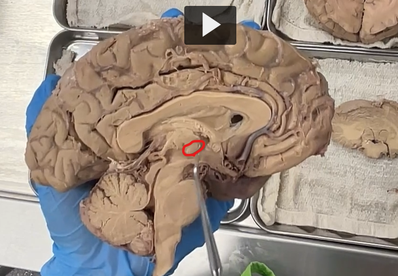
Now, let’s talk about the hypothalamus.
Red: thalamus
Black: hypo-thalamus (under the thalamus)
Hypothalamus — Etymology & Definition
Etymology
Greek roots:
hypo- = “under”
thalamos = “inner chamber” (→ thalamus)
So, hypothalamus literally means “below the thalamus.”
Definition (Neuroanatomy)
The hypothalamus is:
A small but critical region of the brain
Located in the diencephalon
Forms the floor and part of the walls of the third ventricle
Serves as the main regulator of homeostasis


Purple: pituitary stalk (extension of the hypothalamus)
Pituitary Stalk — Etymology & Definition
Etymology
pituitary: from Latin pituita = “phlegm”
(historically, the gland was thought to secrete mucus)stalk: from Old English stalc = “stem” or “support”
So, pituitary stalk literally means “the stem connecting to the pituitary gland.”
Definition (Neuroanatomy)
The pituitary stalk (also called the infundibulum) is:
A thin, funnel-shaped structure
Connecting the hypothalamus to the pituitary gland
Serves as the physical and functional link between the nervous and endocrine systems
Red: mammillary bodies
Mammillary Bodies — Etymology & Definition Etymology
Mammillary comes from Latin mammilla, meaning “little breast” or “nipple.”
Bodies just means rounded structures.
So mammillary bodies literally means “little breast-like bodies”, named for their small rounded shape.
Definition
The mammillary bodies are a pair of small rounded nuclei on the inferior surface of the hypothalamus.
They are part of the limbic system and are involved mainly in memory processing, especially recollective memory.
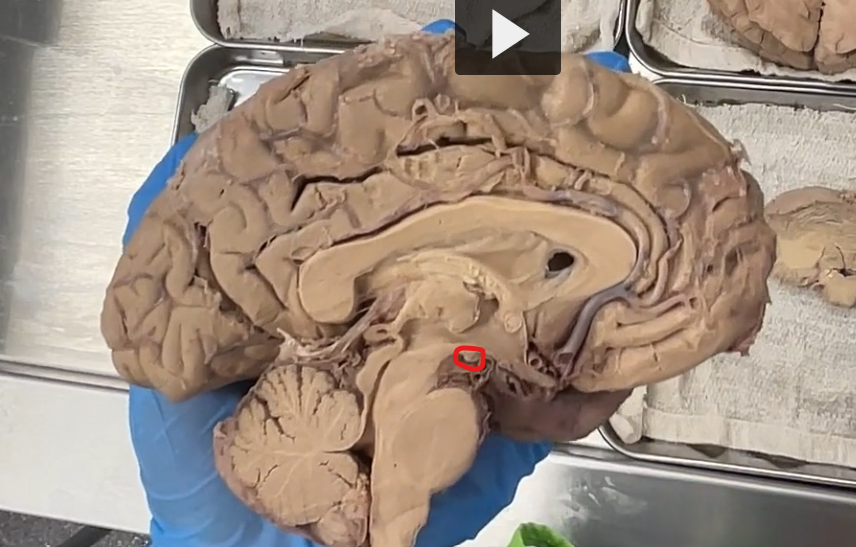
To locate the subthalamus: we want to look posterior and lateral to the hypothalamus.
Since we are in the midline, we are not going to see the subthalamus, but we want to remember that it’s posterior and lateral to the hypothalamus.
Sub-thalamus — Etymology & Definition
Etymology
Greek roots:
sub- = “under”
thalamos = “inner chamber” (→ thalamus)
So, subthalamus literally means “below the thalamus.”
Definition (Neuroanatomy)
The subthalamus is:
A small region of the diencephalon
Located inferior to the thalamus and superior to the midbrain
Functionally linked to the basal ganglia system
red: epithalamus (posterior to the thalamus)
black: pineal gland (projection from the epithalamus)
Epithalamus — Etymology & Definition
Etymology
Greek roots:
epi- = “upon” or “above”
thalamos = “inner chamber” (→ thalamus)
So, epithalamus literally means “upon/above the thalamus.”
Definition (Neuroanatomy)
The epithalamus is:
The posterior–superior part of the diencephalon
Located above and behind the thalamus
Forms part of the roof of the third ventricle
Key Structures (High-Yield)
pineal gland
Habenular nuclei (emotion/reward pathways)
posterior commissure
Functions
Pineal gland:
Secretes melatonin
Regulates circadian rhythms (sleep–wake cycle)
Habenular nuclei:
Involved in emotional processing, reward, and aversion
Posterior commissure:
Coordinates bilateral eye reflexes (especially pupillary light reflex)
Pineal Gland — Etymology & Definition
Etymology
From Latin pinea = “pine cone”
gland = a secreting organ
So, pineal gland literally means “pine cone–shaped gland.”
Definition (Neuroanatomy)
The pineal gland is:
A small endocrine gland
Located in the epithalamus
Positioned posterior to the third ventricle, between the two thalami
Function (High-Yield)
Regulates circadian rhythms via melatonin
Secretes melatonin (not melanin)
Controls:
Sleep–wake cycle
Biological clock
Light exposure ↓ melatonin
Darkness ↑ melatonin

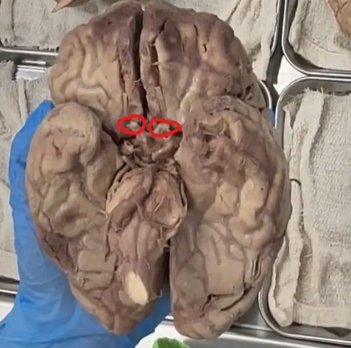
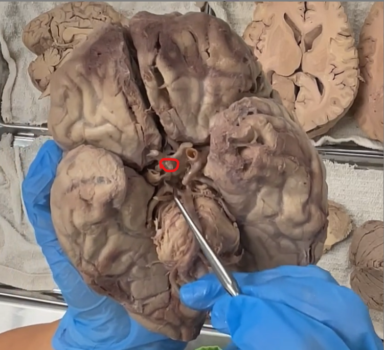
red: optic nerves
where the crossing occurs: optic chiasm
red: mammillary bodies
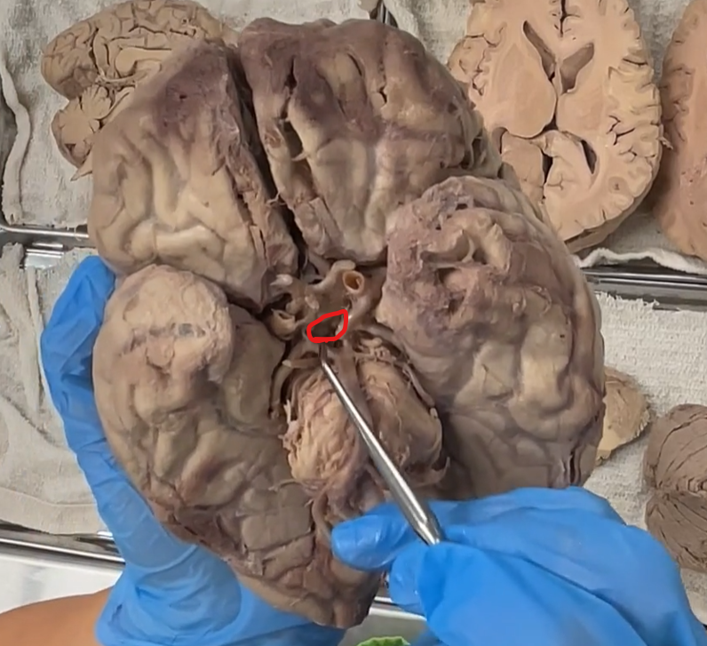

located between the mammillary bodies and the optic chiasm we have a region called the Tuber Cinereum
The Tuber Cinereum is not the stalk, it’s the place between the end of the pituitary stalk and the mammillary bodies.
Etymology
Latin roots:
tuber = “swelling”
cinereum = “ashen” or “gray”
So, tuber cinereum literally means “gray swelling.”
Definition (Neuroanatomy)
The tuber cinereum is:
A region of gray matter in the hypothalamus
Located on the inferior surface of the brain
Forms part of the floor of the third ventricle
Function
Contains the median eminence
Important for:
Hormone release into the hypophyseal portal system
Communication between hypothalamus → anterior pituitary
pituitary stalk

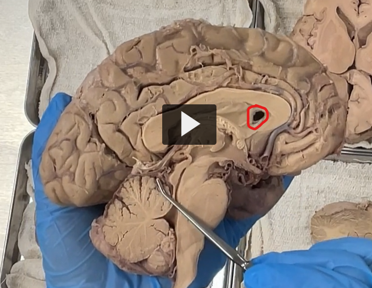
lateral ventricles
Lateral Ventricles — Etymology & Definition
Etymology
Latin roots:
lateralis = “side”
ventriculus = “little belly” or “small cavity”
So, lateral ventricles literally means “side cavities.”
Definition (Neuroanatomy)
The lateral ventricles are:
A pair of large, C-shaped cavities
Located within each cerebral hemisphere
Filled with cerebrospinal fluid (CSF)
Main Parts (High-Yield)
Each lateral ventricle has four key regions:
Anterior (frontal) horn → in frontal lobe
Body → central portion
Posterior (occipital) horn → in occipital lobe
Inferior (temporal) horn → in temporal lobe
Connections
Each lateral ventricle connects to the third ventricle via the
interventricular foramen
Function
Contain and circulate CSF, which:
Cushions the brain
Removes waste
Maintains chemical balance
CSF is produced by the choroid plexus
posterior and lateral to the lateral ventricles is the caudate nucleus
Caudate Nucleus — Etymology & Definition
Etymology
From Latin:
cauda = “tail”
-ate = “having the form of”
👉 So, caudate = “tail-like”
👉 Caudate nucleus = “tail-shaped nucleus”
Definition (Neuroanatomy)
The caudate nucleus is:
A C-shaped mass of gray matter
Part of the basal ganglia
Closely follows the contour of the lateral ventricles
Function (Basal Ganglia Role)
Movement regulation + cognition
Helps:
Initiate and control voluntary movement
Suppress unwanted movements
Participate in learning, habit formation, and cognition
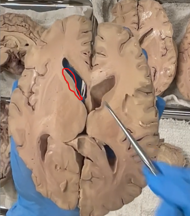

posterior and lateral to the caudate nucleus is the internal capsule.
Internal Capsule — Etymology & Definition
Etymology
Latin roots:
internus = “within”
capsula = “little box” or “container”
👉 So, internal capsule literally means “inner container.”
(Think of it as a bundle of fibers enclosed deep within the brain.)
Definition (Neuroanatomy)
The internal capsule is:
A dense bundle of projection fibers
Located deep within the cerebral hemispheres
white matter that carries information to and from the cerebral cortex
putamen (red)
the caudate nucleus is separated from the putamen by the internal capsule.
Putamen — Etymology & Definition
Etymology
From Latin putamen = “shell” (like the outer covering of a nut)
So, putamen literally means “shell-like structure.”
(This reflects its position as an outer part of the basal ganglia.)
Definition (Neuroanatomy)
The putamen is:
A round mass of gray matter
Part of the basal ganglia
Located lateral to the globus pallidus
Function (Basal Ganglia Role)
Movement regulation (especially execution and modulation)
Helps:
Initiate and smooth voluntary movement
Regulate muscle tone
Suppress unwanted movements
Functional Circuit
Receives input from the cortex
Sends signals to:
globus pallidus
Then → thalamus → cortex
This loop controls movement precision


medial to the putamen is the globus pallidus
Globus Pallidus — Etymology & Definition
Etymology
Latin roots:
globus = “sphere” or “ball”
pallidus = “pale”
So, globus pallidus literally means “pale globe.”
(It looks lighter than surrounding structures due to fewer cell bodies and more myelinated fibers.)
Definition (Neuroanatomy)
The globus pallidus is:
A paired mass of gray matter
Located medial to the putamen
Part of the lentiform nucleus (putamen + globus pallidus)
A key output structure of the basal ganglia
external capsule (in red), the myelinated layer
lateral to the putamen is the external capsule
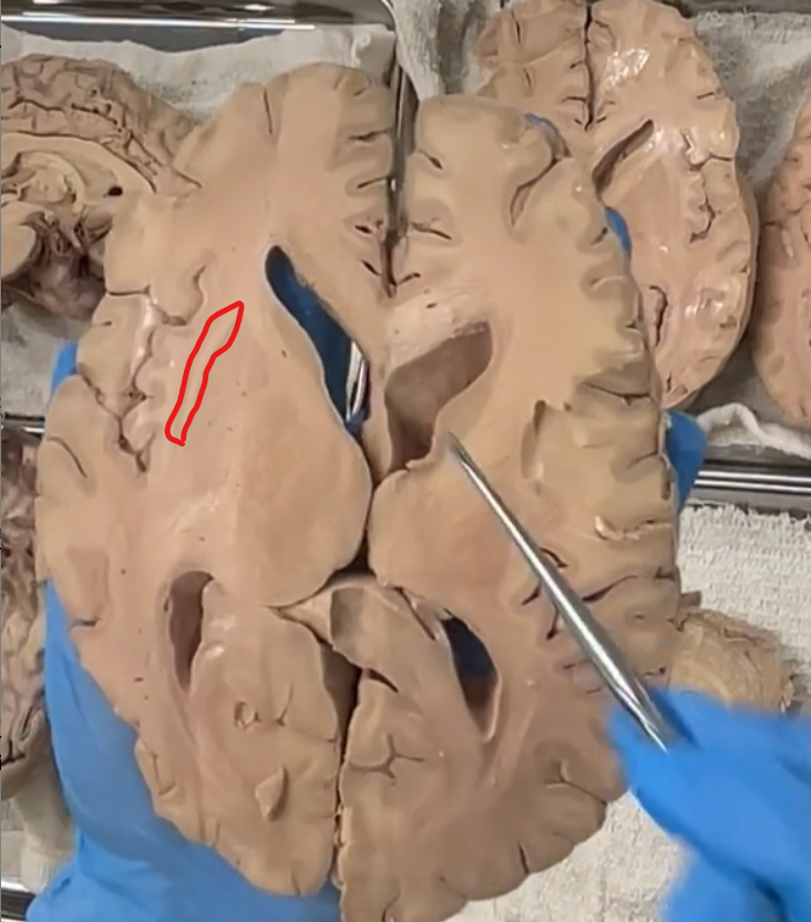
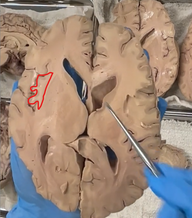
lateral to the external capsule is the collustrum.
the insular cortex is the outer of the collustrum.
medial to the globus pallidus is the thalamus.
third ventricle

horizontal section of the cerebellum at the level of the rostral medulla.
the only cerebellar nucleus we are able to see with the naked eye is the dentate nucleus.
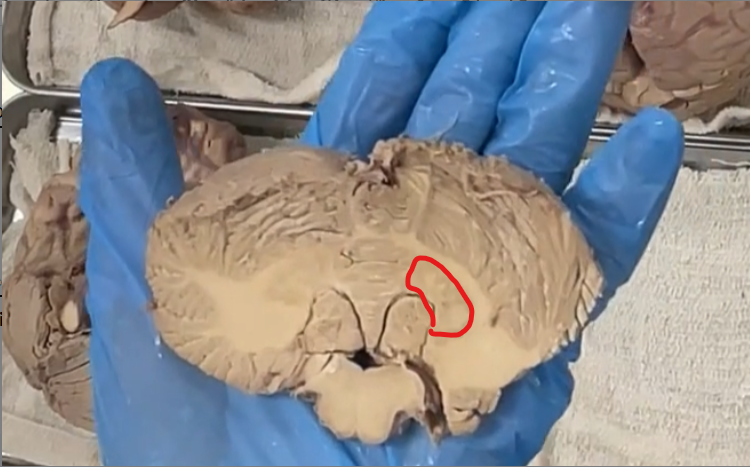
“dont eat greasy foods” this refers to the cerebellar nuclei as we work our way from lateral to medial.
Supplemental image so we can talk about the cerebellar nuclei.
The swiggly green structure is the dentate nucleus that we were able to see grossly.
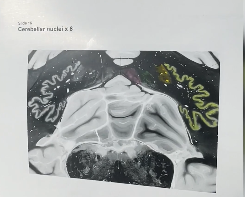
working our way medially, this is the “eat” part of “dont eat greasy foods”.
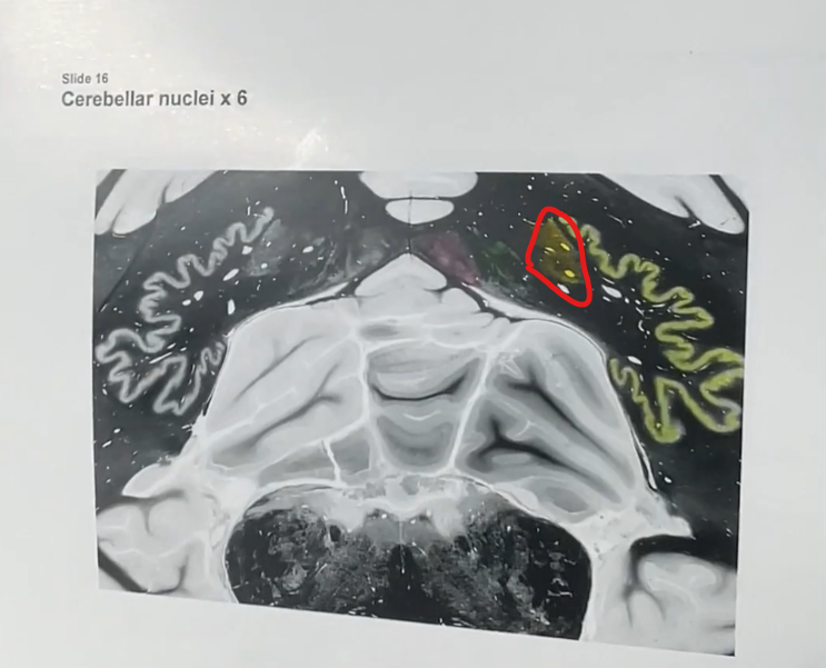
Circled in red is the emboliform nucleus of the cerebellum.
more medially is the globos nucleus is green, “greasy” of “dont eat greasy foods”.
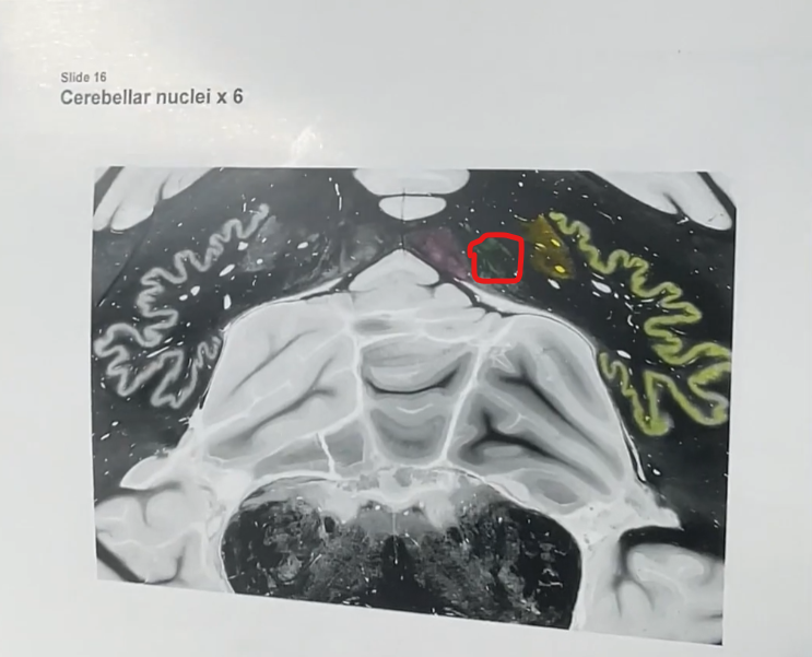
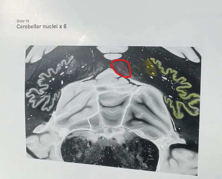
the most medial part, the "foods” in “dont eat greasy foods” is the vestigial nucleus.