development of the gi tract
1/17
There's no tags or description
Looks like no tags are added yet.
Name | Mastery | Learn | Test | Matching | Spaced | Call with Kai |
|---|
No analytics yet
Send a link to your students to track their progress
18 Terms
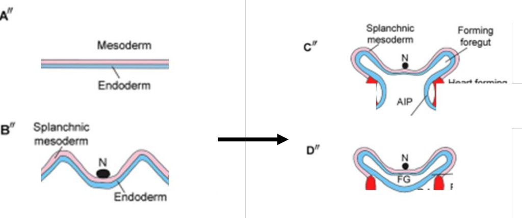
development of the gut tube
endoderm folds and elevates. splachnic mesoderm comes from the LPM
endoderm fold around ventrally. endoderm fuses ventrally
what is the primitive gut tube divided into
foregut
mid gut
hind gut
which membrane is in the oral and anal region
oral- buccopharyngeal
anal- cloacal
layers of the gut- what do endoderm and splanphic mesoderm form
endoderm- epithelial cell adjacent to lumen
splanchnic mesoderm- connective tissue, smooth muscle and mesothelial layers (mesentery and peritoneum)
failure to perforate membranes
persistent buccopharyngeal membrane in human- feeding is impossible, can perforate to treat
persistent cloacal membrane in cow, must be perforated
foregut
forms oesophagus, stomach, liver, gallbladder, bile ducts, pancreas, proximal duodenum
must rotate and twist to elaborate into these structures
rotation of the foregut
what does the greater and lesser omentum contain
what can developmental defects of the omentum causse
greater omentum contains gastrophrenic, gastrocolic, gastrosplenic ligaments
lesser omentum contains hepatophrenic and hepatooesophageal ligaments
developmental defects of the omentum can cause internal hernia
development of the spleen
what type of cells does it form from and what is it also known as
what is it in foetal stages
forms from mesodermal cells in the dorsal mesentery- also called mesogastrium
is a major haemopoetic tissue in foetal stages
what does the midgut consist of
distal half of duodenum
jejunum
ileum
cecum
ascending colon
proximal half of transverse colon
the equine caecum
hindgut fermenter
continues to grow in embryo
in adults roughly 1m in length with a 30L capacity
species differences in ascending colon
horse
ruminant and pig
horse- ascending colon continues to grow forming loop connected by remnants of dorsal mesentery
ruminant and pig- ascending colon continues to grow forming spiral
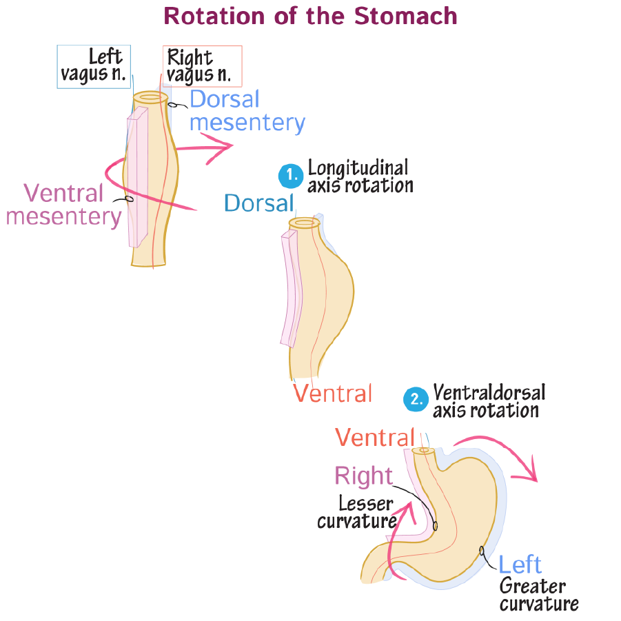
rotation of the stomach
what does it begin as
what attaches it to the body walls
what lies on the ventral and dorsal surfaces
what happens first and what does it do the the other structures
at the end what is the ventral and dorsal mesentery attachment
begins as a spindle shaped tube
ventral and dorsal mesenteries attach tube to body walls
branches of the left and right vagus nerves lie on ventral and dorsal surfaces
90 degree clockwise rotation along the longitudinal axis then occurs
this alters the course of the vagus nerve branches: right now innervates the ventral surface and left lies on the dorsal aspect
as the stomach rotates along the ventral dorsal axis, the caudal end is displaced towards the right and cephalic end to the left
ventral and dorsal mesenteries also displaced to right and left respectively
lesser curvature is the ventral mesentery attachment
greater curvature is the dorsal mesentery attachment
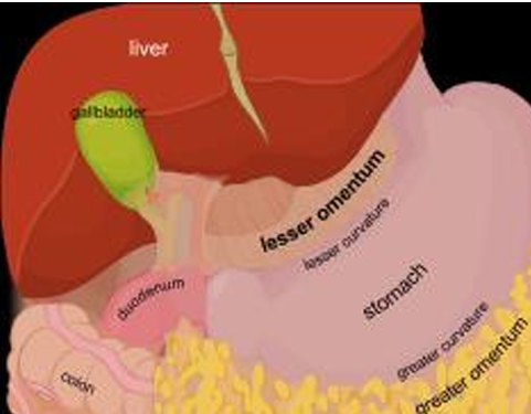
what does the ventral and dorsal mesentery give rise to
ventral to falciform ligament which secures the liver ventrally
also the lesser omentum which connects the liver and stomach and proximal duodenum
dorsal mesentery gives rise to the greater omentum which is an apron like fold of mesentery that attaches to the greater curvature of the stomach and drapes over the small intestine
final arrangement of the biliary and pancreatic ducts step one
what arise as outgrowths of the foregut
what extend ventrally and dorsally
what do liver buds outgrow into
what happens to the proximal duodenum
abdominal accessory digestive organs arise as outgrowths of foregut prior to stomach and duodenal rotation
dorsal pancreatic duct extends dorsally
ventral pancreatic duct, gallbladder and liver buds extend ventrally
liver buds develoop as an outgrowth of the foregut into the septum transversum
proximal duodenum rotates clockwise
part 2
what happens to the dorsal and ventral pancreatic buds and ducts
then where does it go and exttnd
what does the ventral pancreatic bud form
where does the main and accessory pancreatic duct drain into and how
what does the pancreatic duct join to and why
how does the common bile duct form
what direction does the common bile duct wrap around the duodenum
what does it join with
dorsal and ventral pancreatic buds and their ducts fuse
the now formed complete pancreas nestles in the c curve of the duodenum and extends toward the left side of the body
the uncinate process is the portion derived from the ventral pancreatic bud
main pancreatic duct empties into the duodenum via major papilla
accessory drains via minor
the bile and pancreatic ducts join to drain bile and pancreatic juices at the major papilla
liver drains bile into hepatic duct
hepatic and cystic ducts merge to form the common bile duct
due to the rotation of foregut and displacement of papillae the common bile duct wraps posteriorly around the duodenum
it joins with the main pancreatic duct to drain bile at the major papilla to the duodenum
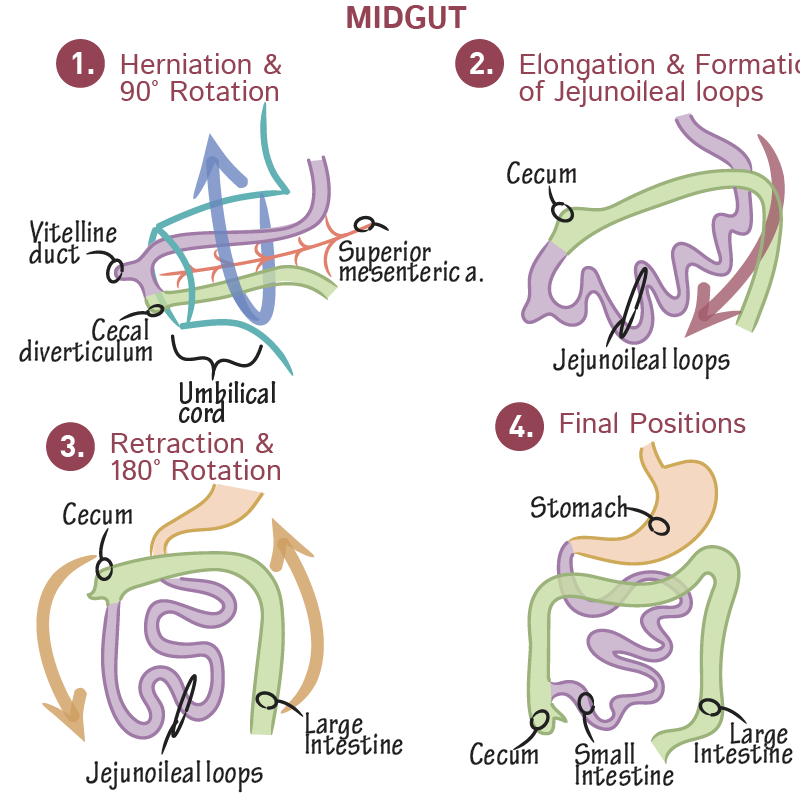
what are the four steps in midgut development
herniation and counterclockwise rotation: counterclockwise rotation about the axis of the superior mesenteric artery. midgut comprises the primary intestinal loop which elongates so rapidly that it temporarily outgrows the abdominal cavity, herniating into the umbilical cord
elongation: the caecum is now on the right hand side, as it has undergone a 270 degree rotation now (in pigs this is 450) . the rotated primary loop elongates rapidly creating the jejunoileal loops
retraction and counterclockwise rotation: loops retract from the umbilical cord into the abdominal cavity, and the intestines rotate 180 degrees counterclockwise as they continue to elongate. caecum now in upper right.
final positions: large intestine frames the small intestine. caecum now in the lower right
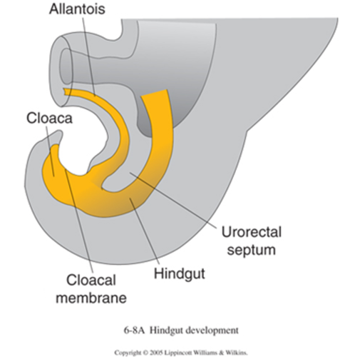
3 steps of hindgut development
what closes off the cloaca
what forms between the allantois and the hindgut
what grows towards the cloacal membrane and what does it separate
what happens to the cloacal membrane
what separates the urogenital and anal membranes
cloaca: initially the cloaca is the common end of the hindgut and urogenital tract. the cloacal membrane (endoderm and ectoderm) close off the cloaca. the urorectal septum forms between the allantois and hindgut
urorectal septum: urorectal septum grows towards the cloacal membrane. it now separates the primitive urogenital sinus which appears as an anterior swelling. cloacal membrane ruptures before the urorectal septum reaches
perineal body: represents the tup of the urorectal septum, separates the urogenital and anal membranes. now completely divides the urinary and digestive tracts so that the urinary bladder lies anteriorly and anorectal canal remains posteirorly