Exam 3 Part B
1/63
There's no tags or description
Looks like no tags are added yet.
Name | Mastery | Learn | Test | Matching | Spaced | Call with Kai |
|---|
No analytics yet
Send a link to your students to track their progress
64 Terms
Nervous System (NS)
the fast-acting internal system of communication involving sensory receptors, networks of nerve cells, and connections to muscles and glands that respond to nerve signals
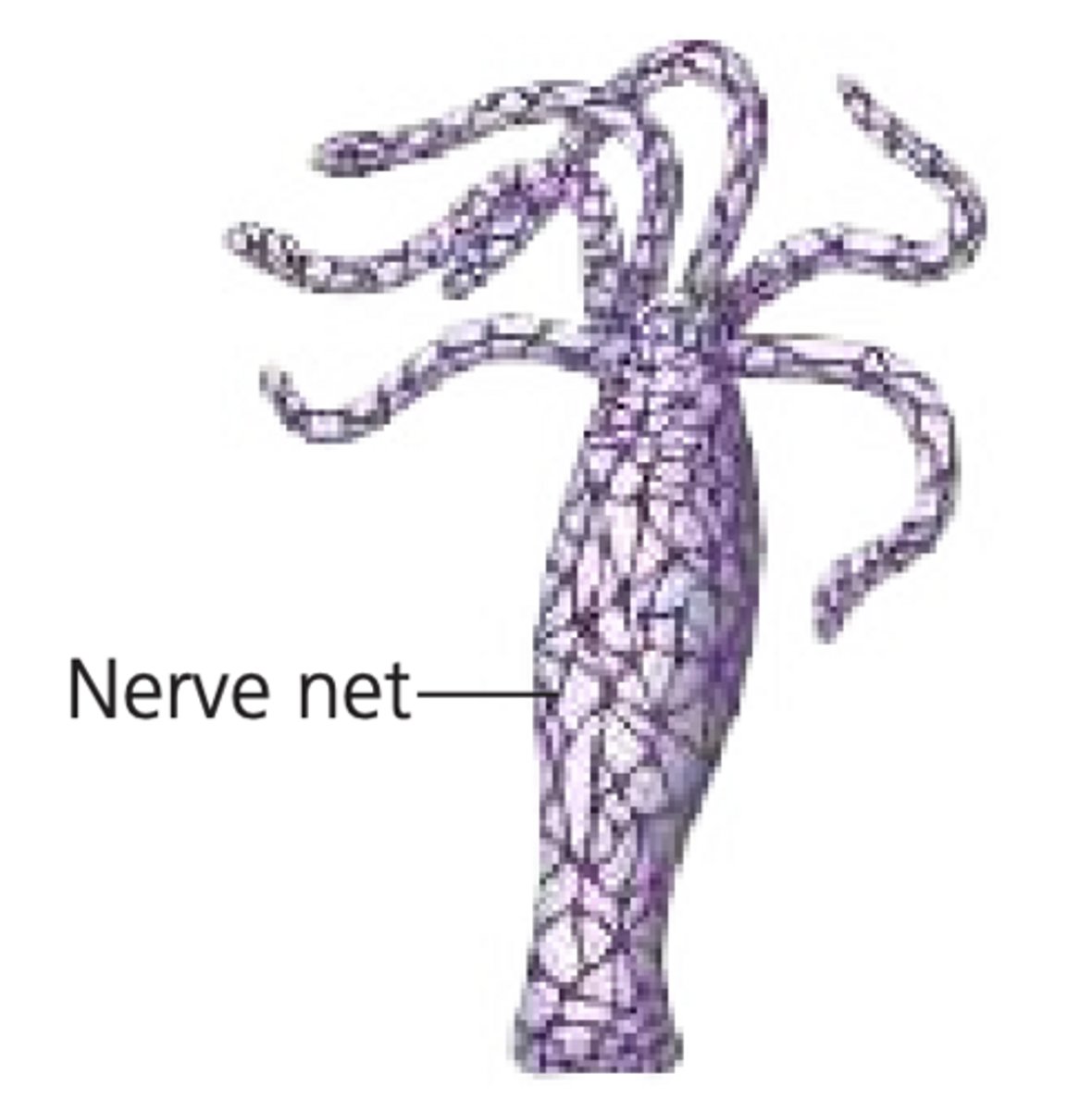
Cnidaria
their nervous system is diffused -> aka a nerve net
-ex: hydra
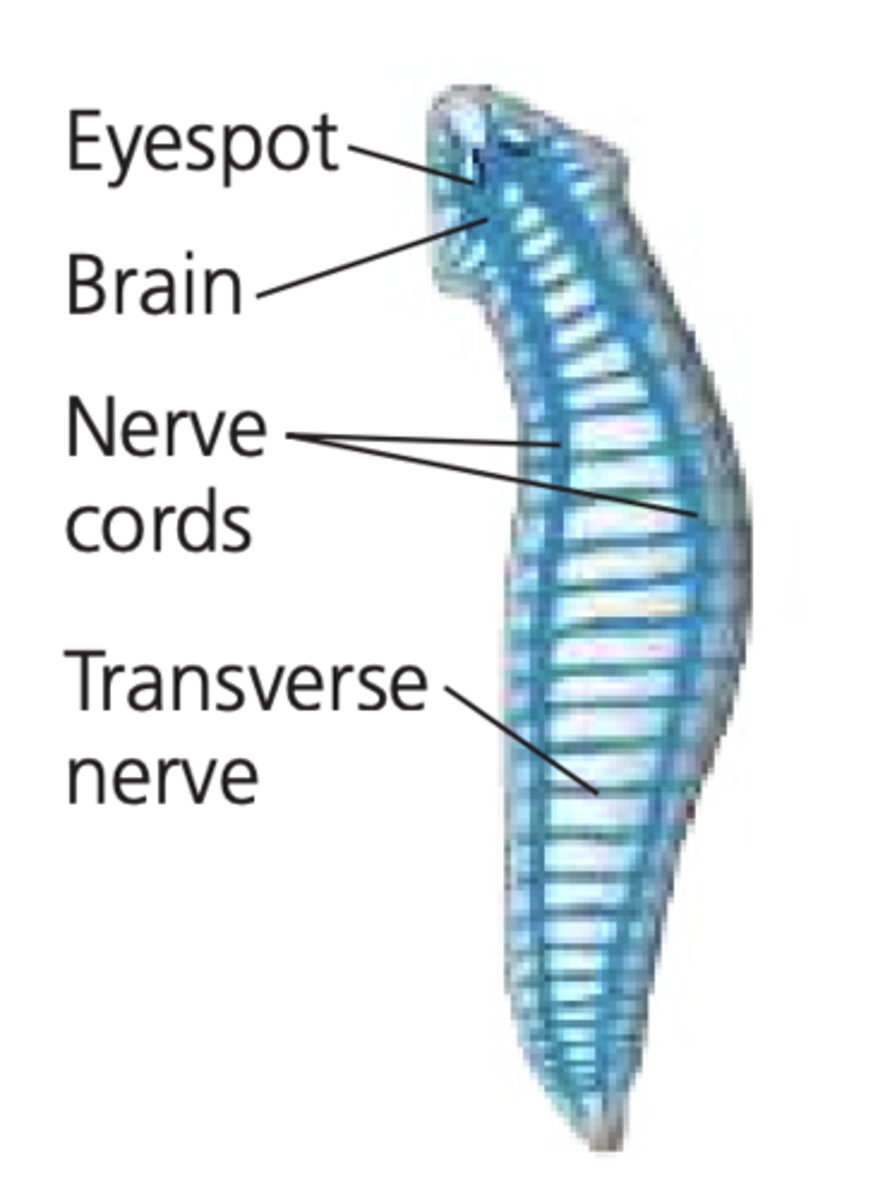
Platyhelminthes
1. pair of ganglia (a group of cell bodies) = the brain
2. 2 nerve cords joined by cross connections
3. cephalization
4. bilateral symmetry
-ex: planaria

Cephalization
formation of a head region
Bilateral Symmetry
an organism in which one plane divides the body into 2 halves that are mirror images of e/o

Peripheral Nervous System (PNS)
1. sends info to CNS
2. transmits messages from CNS to effectors
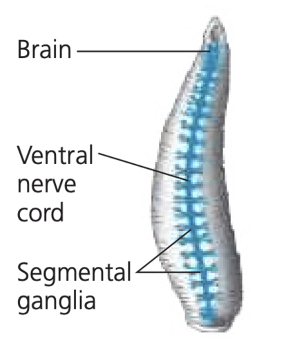
Annelids
1. pair of ganglia (brain) (above pharynx)
2. pair of connectives (arond pharynx)
3. ventral nerve cord with ganglia in ea. segment
4. lateral branching nerves
-ex: earthworm
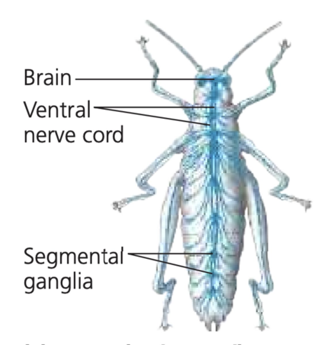
Arthropods
1. pair of ganglia (brain) (above esophagus)
2. pair of connectives
3. double ventral nerve cord with abdominal ganglia
4. lateral branching nerves
-ex: insect
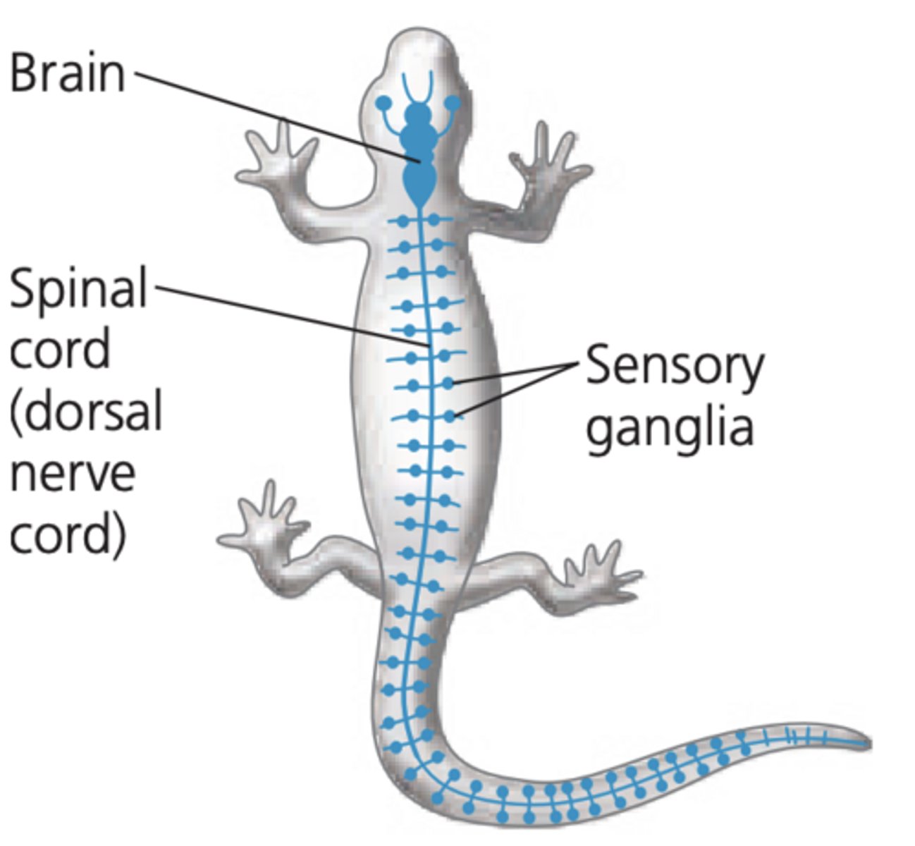
Vertebrates
1. brain -> cranial nerves
2. spinal cord -> spinal nerves
Human Nervous System
1. CNS (brain & SC)
2. PNS (cranial & spinal nerves)
a. Somatic NS (skin + eyes)
i. sensory (afferent)
ii. motor (efferent)
b. Visceral NS (int. organs)
i. sensory
ii. motor/autonomic NS
-parasympathic NS
-sympathetic NS
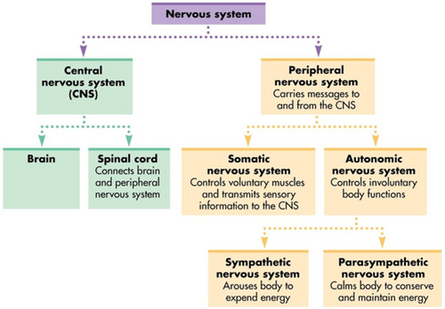
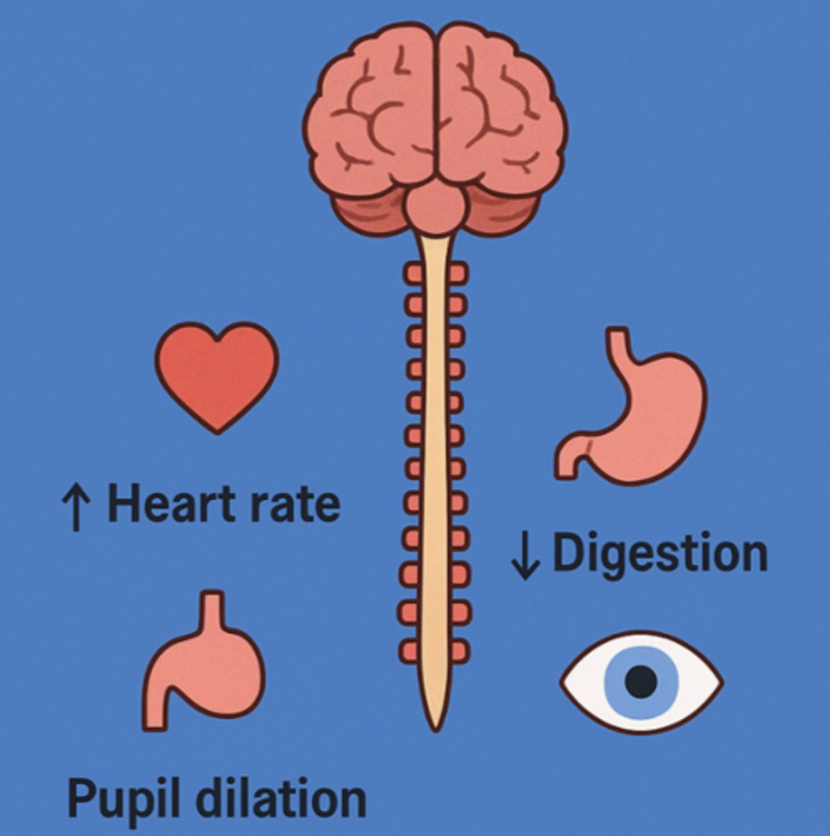
Sympathetic NS
fight or flight; arousal & energy generation
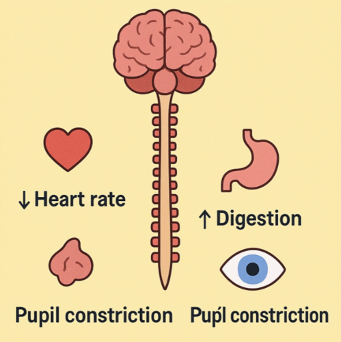
Parasympathetic NS
rest & digest; calm; return to self-maintainance function
How does the NS coordinate activities in the animal body?
it processes information and determines an appropriate response
Stiimulus & NS
1. stimulus goes to receptor on sensory (afferent) neuron
2. stimulus crosses synapse to one of many interneurons
3. stimulus crosses synapse to motor (efferent) neuron
4. stimulus crosses synapse to effector (muscle or glands)
5. response
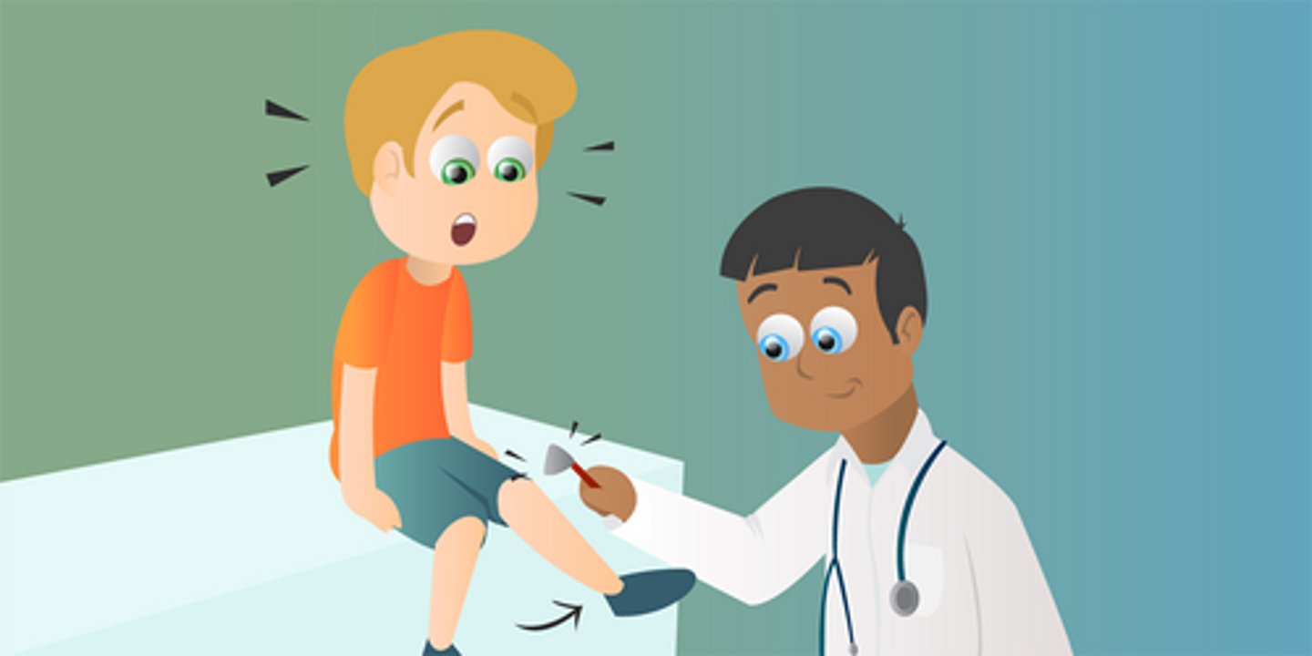
Reflex Action
super fast response to certain stimuli
-ex: placing your hand on a hot stove
Complex Neural Pathways
involve transmission of impulses to and from the brain
Forebrain
activities that include processing of olfactory input, regulation of sleep, learning, and any complex processing
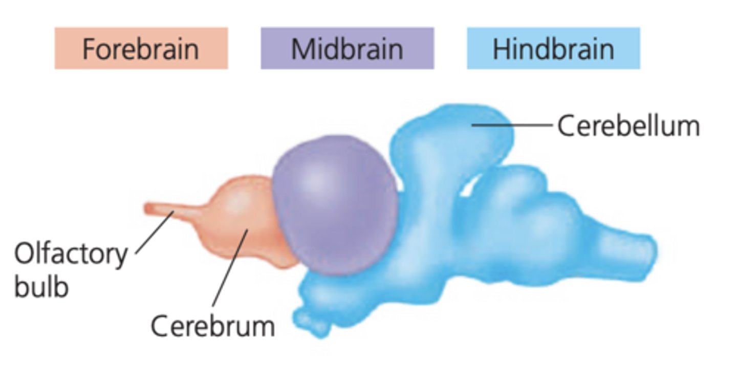
Midbrain
coordinates routing of sensory input
-connects the forebrain and the hindbrain

Hindbrain
controls involuntary activities, such as blood circulation, and coordinates motor activities, such as locomotion

Cerebrum
-controls skeletal muscle contraction
-center for learning, emotion, memory, & perception
-divided into R & L cerebral hemispheres
Cerebral Cortex
-outer layer of the cerebrum
-perception, voluntary movement, and learning
-contralateral organization
Corpus Callosum
a thick band of axons that enables the R & L cerebral hemispheres to communicate
Thalamus
the main relay center thru which sensory info passes to the cerebrum
Hypothalamus
regulates homeostasis and basic suvival behaviors
Suprachiasmatic Nucleus (SCN)
group of neurons in the hypothalamus that acts as the pacemaker for circadian rythms
Posterior Pituitary
stores & releases hormones
Pons and Medulla Oblongata
contains pathways for information traveling btwn the PNS and the cerebrum
Medulla Oblongata
the "vital center" of the brain
-breathing & heart rate
Cerebellum
helps coordinate motor functions
Brainstem
includes the midbrain, pons, and medulla oblongata
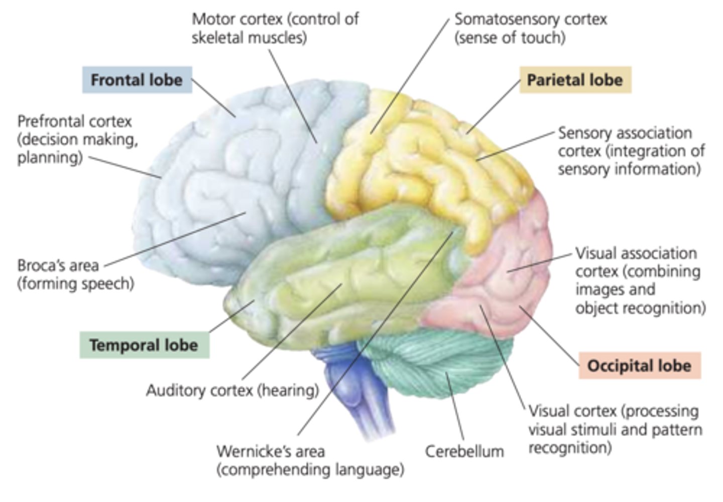
Frontal Lobe
a. prefrontal cortex - decision making & planning
b. motor cortex - control of skeletal muscles

Broca's Area
speech formation

Parietal Lobe
a. somatosensory cortex - sense of touch
b. sensory association cortex - integration of sensory info

Occipital Lobe
a. visual association cortex - combining images & object recog.
b. visual cortex - processing visual stimuli & pattern recog.

Wernicke's Area
language comprehension

Temporal Lobe
a. auditory cortex - hearing
Receptor
specialized peripheral ending of an afferent neuron, or seperate cell intimately associated with it, that detects changes in some aspect of the environment
Classification of Receptors
receptors are classififed based upon the stimuli they respond to
Types of Mechanoreceptors
a. touch and pressure receptors in the skin
-gentle pressure, strong pressure, or gentle vibrations
b. stretch receptors in muscles and tendons
c. haor cells in the ear
Muscle Spindles
provides feedback about the amount and rate of muscle stretch
Golgi Tendon Organs
signal the force that develops in the tendon on muscle contraction
Photoreceptors
respond to light
Chemoreceptors
respond to chemicals
a. taste receptors (taste buds (modified epithelial cells))
b. olfactory receptors (located in olfactory epithelium)
Thermoreceptors
respond to warm and cold
-free nerve endings in the skin
Nociceptors (pain receptors)
respond to excess heat, P, or specific chems
-free nerve endings in the skin
-ex: harmful stimuli that could cause tissue damage
What is within the cochlea?
the organ of corti which contains hair cells
Cupula
structure within the semicircular canals that detects rotational head movement
Rods
detect black, white, and shades of gray (dim light)
Cones
detect color, bright light, and fine detail
Fovea
highly concentrated areas of cones
-site of greatest detail in vision
Optic Disk
aka the blindspot; no photoreceptors present
Motor Mechanisms
-motor neurons of somatic NS -> voluntary muscle (skeletal muscle)
-motor neurons of visceral NS (autonomic NS) -> involuntary muscle (smooth & cardiac muscle) and glands
Types of Effectors
1. Muscles (skeletal, smooth, and cardiac)
2. Glands (oil & digestive)
What stimulates the muscle?
the nervous system stimulates the muscle (this generates a force)
What part of the nervous system stimulates the muscle?
motor neuron but depends on the type of muscle
What are muscles composed of?
muscle fibers or cells that produce myofibrils
Sarcomere
contractile unit of muscle
What happens to sarcomere when the muscle contracts?
myosin globular heads pull the thin (actin) filaments inwards
Sliding-Filament Theory
1. ATP binds → myosin head detaches (low-E)
2. ATP hydrolysis (ADP + Pi) → myosin head = high-E
3. Myosin binds actin → cross-bridge forms
4. ADP + Pi released → thin filament moves twd the center of the sarcomere
Flexion
biceps contracting; triceps relaxing
Extension
biceps relaxing; triceps contracting
Endoskeleton
internal skeleton
How many bones in an adult human?
206
Glands
groups of cells specialized to secrete a substance released from the cells
-ex: oil glands in the skin; glands in the small intestine that release digestive secretions